Method and apparatus for measuring biopotential and mapping ephaptic coupling employing a catheter with mosfet sensor array

a technology of mosfet sensor array and biopotential measurement, which is applied in the field of methods and apparatus for navigating and recording electrical characteristics of electrophysiological signals, can solve the problems of increasing complexity of arrhythmias, increasing the difficulty of detecting ephaptic coupling, and presenting a major challenge, so as to improve the modeling of cellular electrical activity

Inactive Publication Date: 2014-03-20
NEURO KINESIS CORP
View PDF11 Cites 45 Cited by
  • Summary
  • Abstract
  • Description
  • Claims
  • Application Information

AI Technical Summary

Benefits of technology

[0008]The need to improve modeling of cellular electrical activity is central to physiology and electrophysiological studies. Biopotential recording and mapping of such electrical activity enables the physician or researcher to form and fashion his or her understanding of the fundamental data gathering and analysis of such diverse biological activities as sensory perception, communication between neurons, initiation and coordination of skeletal-muscle contraction, synchronization of the heart beat, and the secretion of hormones.

Problems solved by technology

Nevertheless, there is a great and still unsatisfied need for an apparatus and method for measuring biopotential activity with the use of a MOSFET sensor.
However, ablation of more complex arrhythmias, including some atrial tachycardias, many forms of intra-atrial re-entry, most ventricular tachycardias, and atrial fibrillation, continues to pose a major challenge.
This challenge stems in part from the limitations of fluoroscopy and current napping catheter construction and sensory apparatus to locate accurately both the geometry and time domain of the wavefront activity generated by the “avalanche” of the cellular excitable matrix.
The inability of the current electrode technology to account for the cellular biopotential transfer with the resolution and the ionic transfer time depicting the actual energetic event is insufficient in the measurement as well as representing the “avalanche” dynamics of this bioenergetic event.
This is the main drawback of the existing and prior art relating to “electrode technology”.
In summary, the problem of reconstruction of the electrophysiological activity in the prior art is two-fold: At the one hand it is the results of the use of electrodes and its associated electrical circuit design, and on the other hand it is further handicapped by modeling the biopotential activity as a physical phenomenon whereby excitable cells are modeled by employing the “cable theory” with isotropic behavior.
The use of electrodes and cable theory is a good approximation of idealized conditions of such energetic events, but suffers from the inability to associate accurately the intracardiac electrogram with a specific endocardial site which also limits the reliability with which the roving catheter tip can be placed at a site that was previously mapped.
This results in limitations when the creation of long linear lesions is required to modify the substrate, and when multiple isthmuses or “channels” are present.
Additionally, since in conventional endocardial mapping a single localization is made over several cardiac cycles, the influence of beat-to-beat variability on overall cardiac activation cannot be known.
These assumptions, however, may not hold in many systems of biological significance, especially in the central nervous system and cardiac tissue, where micro-histological features may play an essential role in shaping physiological responses.
These methods have limited success in reaching and reporting on an electro-anatomical site and reduces the ability of the operator to provide a clinically optimal resolution to the problem of identifying accurately the source and its location and further, the prior methods and their exemplified apparatus cannot achieve such precision, and hence results in suboptimal successes of remodeling the heart electrical signal propagation, as well as neuromodulation and their intended clinical outcomes.
It is clear to those familiar with the art of electrophysiology mapping, that methods using electrode technologies of all different combinations as noted by the prior art, suffer from the inability to differentiate between signals emanating from “near” and “far fields” as the electrodes in the prior art are typically made of a metal-electrolyte interface.
The difficulty in the prior art of measurement and mapping procedures mainly relates to the degree with which the measuring tools interfere with the measured bioelectric fields and signals, thus affecting the fidelity of the boundary conditions from which the modeling and diagnostic maps are generated.
Other numerical solutions may employ a known differential equation which results in a minimum error for the boundary conditions.
In one embodiment, however, using any of these methods requires accurate boundary condition measurements which produce minimum error due to the measurement itself.

Method used


Image

  • Method and apparatus for measuring biopotential and mapping ephaptic coupling employing a catheter with mosfet sensor array
  • Method and apparatus for measuring biopotential and mapping ephaptic coupling employing a catheter with mosfet sensor array
  • Method and apparatus for measuring biopotential and mapping ephaptic coupling employing a catheter with mosfet sensor array

Examples


Embodiment Construction

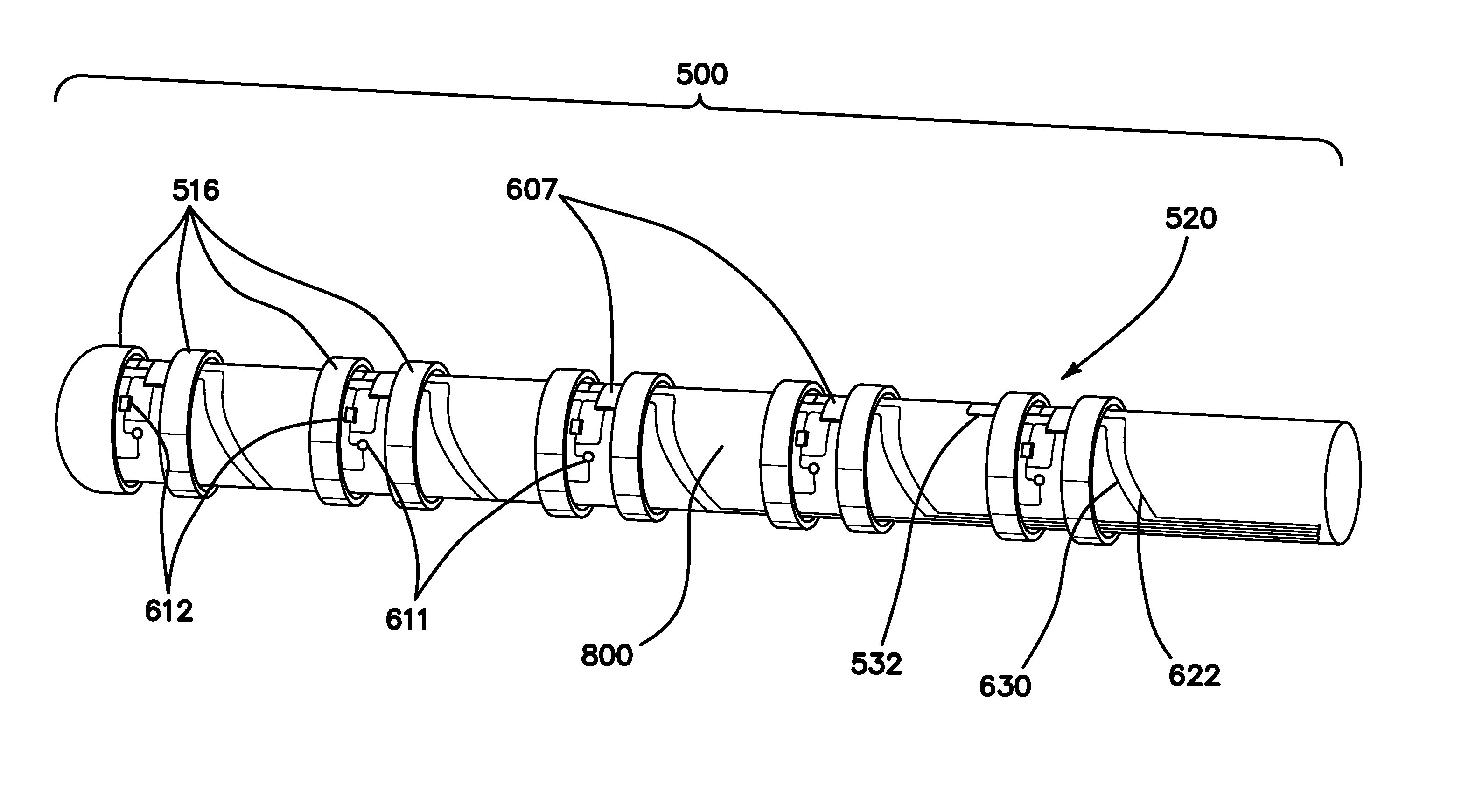
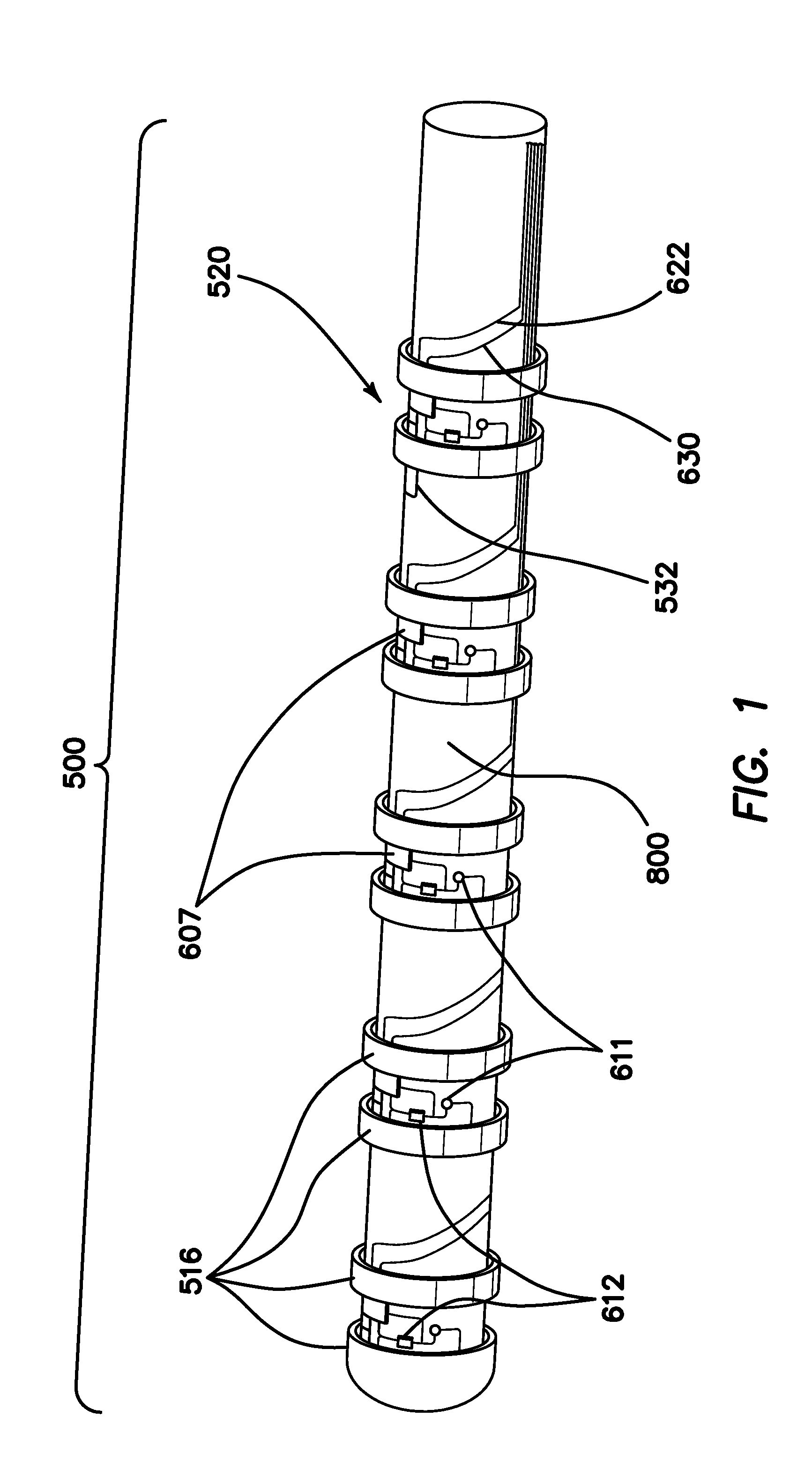
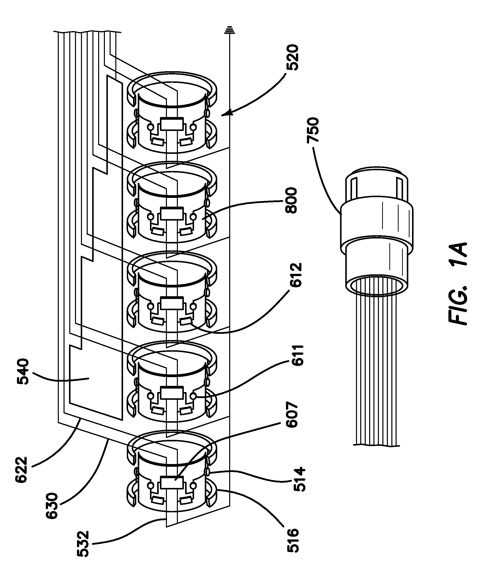
[0071]FIG. 1 is an autographic representation of a decapolar catheter formed with the disclosed MOSFET Sensor Array 500 for measuring bioelectric potential for example, while performing electrophysiological studies within the endocardial tissue. The assembly is comprised of a series of typical electrode rings 516 shown in a decapolar configuration. This layout can be configurable in any combination suitable to those familiar with the art while employing the device while measuring bioelectric potential in a biological media or environment. Electrodes 1A 516 are coupled to a sensor module 520 including a capacitor C1A1611 and resistor R1A2612 which form an electrical unit coupled to a MOSFET 607. The discrete electronic components are mounted on a flexible substrate such as anisotropic conductive film (ACF), mechanically reinforced by the catheter structure 800 formed of polyurethane or silicone, etc. materials commonly used in such construction. The electrical circuit is formed with ...


PUM


Abstract

This invention relates generally to electro-anatomical mapping method and an apparatus using a catheter and more particularly to a mapping catheter having an embedded MOSFET sensor array for detecting local electrophysiological parameters such as biopotential signals within an excitable cellular matrix geometry, for determining physiological as well as electrical characteristics of conduction path and its underlying substrate within the endocardial and epicardial spaces, the arterial structure and in ganglionic plexus. The apparatus with its MOSFET sensor is geometrically configured as a decapolar linear array and optionally with an 8×8 sensor matrix placed on a balloon-like structure.

Description

FIELD OF THE INVENTION[0001]The invention relates to the a method and apparatus for navigating and recording electrical characteristics of electrophysiological signals using a MOSFET sensor guided by a magnetically-deployable mechanism.BACKGROUND[0002]Apparatus for magnetically deployable catheter with MOSFET sensor and method for mapping and ablation,” U.S. Pat. No. 7,869,854, filed on Feb. 23, 2006, published as U.S. Pat. Application No. 2007 / 0197891, assigned to Magnetecs Corporation, which discloses a MOSFET sensor guided by a magnetically-deployable mechanism, is incorporated herein by reference. Corresponding applications include EPO Patent No. 07751190.5; Canadian Patent No. 2637622; and Hong Kong Patent No. 09104053.7.[0003]Nevertheless, there is a great and still unsatisfied need for an apparatus and method for measuring biopotential activity with the use of a MOSFET sensor. This application further improves the efficacy and safety of the procedure by enabling an accurate d...

Claims


Application Information

Patent Timeline
Patent Type & Authority Applications(United States)
IPC IPC(8): A61B5/04A61B5/0478A61B5/308
CPCA61B5/0478A61B5/04001A61B5/6858A61B5/283A61B5/291A61B5/24A61B5/302
Inventor SHACHAR, YEHOSHUAGANG, ELI
Owner NEURO KINESIS CORP
Why Eureka
Try Eureka