Emboli detection in the brain using a transcranial doppler photoacoustic device capable of vasculature and perfusion measurement

a technology of transcranial doppler and photoacoustic device, which is applied in the field of medical imaging, can solve the problems of cerebral ischemia or stroke, many current transcranial imaging devices are severely limited, and the cost of brain blood vessel imaging by magnetic resonance and computed tomography is high, so as to improve the accuracy of diagnosis and treatment, the effect of rapid determination of stroke and related brain insults and injuries

Inactive Publication Date: 2014-07-10
CEREBROSONICS L L C
View PDF33 Cites 98 Cited by
  • Summary
  • Abstract
  • Description
  • Claims
  • Application Information

AI Technical Summary

Benefits of technology

[0067]The present inventors have discovered that mapping blood flow in the brain, including by velocity, enables rapid determination of stroke and related brain insults and injuries. Devices, system and apparatus using such blood flow mapping (captured as resultory medical data) enable rapid classification, transport and selection of treatment options for patients.

Problems solved by technology

Brain blood vessel imaging by magnetic resonance and computed tomographic imaging is expensive (thousands of dollars) and not always reimbursable or accessible.
Additionally, many current transcranial imaging devices are severely limited by the aberrations caused by the skull or intervening tissues.
Both acute and chronic conditions may result in cerebral ischemia or stroke.
However, only 27.4% of patients that are given tPA receive this within the first hour of arriving at the emergency room but in addition to the time of onset and transport emergently; therefore, because of the delays, outcome may be compromised and death risk increased.
There are many logistical and other issues that lead to late delivery or no delivery of TPA to eligible stroke patients within the accepted time windows for treatment.
First, a complex set of events and steps need to occur.
The risk of hemorrhage from clot buster increases in patients with obstruction by acute clot in major brain arteries, including the middle cerebral arteries and the basilar artery.
A stroke that appears on CT may represent a completed stroke and therefore, thrombolytic therapy is not warranted as there is little chance of recovery of the dead tissue and brain hemorrhage risk is increased in this context if thrombolytics are given.
However, this exam may not be available rapidly at many centers.
However, this may be more time consuming due to equipment availability and the time required to perform the test.
The choice between CT perfusion or MR perfusion is variable across stroke centers and may not be available.
Although little time is added by these studies, processing of the images may lead to delays.
These devices or intra-arterial clot buster may lead to reopening or recanalization of acutely blocked blood vessels.
Although intravenous clot buster may reduce mortality and morbidity, it is not always efficacious in removing the clot blocking a vessel.
However, the use of these mechanical reopening devices may have hemorrhage and additional stroke risk that must be balanced with potential benefits for the patient.
Rural sites may allow rapid transit to local Emergency departments, but these facilities may have a CT scanner, but do not have other specialists or diagnostic services.
However, the utility of having an expensive CT scanner in an ambulance, a neurologist or other physician in the ambulance, and the potential lack of vascular imaging may limit this approach in the United States and other venues.
However, stroke telemedicine in ambulances and the like is not routine and can be technically difficult and expensive.
Equipment is not uniformly available, has not been appropriately designed, and logistic issues have not been addressed.
Robots are too large for an ambulance.
These techniques are expensive, inaccessible to rural health care centers, and are time consuming, and the sequence of testing has been discussed above.
Hence, existing modalities to Image the brain and its blood flow require a substantial financial outlay by a healthcare provider and none, including CT and MR testing, can perform real time analysis of blood flow velocity and flow direction, detect and characterize emboli and measure vessel-wall thickness.
In addition to CT and MR testing, positron tomography (PET) and SPECT scanning may provide useful information, but are not accessible and not generally available.
Since this technique can be difficult even in expert hands, its application within the ambulance will involve transducer positioning over the necessary neck and brain blood arteries by remote monitoring and remote control with positioning for quality and maximal signals.
Patient age, gender, race and other factors affect bone thickness, making some examinations more difficult or even impossible.
This results in local heating and thus a pressure wave or sound.
Such pressure waves are in vivo, and cause mechanical disturbances of the media, and manifest themselves partially as broadband ultrasound.
With respect to photoacoustic imaging in the skull, one is limited by the scattering of ultrasound in the skull.
For skull windows, this might result in smearing of the ultrasound resolution by a typical thickness parameter of the skull window, which may be a few millimeters.
Hence, one probably will never be able to attain the same resolution in the brain with ultrasound as one does compared to targets under comparable distances of flesh, but if one is searching for major arteries and veins and large structures, such as a major hemorrhage, such resolution better than a few millimeters is not useful.
It does not, however, help to counteract a decrease in blood pH.
Ventilation, or breathing, may reverse this condition by removal of carbon dioxide, thus causing a shift up in pH.
Typically, because of the much higher resolution afforded by MRI and CT scanning devices, phase array ultrasound has not been used in the brain.
However, when larger structures are imaged, such as major vasculature, and superb resolution is not desired, phased array ultrasound is adequate.

Method used


Image

  • Emboli detection in the brain using a transcranial doppler photoacoustic device capable of vasculature and perfusion measurement
  • Emboli detection in the brain using a transcranial doppler photoacoustic device capable of vasculature and perfusion measurement
  • Emboli detection in the brain using a transcranial doppler photoacoustic device capable of vasculature and perfusion measurement

Examples


Embodiment Construction

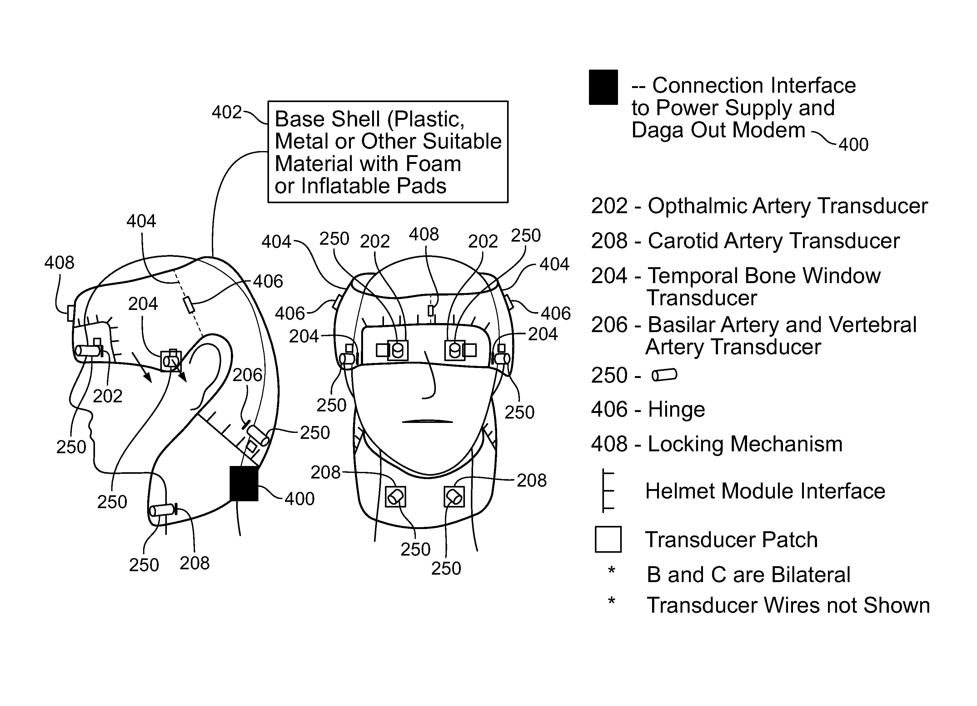
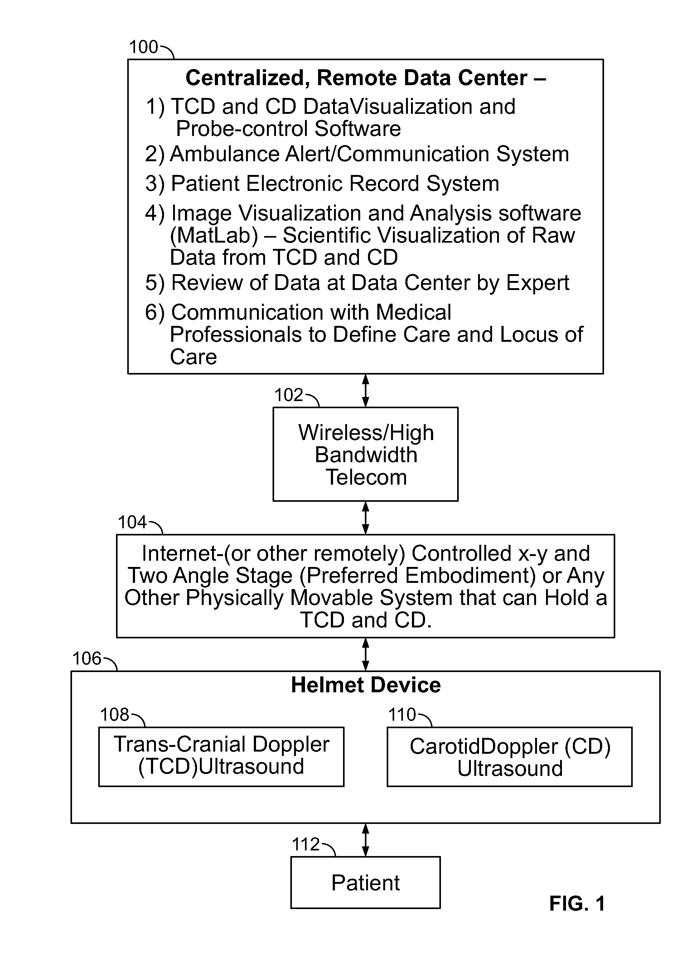
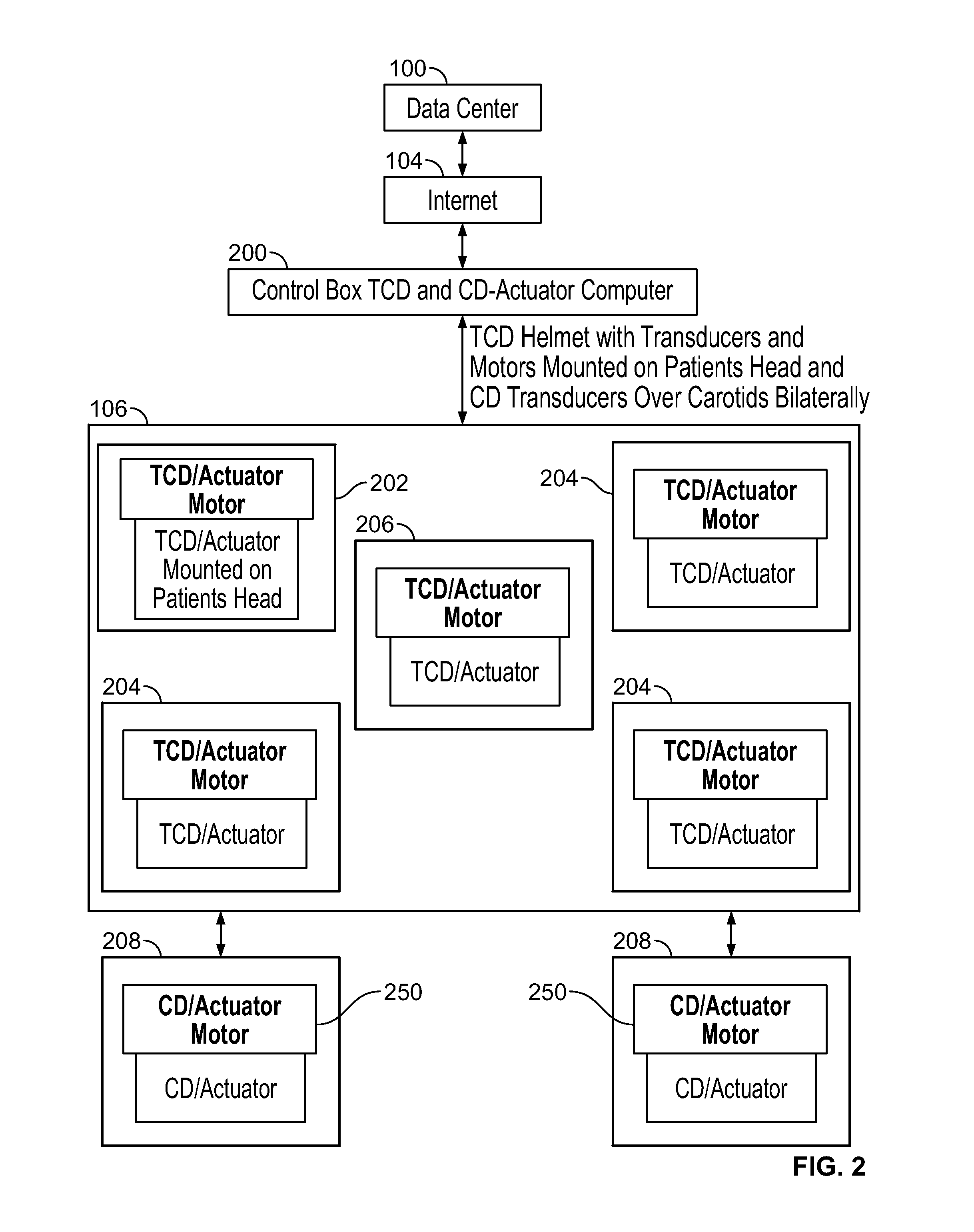
[0083]Briefly stated, a device, method, and system for detecing emboli in the brain is disclosed. The transcranial Doppler photoacoustic device transmits a first energy to a region of interest at an internal site of a subject to produce an image and blood flow velocities of a region of interest by outputting an optical excitation energy to said region of interest and heating said region, causing a transient thermoelastic expansion and produce a wideband ultrasonic emission. Detectors will receive the wideband ultrasonic emission and then generate an image of said region of interest from said wideband ultrasonic emission. A Doppler ultrasound signal will also be deployed to image the region of interest. Another embodiment Doppler would present blood flow. Additionally, a dye can be given to visualize the brain vasculature and a perfusion measurement can be made in various regions of the brain along with the transcranial Doppler and the photoacoustic screening.

[0084]Methods and appara...


PUM


Abstract

A device, method, and system for detecting emboli in the brain is disclosed. A transcranial Doppler photoacoustic device transmits a first energy to a region of interest at an internal site of a subject to produce an image and blood flow velocities of a region of interest by outputting an optical excitation energy to said region of interest and heating said region, causing a transient thermoelastic expansion and produce a wideband ultrasonic emission. Detectors receive the wideband ultrasonic emission and then generate an image of said region of interest from said wideband ultrasonic emission. A Doppler ultrasound signal will also be deployed to image the region of interest. Doppler presents changes in velocity to map blood flow. Additionally, a dye can be given to visualize the brain vasculature and a perfusion measurement can be made in various regions of the brain along with the transcranial Doppler and the photoacoustic screening. Systems are taught using resultory medical data for better triage within an enhanced stroke ecosystem.

Description

FIELD OF THE DISCLOSURE[0001]The present inventions relate to use of medical imaging to classify and support lumen-challenged patients. In particular, the instant disclosure relates to a carotid Doppler, transcranial Doppler, phased array, photoacoustic device that provides remote wireless monitoring and remote control of the sensors or transducers of this device to and from a data / stroke center, staffed by experts, and method that will produce a more complete picture of a traumatic event in the brain, for example, a stroke, or cerebrovascular accident (CVA) or predilection to said condition. In the acute care setting of stroke and particularly in the prehospital situation for patients, in ambulances, helicopter, or airplanes, this invention will aid neurologists, radiologists and stroke teams by simultaneously obtaining rapid blood velocity measurements in neck vessels and brain vessels, determination of neck and brain large blood vessel, acute blockage or narrowing, and obtaining ...

Claims


Application Information

Patent Timeline
Patent Type & Authority Applications(United States)
IPC IPC(8): A61B8/06A61B8/08
CPCA61B8/582A61B5/6803A61B8/06A61B8/085A61B8/488A61B8/4227A61B8/565A61B8/0808A61B5/4064A61B5/0095A61B5/0042
Inventor STEIN, STUARTPENNYPACKER, CARL
Owner CEREBROSONICS L L C
Why Eureka
Try Eureka