Methods and compositions to treat myocardial conditions

a myocardial infarction and composition technology, applied in the field of myocardial infarction treatment, can solve the problems of sudden death in adults, blockage of blood flow, and imbalance between myocardial blood flow and metabolic demand of the myocardium, so as to prevent remodeling, reduce mechanical stress, and strengthen the infarct region

Inactive Publication Date: 2015-01-15
ABBOTT CARDIOVASCULAR
View PDF2 Cites 10 Cited by
  • Summary
  • Abstract
  • Description
  • Claims
  • Application Information

AI Technical Summary

Benefits of technology

[0026]Embodiments herein relate to methods, apparati and compositions for reversing ventricular remodeling using electro-stimulatory therapy in combination with other methods such as structural reinforcement of the area to prevent remodeling and strengthen an infarct region. By pacing sites in proximity to the infarct with appropriately timed pacing pulses, the infarct region is pre-excited in a manner that lessens the mechanical stress to which it is subjected, thus reducing the stimulus for adverse remodeling. Reducing the wall stress of the infarct region also decreases the probability of an arrhythmia arising from the region. Another advantage obtained with resynchronizing the ventricular contraction by pre-exciting a weakened infarct region is the creation of hemodynamically more efficient contraction.
[0028]In one embodiment, a composition is described that is capable of replacing the myocardial cells and may provide reinforcement to the ventricle in addition to the pacing methods describe previously. The replacement cells consist of cells that do not trigger an immune response often seen in graft rejection. In another embodiment, a method is described to increase the compliance of a ventricle by preventing thinning of the infarct region and eliminating the cellular debris created by the infarct. A cellular bolus may be advanced through a delivery device to the infarct zone for reinforcement. In one embodiment, the cellular therapy may be combined with stimulatory therapies created by electrical and / or chemical stimuli. In other embodiments, a treatment agent is delivered via multiple small volumes to the region. These delivery methods may use imaging of the ventricular wall to guide the deposition of the treatment agent to the site of the infarct zone such as deposition of the gel-forming agents.
[0030]In another embodiment, one or more components may be delivered by microparticles harboring the component and / or therapeutic agents or growth factors. Cells that infiltrate the infarct region may be stimulated to proliferate. In other embodiments, cellular treatments may be combined with a peri-infarct treatment such as electrical stimuli by leads in contact with the infarct region that encourages new growth in the infarct area. This modulation of tissue response in the infarct zone is suitable for reinforcing the region and preventing the thinning process of the ventricular wall.
[0031]Other embodiments directed to the prevention of thinning of the infarct region of the ventricle wall are included. Treatment agents that are capable of cross-linking the existing collagen in the infarct region are described. The cross-linked collagen would form a structurally reinforcing wall in the infarct region to bulk-up the infarct zone and reduce the effects of thinning. This may be combined with the cellular replacement and / or electro-stimulatory therapy.
[0033]In another embodiment, a different multi-component treatment of the infarct zone introduces a scaffold system that provides a matrix to facilitate cell growth and attenuate the remodeling event post-MI. In addition, the treatment may include a perfluorinated compound that enhances the re-oxygenation of the tissue. These methods may also be combined with electro-stimulatory therapies in order to enhance the recovery period of a subject (e.g., a subject with heart disease).

Problems solved by technology

Ischemic heart disease typically results from an imbalance between the myocardial blood flow and the metabolic demand of the myocardium.
It is a major cause of sudden death in adults.
Thus, the flow of blood is blocked and downstream cellular damage occurs.
This damage can cause irregular rhythms that can be fatal, even though the remaining muscle is strong enough to pump a sufficient amount of blood.
Even though relatively effective systemic drugs exist to treat MI such as ACE-inhibitors and Beta-blockers, a significant portion of the population that experiences a major MI ultimately develops heart failure.
In other areas, remote regions experience hypertrophy (thickening) resulting in an overall enlargement of the left ventricle.
These changes also correlate with physiological changes that result in increase in blood pressure and worsening systolic and diastolic performance.
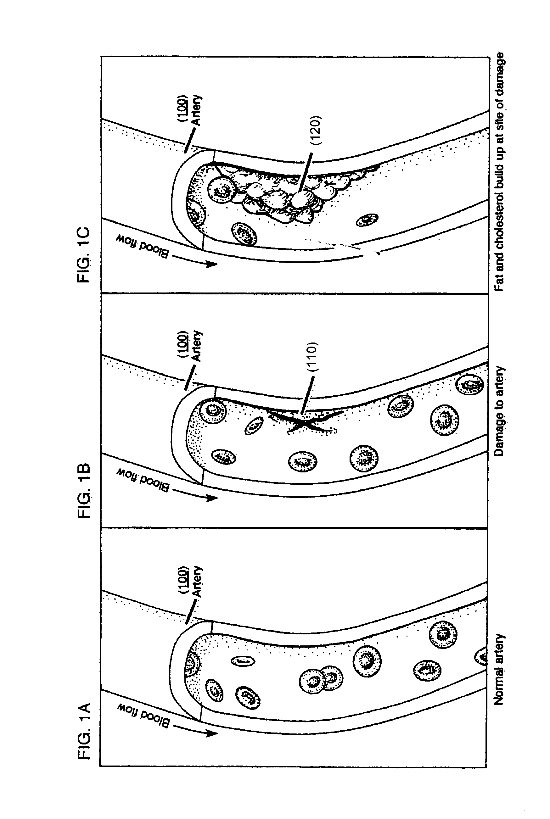
Fat and cholesterol build up at the site of damage.
Thus the T-cells, platelets, fibrin and multiple other factors and cells are blocked from progression through the blood stream and the result is an inadequate vascular supply as seen.
This leads to myocyte death.
Myocyte death, in addition to fibrosis in the form of collagen deposition, can lead to a compromised left ventricle and overload on the remaining myocytes.
This process is further complicated by compensation of the remaining myocytes that hypertrophy (enlarge).
This can cause the left ventricle to enlarge and if the cycle continues can result in eventual heart failure.
This damage may include extensions into the right ventricular wall.
This ultimately leads to fibrosis and a scar is formed by about 7 weeks post MI.
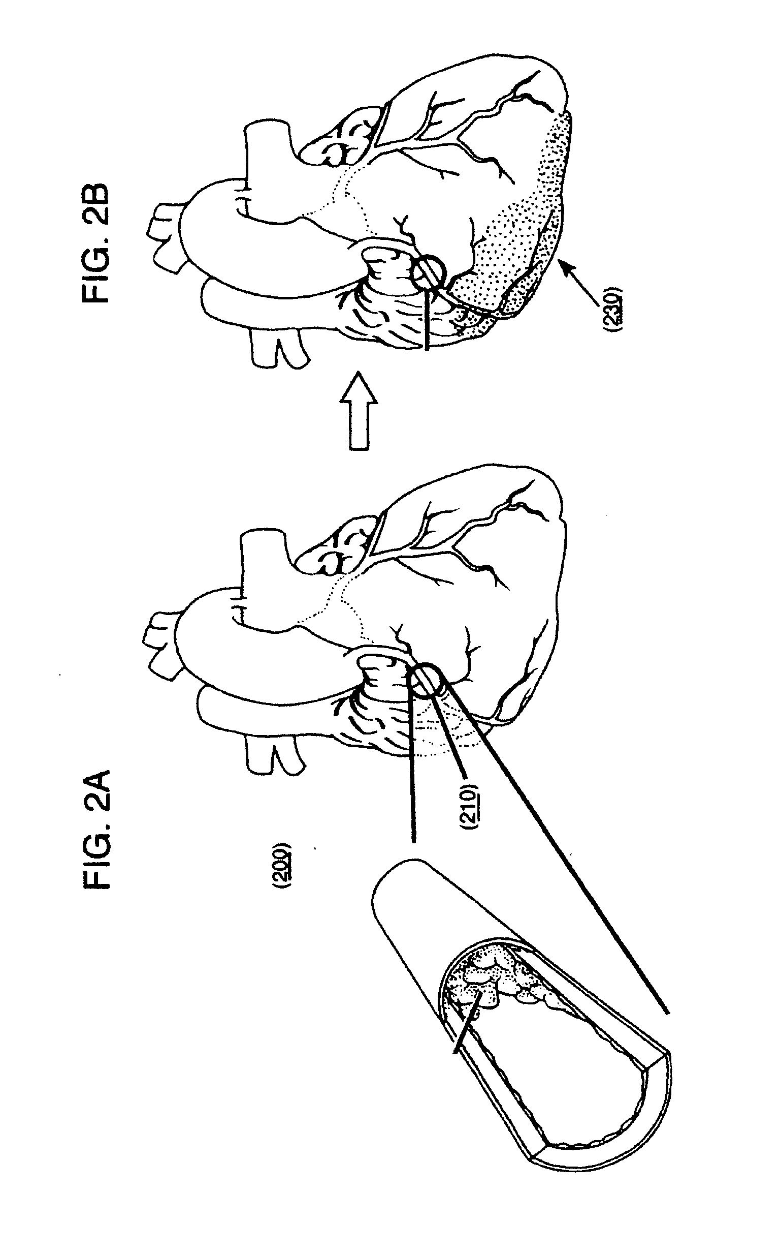
Thus, the obstructed artery may lead to damage observed in a ventricle of an MI subject.
A complication of an MI is an aneurysm that looks like a bulge in the left ventricular wall.
Additionally, parts of this mass can form a mural thrombus that can break off and embolize to the systemic circulation.
The E type prostaglandin, a powerful dilator of blood vessels, has been found in animal experiments to reduce high blood pressure—another cause of heart attacks and stroke.
Reducing the wall stress of the infarct region also decreases the probability of an arrhythmia arising from the region.

Method used


Image

  • Methods and compositions to treat myocardial conditions
  • Methods and compositions to treat myocardial conditions
  • Methods and compositions to treat myocardial conditions

Examples


example 1

[0213]In one example a 2-component gel may be injected via a dual needle catheter to an infarct region. One possible 2-component gel material may include Na-Alginate (component 1) which will likely ionically crosslink (often within seconds) when added to a soluble solution of calcium, barium and / or strontium (component 2). One potential crosslinker for the components may be CaCl2 (calcium chloride). In one example, covalently conjugating a peptide or protein to some of the acid groups of the alginate may enhance a cellular response. For example, conjugation of RGD groups or gelatin may promote cell adhesion, since these are binding sites for cells. The amine (N) terminus of gelatin or RGD may be conjugated to the acid groups of alginate via carbodiimide chemistry, forming an amide. The reaction may be mediated to higher yield with fewer side products (such as the inactive N-acyl urea) by first forming an active ester with, for example, 1-hydroxybenzotriazole or N-hydroxy succinimide...

example 2

[0215]In one example, a person with an mild or severe desynchrony (such as left bundle branch block), detectable through QRS duration, echocardiography, or other means, and with a history of a previous myocardial infarction, may first receive a catheter delivered injection of micronized porcine urinary bladder matrix (UBM) particulate (cryogenically ground UBM and resuspended at 5% weight / volume in PBS) to achieve alterations in the mechanical properties of the previous / scarred region. Following injection(s), implantation of a cardiac rhythm management device may be used to provide cardiac resynchronization therapy (CRT) to alter the desynchronized condition and alter the patient's progression to heart failure.


PUM

PropertyMeasurementUnit
volumeaaaaaaaaaa
volumeaaaaaaaaaa
volumeaaaaaaaaaa

Abstract

Methods, devices, kits and compositions to treat a myocardial infarction. In one embodiment, the method includes the prevention of remodeling of the infarct zone of the ventricle using a combination of therapies. The method may include the introduction of structurally reinforcing agents. In other embodiments, agents may be introduced into a ventricle to increase compliance of the ventricle. The prevention of remodeling may include the prevention of thinning of the ventricular infarct zone. Another embodiment includes the reversing or prevention of ventricular remodeling with electro-stimulatory therapy. The unloading of the stressed myocardium over time effects reversal of undesirable ventricular remodeling. These therapies may be combined with structurally reinforcing therapies. In other embodiments, the structurally reinforcing component may be accompanied by other therapeutic agents. These agents may include but are not limited to pro-fibroblastic and angiogenic agents.

Description

CROSS REFERENCE TO RELATED APPLICATIONS[0001]This application is a continuation of U.S. patent application Ser. No. 11 / 938,752, filed Nov. 12, 2007, U.S. Pat. No. 8,795,652, which application is a divisional of U.S. patent application Ser. No. 10 / 802,955, filed Mar. 16, 2004, U.S. Pat. No. 7,294,334, which is a continuation-in-part of U.S. patent application Ser. No. 10 / 414,602, filed Apr. 15, 2003, U.S. Pat. No. 8,383,158.FIELD OF THE INVENTION[0002]The treatment of myocardial infarction, and more particularly, in one embodiment, to the reinforcement of the infarct regional wall of a heart chamber using targeted cell delivery and / or the inhibition of the thinning of the infarct regional wall of a heart chamber using cell delivery in combination with other therapies such as electrostimulation. In further embodiments, to combine reinforcement of the infarct regional wall and / or cellular replacement with stimulating the heart using a pulsing and / or pacing device. In addition, this inv...

Claims


Application Information

Patent Timeline
Patent Type & Authority Applications(United States)
IPC IPC(8): A61N1/362A61L27/54A61L27/50A61N1/365A61L27/20A61K31/00A61K31/728A61K31/78A61K31/79A61K38/40A61K41/00A61K45/06A61K47/48A61K49/04A61L24/04A61P9/04A61P9/10A61P43/00
CPCA61N1/36521A61N1/3625A61L27/50A61L27/20A61L27/54A61K9/0024A61K31/00A61K31/728A61K31/78A61K31/79A61K36/886A61K38/40A61K41/0042A61K45/06A61K47/34A61K49/0404A61L24/043A61L2430/20A61K47/60A61K47/62A61P43/00A61P7/02A61P7/04A61P9/04A61P9/10A61P9/14A61K2300/00A61K35/34A61K38/1825A61K38/1841A61K38/1866A61K38/1858A61K38/1891A61K38/18A61K31/732A61K31/734
Inventor MICHAL, EUGENE T.ROSS, JEFFREY
Owner ABBOTT CARDIOVASCULAR
Why Eureka
Try Eureka