Biocompatible implantable sensor apparatus and methods

a sensor and biocompatible technology, applied in the field of biocompatible implantable sensor apparatus and methods, can solve the problems of not being realistically expected, precluding warning strategies that rely, and not being able to meet the needs of patients, and achieve the effects of not and reducing the risk of infection

Active Publication Date: 2017-12-07
GLYSENS
View PDF4 Cites 12 Cited by
  • Summary
  • Abstract
  • Description
  • Claims
  • Application Information

AI Technical Summary

Benefits of technology

[0037]In yet a further aspect of the disclosure, a sensor element configuration is disclosed. In one embodiment, the sensor element is used as part of an implantable sensor apparatus, and the configuration includes an enzyme-free outer layer or portion which prevents tissue of the host being from contacting immobilized underlying enzymes (e.g., glucose oxidase and catalase) within the sensor element when the sensor element is in vivo, thereby both (i) mitigating or eliminating a foreign body response (FBR) within the tissue due to lack of exposure to the enzymes or byproducts of the underlying reaction (e.g., peroxide), and (ii) ultimately enhancing operation and longevity of the sensor apparatus due to, inter alia, the aforementioned lack of FBR and the (excess) immobilized enzymes. In one variant, the enzyme-free outer layer or portion comprises a soluble protein-based (e.g., albumin-based) material which is cured using a cross-linking agent, and which is configured so as not to encourage blood vessel growth into the membrane after implantation of the sensor element into a living host.
[0038]In a further aspect of the disclosure, a method of mitigating foreign body response (FBR) associated with an implanted analyte detector element is disclosed. In one embodiment, the method includes creating an enzyme-free buffer zone between a reactive enzymatic portion of the detector element using a non-enzymatic membrane or layer that is permeable to both the analyte (e.g., glucose) and oxygen. In one variant, the enzymatic portion comprises a mechanically stable matrix, such that the enzymes of the matrix are substantially immobilized and do not migrate outward toward the host's tissue through the non-enzymatic membrane or layer, but the analyte and oxygen from the tissue can migrate inward. In one implementation, the non-enzymatic membrane material is selected so as to not exacerbate the FBR, thereby mitigating fibrous tissue formation and making subsequent explants of the detector element easier due to limited FBR-induced encapsulation.
[0046]In one implementation, at least one of (i) a shape or dimension of the aperture, (ii) a thickness of the non-enzyme yet permeable substance, and / or (iii) a shape or dimension of the cavity, for the at least one first detector element is different from that for the second at least one detector element, thereby enabling the at least one first detector element and the at least one second detector element to have a different operational characteristic from the other.

Problems solved by technology

This procedure has several disadvantages, including: (1) the discomfort associated with fingersticking, which should be performed repeatedly each day; (2) the near impossibility of sufficiently frequent sampling (some blood glucose excursions require sampling every 20 minutes, or more frequently, to accurately treat); and (3) the requirement that the user initiate blood collection, which precludes warning strategies that rely on automatic early detection.
Using the extant fingersticking procedure, the frequent sampling regimen that would be most medically beneficial cannot be realistically expected of even the most committed patients, and automatic sampling, which would be especially useful during periods of sleep, is not available.
Partially implanted sensors (discussed in greater detail below) are not viable for long-term use, particularly due to the undesirability of having an essentially open wound on the body for an extended period, and all of the attendant problems associated therewith (including greater risk of infection, the body's natural response to attempt to expel the percutaneous or “through the skin” portion of the implant, etc.).
Although this sensor provided direct recording of blood glucose, which is most advantageous for clinical applications, the described implantation at a central venous site poses several risks and drawbacks, including risk of blood clot formation and vascular wall damage.
Light-based systems using either absorption of light, or emission of light when glucose is “excited” by light have not proven to be accurate since there is no specific light absorption or emission spectrum for glucose.
Furthermore, numerous other chemicals or interfering substances in the blood overlap in spectrum with glucose, causing optical methods to be insufficiently specific for glucose monitoring.
However, this requirement gives rise to a sensor design problem related to “oxygen deficit,” since the concentration of oxygen in subcutaneous tissue is significantly less than that of glucose.
As the chemical reaction, and thus, the sensor signal, is limited by the reactant that is present in the sensor's reaction zone at the lowest concentration, an implanted sensor of simple construction would remain limited by oxygen, and would therefore be insensitive to the metabolite of interest (e.g. glucose).
However, one problem with the use of a macroporous or microporous membrane relates to exposure of the sensing element of the sensor to the environment of the body, which can result in “fouling” or other deleterious effects.
However, a major disadvantage of vascular implantation is the possibility of eliciting blood clots or vascular wall damage, as noted supra.
However, because the long axis of the “pocket” is oriented parallel to the electrode surface, this design may be less amenable to miniaturization for tissue implantation, and may suffer from yet other disabilities relating thereto.
However, one salient disadvantage of this approach relates to the requirement that the amount of hydrophobic polymer phase must be relatively large to allow for adequate oxygen permeability.
This substantially reduces the hydrophilic volume available for enzyme inclusion sufficient to counter inactivation during long-term operation.
For implanted devices that depend on diffusive transport of solutes to or from the bloodstream (e.g. implanted chemical sensors), such responses can negatively impact device operation due to an increase in mass transfer resistance between the bloodstream and active portions of the device surface resulting from an FBR-mediated development of fibrous tissue surrounding the device.
The FBR also can complicate explants of the implanted device (due to, e.g., the FBR causing significant encapsulation of the implanted device, thereby increasing its effective size when explanted), and result in yet other disabilities.
Such approaches have typically used materials for such layer(s) which are designed to encourage blood vessel growth and perfusion in the vicinity of the sensor or into the layer(s), which is undesirable, because such modulated responses are often not predictable and furthermore may not be sustainable for extended durations.

Method used


Image

  • Biocompatible implantable sensor apparatus and methods
  • Biocompatible implantable sensor apparatus and methods
  • Biocompatible implantable sensor apparatus and methods

Examples


Embodiment Construction

[0060]Reference is now made to the drawings, wherein like numerals refer to like parts throughout.

Detailed Description of Exemplary Embodiments

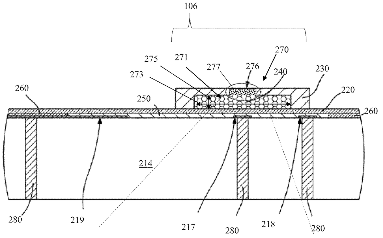
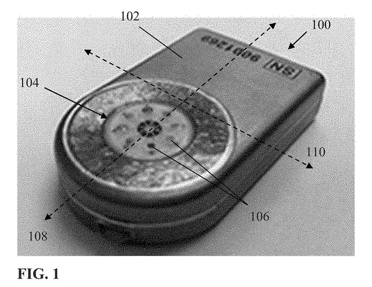
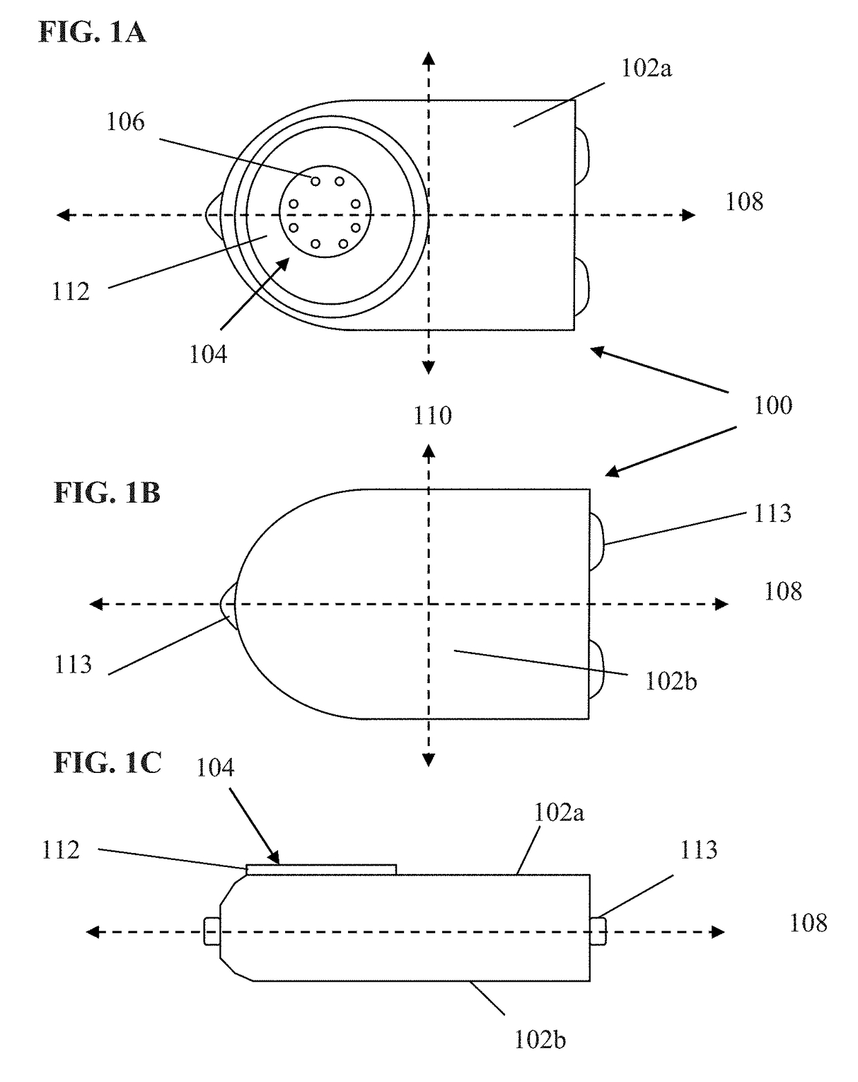
[0061]Exemplary embodiments of the present disclosure are now described in detail. While these embodiments are primarily discussed in the context of a fully implantable glucose sensor, such as those exemplary embodiments described herein, and / or those set forth in U.S. Patent Application Publication No. 2013 / 0197332 filed Jul. 26, 2012 entitled “Tissue Implantable Sensor With Hermetically Sealed Housing;” U.S. Pat. No. 7,894,870 to Lucisano et al. issued Feb. 22, 2011 and entitled “Hermetic implantable sensor;” U.S. Patent Application Publication No. 20110137142 to Lucisano et al. published Jun. 9, 2011 and entitled “Hermetic Implantable Sensor;” U.S. Pat. No. 8,763,245 to Lucisano et al. issued Jul. 1, 2014 and entitled “Hermetic feedthrough assembly for ceramic body;” U.S. Patent Application Publication No. 20140309510 to Lucisano et al. pu...


PUM


Abstract

Enzymatic and non-enzymatic detectors and associated membrane apparatus, and methods of use, such as within a fully implantable sensor apparatus. In one embodiment, detector performance is controlled through selective use of membrane configurations and enzyme region shapes, which enable accurate detection of blood glucose level within the solid tissue of the living host for extended periods of time. Isolation between the host's tissue and the underlying enzymes and reaction byproducts used in the detectors is also advantageously maintained in one embodiment via use of a non-enzyme containing permeable membrane formed of e.g., a biocompatible crosslinked protein-based material. Control of response range and / or rate in some embodiments also permits customization of sensor elements. In one variant, heterogeneous detector elements are used to, e.g., accommodate a wider range of blood glucose concentration within the host. Methods of manufacturing the membranes and detectors, including methods to increase reliability, are also disclosed.

Description

RELATED APPLICATIONS[0001]This application is related to co-owned and U.S. patent application Ser. No. 13 / 559,475 filed Jul. 26, 2012 entitled “Tissue Implantable Sensor With Hermetically Sealed Housing,” and co-pending and co-owned U.S. patent application Ser. No. 14 / 982,346 filed Dec. 29, 2015 and entitled“Implantable Sensor Apparatus and Methods,” each of the foregoing incorporated herein by reference in its entirety. This application is also related to U.S. patent application Ser. No. 10 / 719,541 filed Nov. 20, 2003, now issued as U.S. Pat. No. 7,336,984 and entitled “Membrane and Electrode Structure for Implantable Sensor,” also incorporated herein by reference in its entirety.GRANT INFORMATION[0002]This invention was made in part with government support under NIH Grant No. DK-77254. The United States government has certain rights in this invention.COPYRIGHT[0003]A portion of the disclosure of this patent document contains material that is subject to copyright protection. The co...

Claims


Application Information

Patent Timeline
Patent Type & Authority Applications(United States)
IPC IPC(8): A61B5/1486A61B5/00A61B5/1473A61B5/145A61B5/07
CPCA61B5/14865A61B5/0031A61B5/076A61B2562/04A61B5/1473A61B5/14542A61B2562/0209A61B5/14532A61B5/6861A61B17/0206A61B17/0218A61B17/1611A61B17/1757A61B17/7037A61B17/708A61B17/7082A61F2/4455A61F2002/30131A61F2002/30772A61F2002/4485A61F2310/00359A61B17/025A61B17/7032A61B17/7077A61B17/7079A61B17/7083A61B17/88A61B2017/00473A61B2017/0256A61B2017/564A61B2017/681A61F2/442A61F2/4611A61F2002/4435A61F2002/4635
Inventor LUCISANO, JOSEPHJAVIDI, BAHMANKURBANYAN, LEVLIN, JOEROUTH, TIMOTHYWALKER, BRADLEY
Owner GLYSENS
Why Eureka
Try Eureka