Nucleic acid probes and methods for detecting clinically important fungal pathogens

a technology of nucleic acid and pathogens, applied in the field of detection and identification of clinically important fungi, can solve the problems of increasing the incidence of infectious diseases, complicating the course, and affecting the survival rate of patients, and achieve the effect of enhancing the functionality of the diseas

Inactive Publication Date: 2005-02-22
ENTERPRISE IRELAND +2
View PDF23 Cites 66 Cited by
  • Summary
  • Abstract
  • Description
  • Claims
  • Application Information

AI Technical Summary

Benefits of technology

[0043]The term “isolated” as used herein means that the oligonucleotides of the current invention are isolated from the environment in which they naturally occur. In particular, it means that they are not anymore part of the genome of the respective fungal species, and thus liberated from the remaining flanking nucleotides in the ITS region of said fungal species. On the contrary, new (=heterologous) flanking regions may be added to the 3′ and / or 5′ end of the probe, in order to enhance its functionality. Functional characteristics possibly provided by said heterologous flanking sequences are e.g. ease of attachment to a solid support, ease of synthesis, ease of purification, labelling function etc.
[0044]The term “complementary” nucleic acid as used in the current invention means that the nucleic acid sequences can form a perfect base-paired double helix with each other.

Problems solved by technology

Fungal infections are becoming an increasingly significant cause of morbidity and mortality.
Fungal infections account for a large number of AIDS-defining diagnoses and complicate the course of most patients with HIV disease.
Superficial mycoses, caused by a fungus such as Epidermophyton, Microsporum, Trichophyton or Sporothrix growing on the body surface (skin, nails or hair) are unpleasant but usually mild infections.
Deep mycoses, involving the internal organs, are often life-threatening.
The fungi Aspergillus, Blastomyces, Candida, Coccidioides, Cryptococcus, Histoplasma are responsible for deep mycoses, and pose tremendous challenges for clinicians.
Pneumocystis carinii is a major opportunistic infectious agent in immuno-compromised patients, causing pneumonia which has a high mortality rate if the patient is not properly treated (Stringer J. R., 1996).
Indeed, in the case of chronic hyperplastic candidosis, reporting is usually limited to the presence of structures consistent with candidal hyphae following histological examination of lesional tissue.
These techniques are time-consuming, and their reliance on phenotypic expression makes Them potentially unreliable.
The clinical diagnosis of pulmonary cryptococcosis in patiens without underlying diseases is generally difficult.
However, evaluations of serological assays for detecting cryptococcal antigen showed false-positive reactions with sera from patients infected with Trichosporon beigelii (Kohno et al., 1993).
Unfortunately, the diagnosis of IA remains difficult and sometimes is confirmed only at autopsy.
Serological tests such as those involving the detection of antibodies for Aspergillus species are less helpful because of the poor antibody responses in immunocompromised patients.
In addition, the methods used for detecting circulating Aspergillus antigens, such as radioimmunoassay, immunoblotting assay, enzyme imununoassay, and the latex agglutination test, have poor sensitivity (Rogers et al., 1990; Sabetta et al., 1985).
Laboratory diagnosis of fungal infections is often problematic.
Fungi are often difficult to culture from readily accessible samples, such as patient's urine, blood or sputum.
And because fungi are ubiquitous in nature, a single positive culture from urine or sputum is of limited clinical value.
However, the sensitivity of these assays is very low—often less than 50%—as many immunocompromised patients have difficulty raising an adequate immune response.
Because of its poor sensitivity, inmmunodiagnosis of fungal infection is not cost-effective.
It often gives a false negative result, and may also lead to fungal infections being diagnosed where none exist, leading to the inappropriate administration of antifungal drugs.
A major problem is the transient nature of antigens in serum.
Moreover, none of the above-cited methods allows the identification of the fungus up to the species level.
Moreover, methods for simultaneous detection and differentiation of a wide variety of fungal species with clinical importance have not been described yet.

Method used


Image

  • Nucleic acid probes and methods for detecting clinically important fungal pathogens
  • Nucleic acid probes and methods for detecting clinically important fungal pathogens

Examples


examples

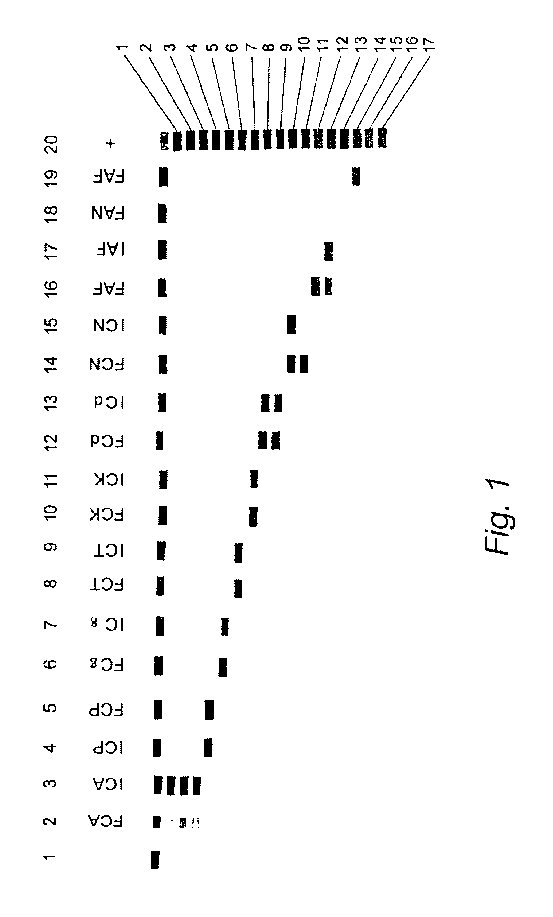
1. Specificity Testing of the Oligonucleotide Probes

[0280]1.1. Nucleic Acid Extraction.

[0281]A rapid extraction method based on physical disruption of the fungal cells followed by crude separation of the cell debris from the genomic DNA was used for the production of DNA from single colonies of yeasts (Roberts, 1997). For filamentous fungi, a more elaborate sample preparation method based on a combination of beadbeating and lysis with a GuSCN buffer followed by capturing of the DNA on silica was used.

[0282]1.2. PCR Amplification

[0283]20-50 ng genomic DNA or 5 μl of DNA extracted by the rapid extraction procedure described above were included in the PCR reaction. PCR reactions contained per 100 μl reaction: 200 μM of each dNTP's, 1×Taq buffer, 3 mM MgCl2, 15% glycerol, 40 pmol of each biotinylated primer (ITS5 and ITS4 for amplification of the full ITS region, ITS5 and ITS2 for amplification of ITS-1 only), 1 U Uracil N glycosylase and 2.5 U Taq polymerase. PCR thermal cycling condit...


PUM

PropertyMeasurementUnit
temperatureaaaaaaaaaa
timeaaaaaaaaaa
volumeaaaaaaaaaa

Abstract

The current invention relates to the field of detection and identification of clinically important fungi. More particularely, the present invention relates to species specific probes originating from the Internal Transcribed Spacer (ITS) region of rDNA for the detection of fungal species such as Candida albicans, Candida parapsilosis, Candida tropicalis, Candida kefyr, Candida krusei, Candida glabrata, Candida dubliniensis, Aspergillus flavus, Aspergillus versicolor, Aspergillus nidulans, Aspergillus fumigatus, Cyptococcus neoformans and Pneumocystis carinii in clinical samples, and methods using said probes.

Description

[0002]This application is a continuation of PCT / EP00 / 04714, filed May 24, 2000, and claims the benefit of U.S. provisional Application Ser. No. 60 / 138,621, filed Jun. 11, 1999, the entire contents of each of which being incorporated herein by reference.FIELD OF THE INVENTION[0003]The current invention relates to the-field of detection and identification of clinically important fungi. More particularely, the present invention relates to species specific probes originating from the Internal Transcribed Spacer (ITS) region of rDNA for the detection of fungal species such as Candida albicans, Candida parapsilosis, Candida tropicalis, Candida kefyr, Candida krusei, Candida glabrata, Candida dubliniensis, Aspergillus flavus, Aspergillus versicolor, Aspergillus nidulans, Aspergillus fumigatus, Cyptococcus neoformans and Pneumocystis carinii in clinical samples, and methods using said probes.BACKGROUND OF THE INVENTION[0004]Fungal infections are becoming an increasingly significant cause of...

Claims


Application Information

Patent Timeline
Patent Type & Authority Patents(United States)
IPC IPC(8): C12Q1/68C12N15/09
CPCC12Q1/6895
Inventor SMITH, TERRYMAHER, MAJELLAMARTIN, CARAJANNES, GEERTROSSAU, RUDIVAN DER WEIDE, MARJO
Owner ENTERPRISE IRELAND
Why Eureka
Try Eureka