Drugs for the diagnosis of tissue-reproductive activity or the treatment of proliferative diseases

a tissue-reproductive activity and drug technology, applied in the field of radiolabeled nucleoside derivatives, can solve the problems of difficult non-invasive evaluation of the proliferation activity of tumors, inability to become popular as a diagnostic technique of nuclear medicine imaging, and use of carbon-11-labeled thymidine in particular, so as to achieve local proliferative activity definition

Inactive Publication Date: 2006-05-16
NIHON MEDI PHYSICS CO LTD
View PDF12 Cites 7 Cited by
  • Summary
  • Abstract
  • Description
  • Claims
  • Application Information

AI Technical Summary

Benefits of technology

[0013]The radiolabeled compounds of the present invention are stable in vivo, and can retain in cells after being phosphorylated by mammalian thymidine kinase or reflect the DNA synthesis activity after being incorporated in DNA. Therefore, they realize effective diagnosis of tissue proliferation activity and treatment of proliferative disease, and are particularly useful as diagnostic radioactive imaging agents for diagnosis of tissue proliferation activity or as radioactive therapeutic agents for treatment of proliferative disease in accordance with internal radiotherapy, local radiotherapy or the like.
[0029]Another useful method for labeling the compounds represented by the above formula at the “5” position with a radioactive halogen is to allow a compound (5-trialkyltin compound), in which the pyrimidine base is substituted by a trialkylstannyl group at the “5” position as represented by Formula 11 in FIG. 1, Formula 21 in FIG. 2, Formula 28 in FIG. 3, Formula 40 in FIG. 4, Formula 50 in FIG. 5 or Formula 58 in FIG. 6, to react with 0.1N sodium hydroxide solution of a radioactive halogen in an appropriate solvent such as chloroform, so that the trialkylstannyl group at the “5” position is converted into a radioactive halogen-substituent. This labeling method, which uses a 5-trialkyltin compound, is preferable as it does not suffer such problems as with the above three labeling methods. Specifically, this method requires only a relatively short reaction time, and it does not produce “5-chloro” compounds or need heating as the reaction readily proceeds at room temperature. The resulting labeled compounds are free of carriers, and if a lower specific activity is desired, a labeled compound with a fixed specific activity can be readily prepared by adding a carrier. This method is also featured in that purification after the reaction is easy to operate. Specifically, 5-trialkyltin compounds are largely different from the corresponding radioactive halogen-labeled compounds in terms of overall molecular polarity as the electrical properties at the “5” position differ between them. Owing to the difference in the molecular polarity, labeled compounds and unreacted precursors can be separated easily by using a commercial reverse-phase silica gel cartridge after the labeling reaction. This permits elimination of the need of troublesome high performance liquid chromatographic purification.
[0048]The agent for diagnosis of tissue proliferation activity of the present invention can serve for quantitative determination of local proliferative activity based on appropriate model analysis. Furthermore, if non-proliferation tissue is used as a control, local proliferative activity can be defined easily in a semi-quantitative way.
[0049]The agent for treatment of proliferative disease of the present invention, when a beta-emitter such as I-131 is used therein, can serves to decrease large tumors of 1 cm or more in diameter, depending on the range of the ray. When an alpha-emitter such as At-211 is used, they can work on small lesions of 0.1 mm or less in diameter more effectively than beta-emitter, and therefore, they are expected to serve for treatment of micrometastasis over the body. Furthermore, nuclides that emit Auger electrons, such as I-125, can have antitumor effects due to DNA breakage, only after labeled compounds have gathered around the DNAs. Therefore, suitable label nuclides for treatment of systemic tumor foci including metastatic ones include alpha-emitter such as At-211, and beta-emitter such as I-131 that can have effect on portions around the foci depending on the range. The most effective method is the cocktail therapy which uses a mixture of a compound labeled with an alpha-emitter and a compound labeled with a beta-emitter.

Problems solved by technology

These radiolabeled compounds, however, are decomposed rapidly in vivo, making it difficult to perform non-invasive evaluation of the proliferation activity of the tumor.
The method using carbon-11-labeled thymidine, in particular, requires very complicated mathematical model analysis, and cannot become popular as a diagnostic technique of nuclear medicine imaging.
If the C—N glycosidic bonds are cleaved, the compound loses its affinity to tumors, thereby decreasing in accumulation of radioactivity in tumors, while the radioactive metabolites increase background radioactivity, thereby making imaging of the tumors difficult.
Though this radiolabeled compound is stable in vivo, it is a radio-labeled compound with a short-life positron emitter, and therefore a cyclotron is required in the hospital, limiting the usage of the compound.

Method used


Image

  • Drugs for the diagnosis of tissue-reproductive activity or the treatment of proliferative diseases
  • Drugs for the diagnosis of tissue-reproductive activity or the treatment of proliferative diseases
  • Drugs for the diagnosis of tissue-reproductive activity or the treatment of proliferative diseases

Examples


example 1

Synthesis of 5-trimethylstannyl-4′-thio-2′-deoxyuridine (Compound 11)

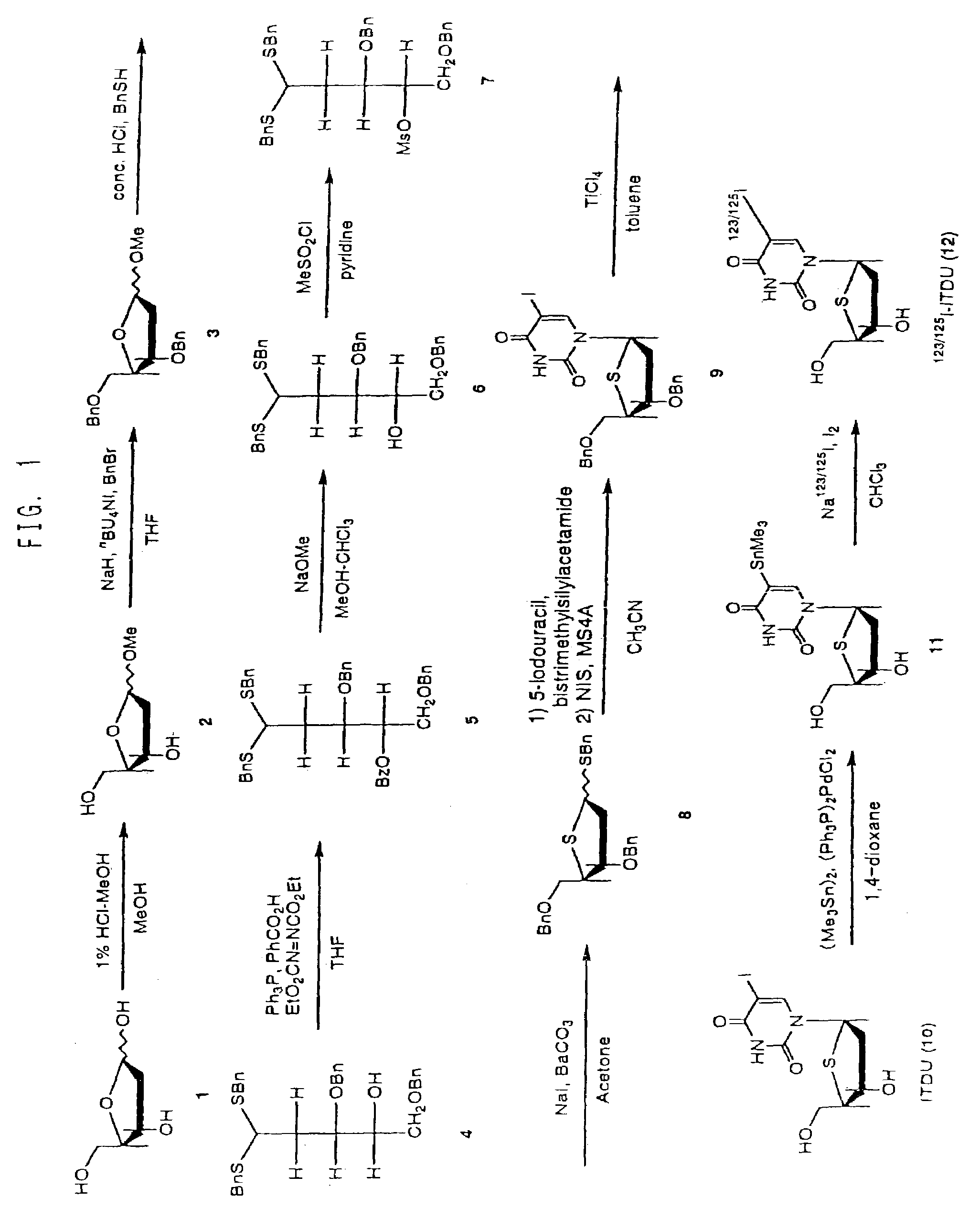
[0063]As shown in FIG. 1, benzyl-3,5-di-O-benzyl-2-deoxy-1,4-dithio-α,β-D-erythro-pentofuranoside (Compound 8) was synthesized, using 2-deoxy-D-erythro-pentose (Compound 1) as starting material, according to the method of Dyson M R et al. (Carbo. Res. 216, p. 237 (1991)). Further, 5-iodo-4′-thio-2′-deoxyuridine (ITDU: Compound 10) was produced from Compound 8 according to the method of Oivanen M et al. (J. Chem. Soc., Perkin Trans. 2, p. 2343 (1998)). Compound 10 was then used as a starting material to produce 5-trimethylstannyl-4′-thio-2′-deoxyuridine (Compound 11) according to the following procedure.

[0064]Compound 10 (9.5 mg, 0.026 mmol), bis(trimethyltin) (17.3 mg, 0.052 mmol) and bis(triphenylphosphine)palladium(II) chloride (5 mg) were dissolved in anhydrous 1,4-dioxane (3 mL) under argon atmosphere, and after heating at reflux for 3 hours, concentrated under reduced pressure. The residue was purified by sili...

example 2

Synsthesis of [I-125]-5-iodo-4′-thio-2′-deoxyuridine ([I-125] ITDU: Compound 12)

[0066]To 0.1N sodium hydroxide solution (50 μL) of [I-125]-sodium iodide (33 MBq), water (1 mL) and chloroform (1 mL) were added, and then chloroform solution (4.7 μL) of iodine (60 μg, 0.47 μmol) was added, and shaken for 10 seconds. After removing only the aqueous layer, ethyl acetate solution (100 μL) of Compound 11 (100 μg, 0.25 μmol) was added, and the resulting solution was left to stand at room temperature for 2 hours. One drop of 1N sodium thiosulfate solution was added, and chloroform was evaporated. After adding water (1 mL), the solution was passed through a Sep-Pak Plus QMA cartridge column. The column was washed with water (0.5 mL×2), and the resulting aqueous solution was combined to produce I-125-labeled Compound 12 (7.3 MBq, 22%).

example 3

Synthesis of [I-123]-5-iodo-4′-thio-2′-deoxyuridine ([I-123] ITDU: Compound 12)

[0067]To 0.1% ammonium iodide solution (1 mL) containing [I-123]-ammonium iodide (2.0 GBq), 1N hydrochloric acid (0.1 mL) and chloroform (1 mL) were added, and then chloroform solution (4.7 μL) of iodine (60 μg, 0.47 μmol) was added, and shaken for 10 seconds. After removing only the aqueous layer, ethyl acetate solution (100 μL) of Compound 11 (100 μg, 0.25 μmol) was added and left to stand at room temperature for 2 hours. One drop of 1N sodium thiosulfate solution was added, and chloroform was evaporated. After adding water (1 mL), the solution was passed through a Sep-Pak Plus QMA cartridge column. The column was washed with water (0.5 mL×2), and the resulting aqueous solution was combined to produce I-125-labeled Compound 12 (228 MBq, 15%).


PUM

PropertyMeasurementUnit
diameteraaaaaaaaaa
diameteraaaaaaaaaa
radioactiveaaaaaaaaaa

Abstract

An agent which comprises, as an active ingredient, a radiolabeled compound as represented by the following formula or a pharmaceutically acceptable salt thereof:wherein R1 denotes hydrogen, or a linear- or branched-chain alkyl group having 1–8 carbon atoms, R2 denotes hydrogen, hydroxyl or a halogen substituent, R3 denotes hydrogen or fluorine substituent, R4 denotes oxygen, sulfur, or a methylene substituent, and R5 denotes a radioactive halogen substituent.The agent is stable in vivo, and either stays in cells or is incorporated in DNA, thus serving for diagnosis of tissue proliferation activity or treatment of proliferative disease.

Description

CROSS-REFERENCED APPLICATIONS[0001]This application is a National phase of International Application PCT / JP02 / 00408, filed 22 Jan. 2002, which designated the U.S. and that International Application was not published under PCT Article 21(2) in English.TECHNICAL FIELD[0002]The present invention relates to use of radiolabeled nucleoside derivatives for diagnosis of tissue proliferation activity and treatment of proliferative diseases.BACKGROUND ART[0003]If proliferation activity of tumor cells can be determined non-invasively by image diagnosis, it will be help for evaluation of growth rate and malignancy of the tumor. Detection of the most rapidly growing regions of a tumor by image diagnosis will be useful in preparing plans for radiation fields in radiotherapy and identifying suitable portions for biopsy. Such methods will permit an early and accurate evaluation of therapeutic effects, which is difficult to identify by CT- or MRI-based anatomical evaluation or PET-based measurement ...

Claims


Application Information

Patent Timeline
Patent Type & Authority Patents(United States)
IPC IPC(8): A61K51/00A61M36/14A61K51/04A61P31/12A61P35/00C07H19/06
CPCC07H19/06A61K51/0491A61P29/00A61P31/12A61P35/00A61K51/00
Inventor TOYOHARA, JUNHAYASHI, AKIO
Owner NIHON MEDI PHYSICS CO LTD
Why Eureka
Try Eureka