

Method for detecting microorganisms
- Summary
- Abstract
- Description
- Claims
- Application Information
AI Technical Summary
Benefits of technology
Problems solved by technology
Method used
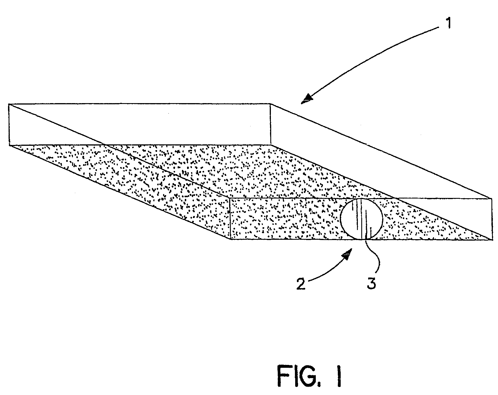
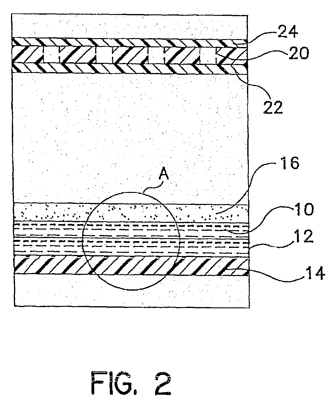
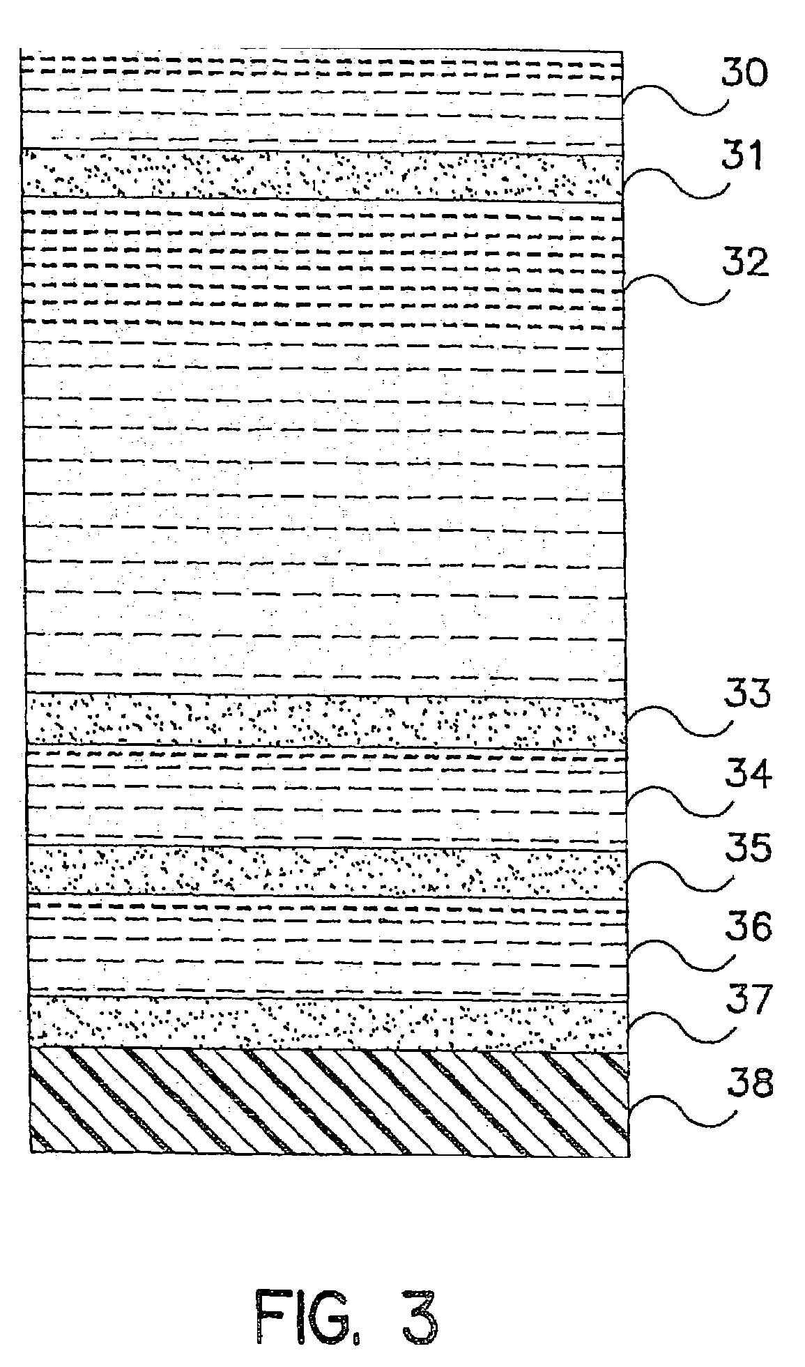
Image

Examples
example 1
CMC Dried Film
[0080]A solution of 1.5% (w / v) carboxymethyl cellulose (CMC, Aldrich Chemical Company, average MW of 700,000) was autoclaved at 121° C. for 15 minutes in liquid cycle. After cooling, the solution was added in 10 ml portions to 60 mm (52 mm inside diameter, 21.2 cm2) petri dishes and dried overnight at 50° C. The temperature was then increased to 80° C. for 1 hour to reduce any bacterial load from contamination.
[0081]Sheep blood was spiked with S. aureus (ATCC#25923) at a concentration of approximately 10 CFU / ml, and was lysed by the addition of saponin to a final concentration of 0.5% (w / v). The dried CMC films were inoculated with 2.5 ml of the lysed blood solution, covered and incubated overnight at 35° C.
[0082]After the overnight incubation, all fluid had been absorbed into the gel and mutually isolated colonies of S. aureus were easily observed on, and harvested from, the surface of the gel. There was no visible penetration of the bacteria into the gel, nor were an...
example 2
[0083]A pre-polymerization solution for forming light initiated polyacrylamide gels was made with the following composition; Acrylamide / BisAcrylamide (10% T, 0.4% CO, TEMED (0.04% v / v), riboflavin phosphate (0.0025% w / v), and sodium phosphate (10 mm pH 6.5, final concentration). The solution was added in 20 ml portions to 90 mm (86 mm inside diameter), 58.1 cm2 petri dishes. The petri dishes were stacked and sealed in an anaerobic culture jar with an anaerobic atmosphere generator pack. The anaerobic jar was then placed in the dark for 30 minutes to allow time for the oxygen to be removed before polymerization.
[0084]The plates in the jar were then illuminated under a 15W fluorescent desk lamp at a distance of approximately 10 cm, overnight at approximately 23° C. After polymerization, 5 ml of 10% Glycerol was added on top of each gel, then the gels were, dried overnight at 35° C., and baked for 1 hour at 65° to reduce any bacterial contamination.
[0085]The gels were par...
example 3
Xanthan / Guar Paste with Support
[0087]A paste was made by combining 10 g of pre-hydrated xanthan gum and 2.5 g of pre-hydrated guar gum, with 50 ml to tryptic soy broth CTSB). Portions of the paste were sandwiched between two layers of a woven nylon support mesh and pressed between plastic plates until 1.5 g of paste formed approximately a 60 mm diameter circles. A 51 mm diameter punch was used to cut our circles of the paste sandwich, which were transferred to a polycarbonate sheet and sterilized by autoclaving. After cooling, the paste circles were transferred to a 60 mm (52 mm inside diameter) petri dishes along with 0.5 ml of TSB to partially hydrate the paste and aid in placement of the disks.
[0088]Human blood was collected with SPS as an anticoagulant and spiked with S. aureus (ATCC#25923) or E. coli (ATCC#25922) at a concentration of approximately 25 CFU / ml. The spiked blood was lysed by the addition of an equal quantity of TSB with 2.0% saponin (w / v). The gum paste disks were...
PUM
 Abstract
Description
Claims
Application Information