Device and method for manual retinal vein catheterization

Inactive Publication Date: 2007-05-15
THE JOHN HOPKINS UNIV SCHOOL OF MEDICINE
View PDF10 Cites 64 Cited by
  • Summary
  • Abstract
  • Description
  • Claims
  • Application Information

AI Technical Summary

Benefits of technology

[0026]In a particularly preferred embodiment, the microcatheter system has an intraocular portion that is composed of a hybrid of two different size cannulae. In addition to the above-described flexible cannula that is inserted into the retinal vein lumen, the microcatheter system further comprises a larger cannula encasing a portion of the small cannula. The larger cannula is less flexible than the smaller cannula, and may be grasped by forceps during manipulation and insertion of the cannula within the retinal vein, thereby protecting the flexible cannula. In a preferred embodiment, the larger cannula forms the part of the microcatheter system that is inserted just inside the eye and, as such, it provides the rigidity necessary to pass it into the eye without damage. The larger cannula preferably has an outer diameter ranging from about 400 μm to about 800 μm, and more preferably, about 556 μm. This larger cannula is sized so as to allow it to be easily attached to the tip of a syringe loaded with infusion fluid. The plunger of the syringe loaded with fluid may then be attached to a motoriz

Problems solved by technology

Retinal vascular occlusions are a leading cause of blindness in elderly patients.
Occlusion in any of these retinal vessels leads to a build-up of pressure, which may lead to hemorrhages and also leakage of fluid and other constituents of blood.
When retinal vessels are blocked, the area of the retina supplied by that vessel has poor blood flow, which results in sub-optimal performance by the local ganglion cells and even death of these local ganglion cells.
In some severe cases, poor blood flow resulting from retinal vascular occlusions can also lead to the development of abnormal new vessels (neovascularization).
These new vessels are fragile and leak fluid and blood, which damages cells in the retina.
In severe cases of neovacularization, the new abnormal blood vessels may pull on the retina, leading to retinal detachment.
In very serious cases, the eye may develop neovascular glaucoma, in which fluid outflow channels in the eye are blocked, placing the eye is under very high pressure.
This may result in severe vision loss, pain and even loss of the eye itself.
Although retinal venous occlusive disease is a highly prevalent disease with serious consequences, treatment options remain limited.
This approach does not attempt to re-establish the normal venous return pathway from the retina.
Moreover, it has had limited success and can lead to uncontrolled choroidal neovascularization resulting in vitreous hemorrhage and tractional retinal detachment.
However, studies involving the systemic use of thrombolytics to treat central retinal vein occlusions have not been substantiated.
When presented with a static column of blood, thrombolytic agents delivered systemically are not able to diffuse through the vessels and

Method used


Image

  • Device and method for manual retinal vein catheterization
  • Device and method for manual retinal vein catheterization
  • Device and method for manual retinal vein catheterization

Examples


example 1

Enucleated Porcine Eves Testing—Testing The Polyimide Tubing

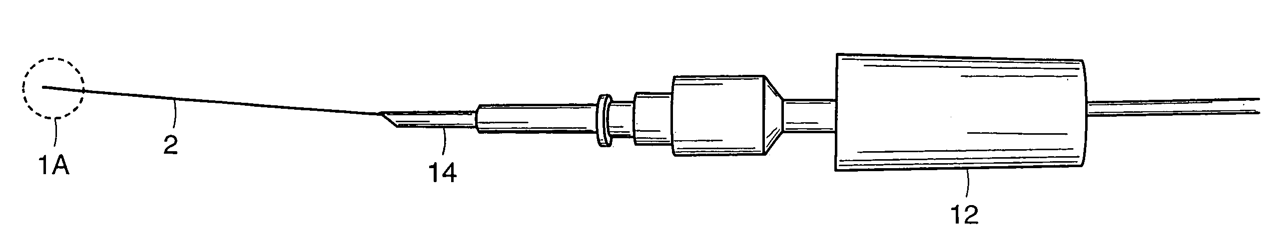
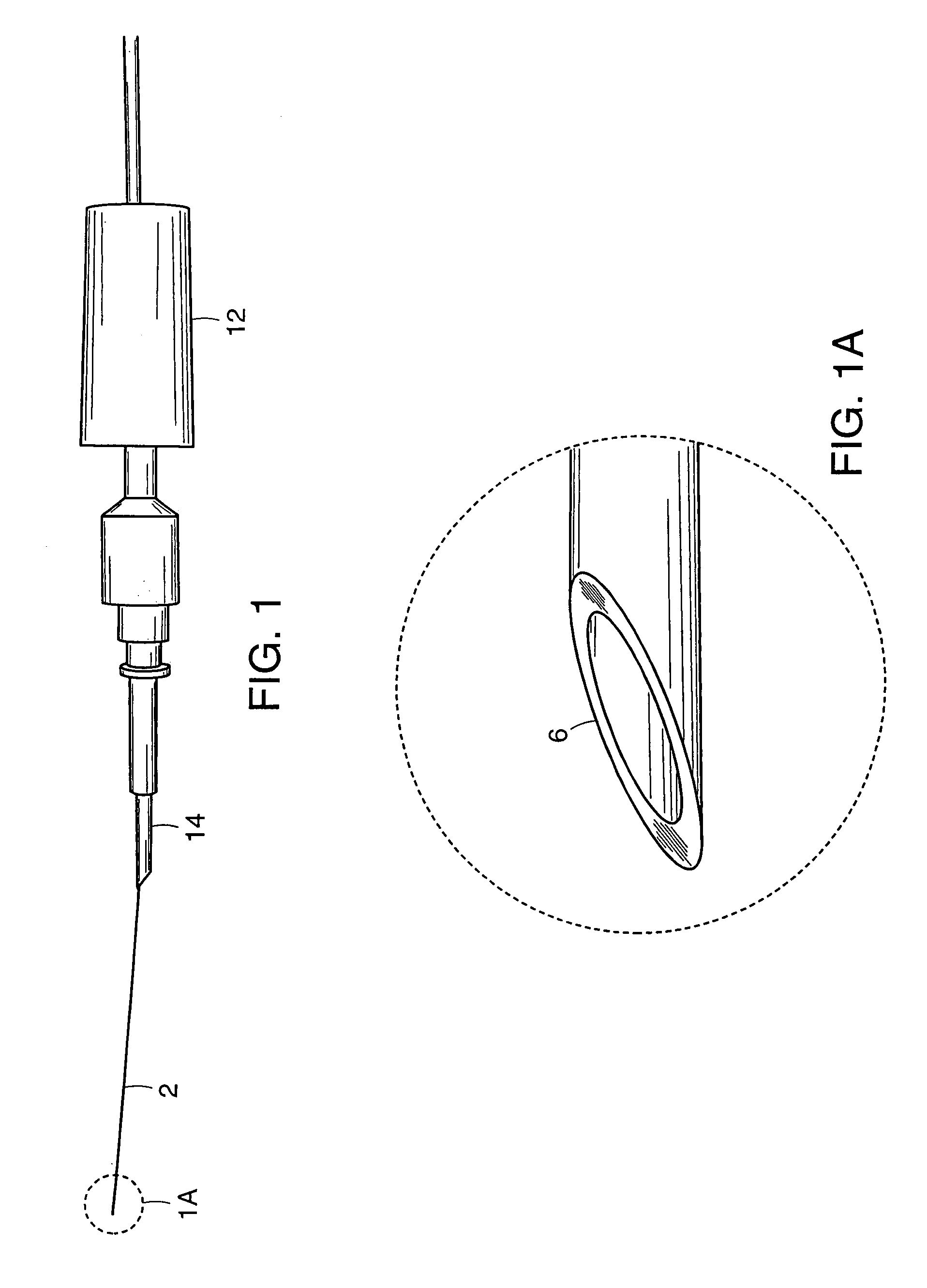
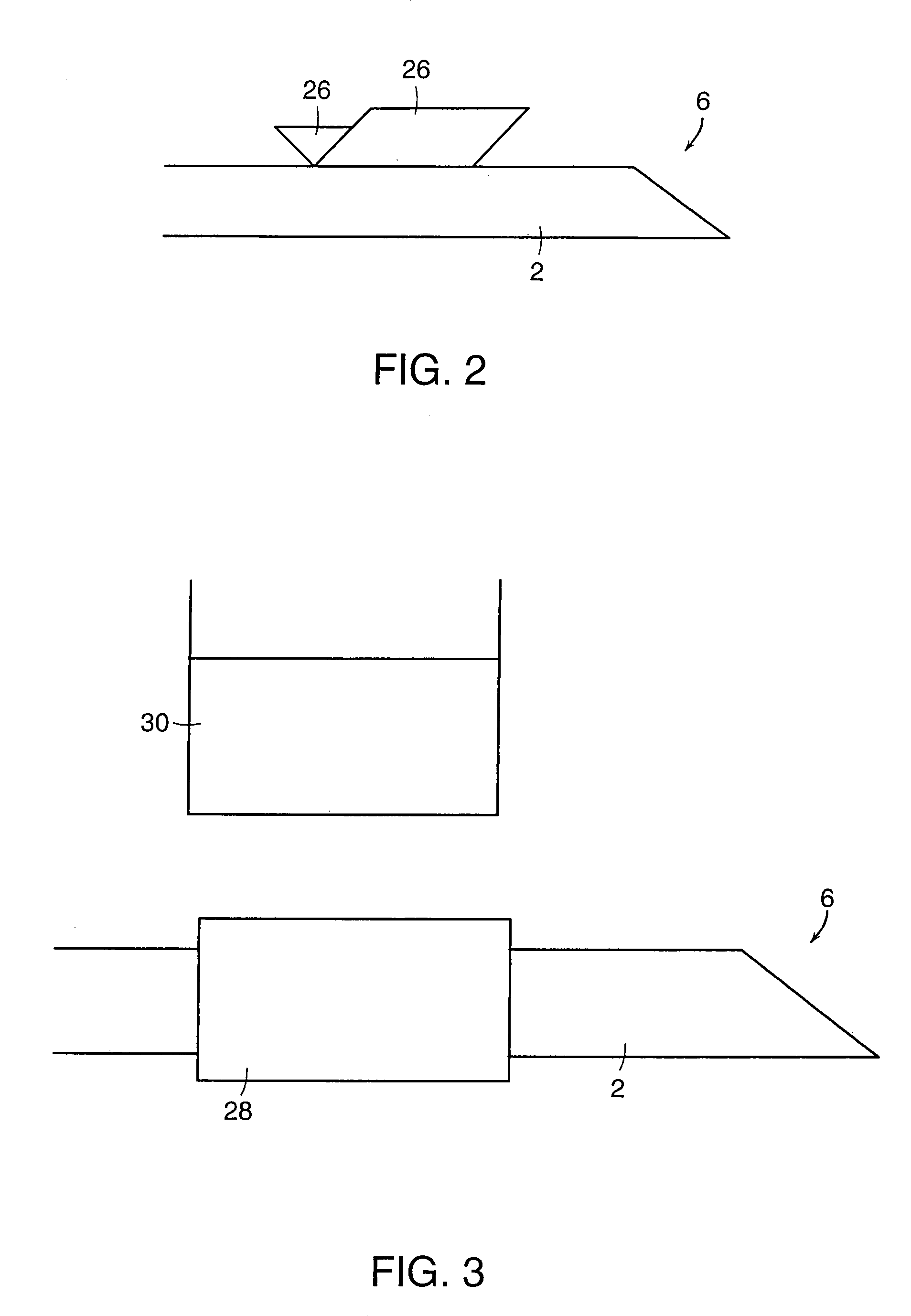
[0064]In order to test the distal end 6 of cannula 2, enucleated porcine eyes were used. Twenty-two catheterization procedures were performed in twelve cadaveric porcine eyes. During these experiments, the anterior segment and the vitreous gel were removed. Four cuts were made in the sclera of the remaining posterior half of the globe and the sclera was pinned down to flatten the retina. The cannula was grasped at approximately 0.5 mm from the distal end 6 to be inserted into the vein. In order to make the distal end 6 of the cannula 2 sharp enough to penetrate the vein, the distal end 6 was cut using microscissors so that the tip would approximate a 30° ramp-like configuration. Under microscopic guidance, the distal end 6 of the cannula 2 punctured the vein wall and was advanced slightly within the lumen of the vein. The cannula was then released and the infusion pump activated to ascertain the intravascular status of the ...

example 2

In-Vivo Dog Eyes Testing—Developing the Microcatheter System & Surgical Technique

[0067]In order to develop the surgical technique and delivery system to insert the microcatheter system inside the eye, seven eyes of seven dogs were operated on. The dog was selected as an animal model because the caliber of its retinal blood vessels and because the manner in which the vessels emanate from the optic disc are very similar to humans. All animal procedures were performed in accordance with both Johns Hopkins University Animal Care and Use Committee and the ARVO Statement for the Use of Animals in Ophthalmic and Vision Research.

[0068]The dogs were anesthetized initially with Telasol (Tiletamine HCl and Zolazepam HCl, Fort Dodge Animal Health; 4–7.5 mg / kg IM) and then they received a gas mixture with oxygen and halothane (98–99% oxygen and 1–2% halothane at a rate of 1.5–2 l / min). Throughout the surgery the dog's EEG, respiration rate and body temperature were monitored. The operated eye wa...


PUM


Abstract

A microcatheter system that allows for vascular infusion into retinal veins for extended periods of time. The microcatheter system includes a flexible cannula that is inserted into the retinal vein lumen and that remains stably within the retinal vein lumen without being held by a robot, micromanipulator or similar holding devices. The microcatheter system is particularly suitable for the treatment of retinal venous occlusive disease.

Description

[0001]The present application claims the benefit of U.S. provisional application No. 60 / 174,260, filed on Jan. 3, 2000; and U.S. provisional application No. 60 / 217,468, filed on Jul. 11, 2000, both of which applications are incorporated herein by reference in their entirety.FIELD OF THE INVENTION[0002]The present invention relates to improved microcatheter devices. More particularly, the present invention relates to a flexible microcatheter instrument and surgical technique that allows for vascular infusion into retinal veins for extended periods of time. The device and method are particularly suitable for the treatment of retinal venous occlusive disease.BACKGROUND OF THE INVENTION[0003]Retinal vascular occlusions are a leading cause of blindness in elderly patients. While younger patients may develop this disease (referred to as papillophlebitis in younger patients), most occlusions occur in individuals over the age of 50.[0004]A retinal vascular occlusion is characterized by a fu...

Claims


Application Information

Patent Timeline
IPC IPC(8): A61M25/00A61M5/32A61B17/00A61B19/00A61F9/00A61F9/007
CPCA61B5/6848A61B5/6885A61F9/0017A61M25/00A61B2017/00345A61B2019/2292A61F9/007A61M2210/0612A61B34/76
Inventor HUMAYUN, MARK S.JENSEN, PATRICK S.SHELLEY, TERRY H.FUJII, GILDO Y.HAMZA, HANY S.BARNES, AARON C.DEJUAN, JR., EUGENE
Owner THE JOHN HOPKINS UNIV SCHOOL OF MEDICINE
Why Eureka
Try Eureka