Methods for cardiac gene transfer

a gene transfer and gene technology, applied in the direction of dsdna viruses, peptide/protein ingredients, drug compositions, etc., can solve the problems of affecting the practical application of gene transfer in vivo, affecting the effect of cardiac gene transfer, and uneven gene expression, etc., to achieve high level of in vivo cardiotopic gene transfer, high consistency, and efficient gene transfer and expression

Inactive Publication Date: 2008-07-15
RGT UNIV OF CALIFORNIA
View PDF5 Cites 5 Cited by
  • Summary
  • Abstract
  • Description
  • Claims
  • Application Information

AI Technical Summary

Benefits of technology

[0018]The invention is a method for efficient gene transfer and expression in cardiac muscle cells. The invention overcomes limitations in previous methods of achieving a high level of in vivo cardiotopic gene transfer with high consistency (average 60-70% of cardiac myocytes) in normal and cardiomyopathic animal models. The invention involves increasing the dwell time of the viral vector, either Ad or AAV containing the gene of interest, in the heart by the induction of hypothermia, isolation of the heart from circulation, and near or complete cardiac arrest. Permeablizing agents are an essential component of the invention and are used during the administration of the virus to increase the uptake of the virus by the cardiac cells. The gene expression via AAV vectors is highly restricted to cardiac muscle and maintained long-term, with no signs of myocardiac inflammation.

Problems solved by technology

There have been several critical limitations that have hindered the practical application of gene transfer in vivo.
However, in these reports, either the extent or the specificity of gene transfer was not described properly, or the gene expression was patchy in distribution.
Activity of the sarcoplasmic reticulum Ca2+ ATPase (SERCA2a) is reduced in failing hearts, resulting in abnormal Ca2+ handling, and eventually leading to contractile failure.
Intracardiac injection of an Ad-SERCA2a viral vector in rats was sufficient to induce some physiological improvement; however, there were a number of limitations.
The delivery method produced diffuse, multichamber myocardial expression, and cardiac function was improved; however, neither the efficiency nor the specificity of gene transfer was discussed in the report.
Thus, the demonstration of the therapeutic effect of cardiac somatic gene transfer has been hampered by the lack of in vivo gene delivery strategies to effect long-term, high-efficiency, cardiotropic expression in the intact heart.
To date, there are no reports of stable, high efficiency transfer of genes into cardiac tissue.
This mutation results in decreased stable expression of all of the sarcoglycan genes (α, β, and γ), resulting in decreased structural integrity in all muscle cells.
In the absence of δ-sarcoglycan, the complex collapses and is no longer able to stabilize the cells, making them more susceptible to mechanical stress.
Additionally, the isolation of the heart from circulation by tourniquet to increase exposure of the virus to the tissue of interest would clearly be problematic; therefore, the same method could not be used for cardiac tissue.
Efficient gene delivery to the heart presents a greater problem than delivery to striated muscle tissue due to structural differences of the tissues.
These manipulations achieved local gene delivery due to a lack of intense viral vector diffusion; however, the outcome of these methods is highly restricted gene expression with local tissue damage.
Transcoronary delivery of AAV was attempted in porcine myocardium; however, extremely low gene transfer efficiency was observed (0.2%) (Kaplitt et al., 1996).
To date, there is no report of high efficiency in vivo cardiotropic gene delivery system with long-term sustainable expression.

Method used


Image

  • Methods for cardiac gene transfer
  • Methods for cardiac gene transfer
  • Methods for cardiac gene transfer

Examples


example 1

[0030]Construction of viral vectors. A replication deficient (E1A and E1B deleted) Ad vector was constructed that contains the β-galactosidase gene with a nuclear localization signal sequence (Ad.CMV LacZ) or the hamster δ-sarcoglycan gene (Ad.CMV δ-sarcoglycan) driven by the CMV promoter. The viral constructs were generated by the use of the shuttle vector PACCMV.PLPA by the method of Graham et al., 1995 (incorporated herein by reference). Ad vectors generated by in vitro recombination were amplified in 293 cells. Cells were harvested and subjected to three freeze-thaw cycles to release the virus particles. Virus was purified through two consecutive CsCl gradients. The average titers of the viruses were 1.27×10¹¹ pfu / ml for AdV.CMV LacZ and 5.5×10⁹ pfu / ml for Ad.CMV δ-sarcoglycan.

[0031]AAV vectors are based on the non-pathogenic and defective parvovirus type 2 and were constructed essentially as described by Xiao, et al. 1998 (incorporated herein by reference). Briefly, the three p...

example 2

[0032]In vivo transduction. Hamsters were anesthetized with sodium pentobarbitol (85 mg / kg, i.p.), intubated and ventilated, ECG electrodes were placed on the limbs and a 6F thermistor catheter was inserted into the rectum. The chest was shaved and a small (4-5 mm) left anterior thoracotomy performed in the second intercostal space; braided silk ligature were looped around the ascending aorta and the main pulmonary artery and threaded through plastic occluder tubes. Through a midline neck incision, the right carotid was exposed, cannulated with flame-stretched PE-60 tubing and the tip advanced into the ascending aorta just above the aortic valve (below the aortic snare) for measurement of arterial pressure and later injections of viral particles.

[0033]Bags filled with ice water were placed around the supine animal, including the head, and the heart rate and temperature were monitored every three minutes until the core temperature reached 18° C. (average time 40 minutes in the normal...

example 3

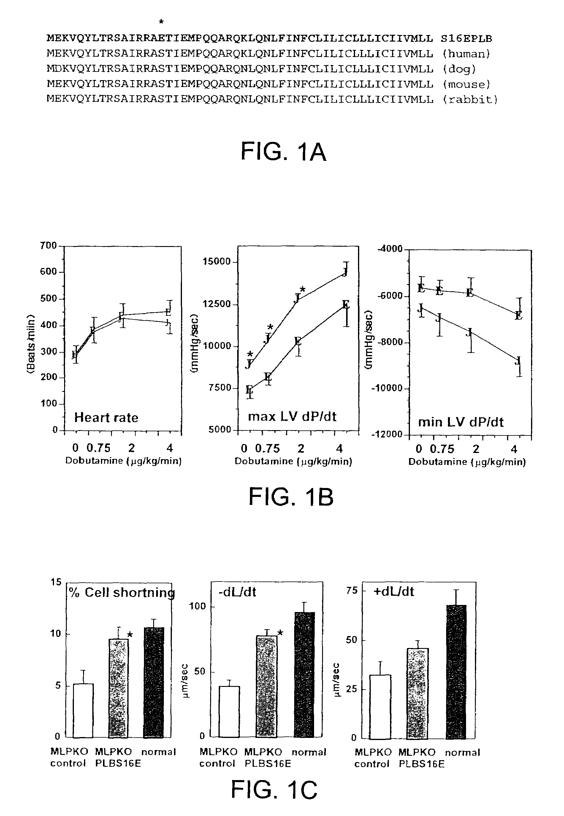
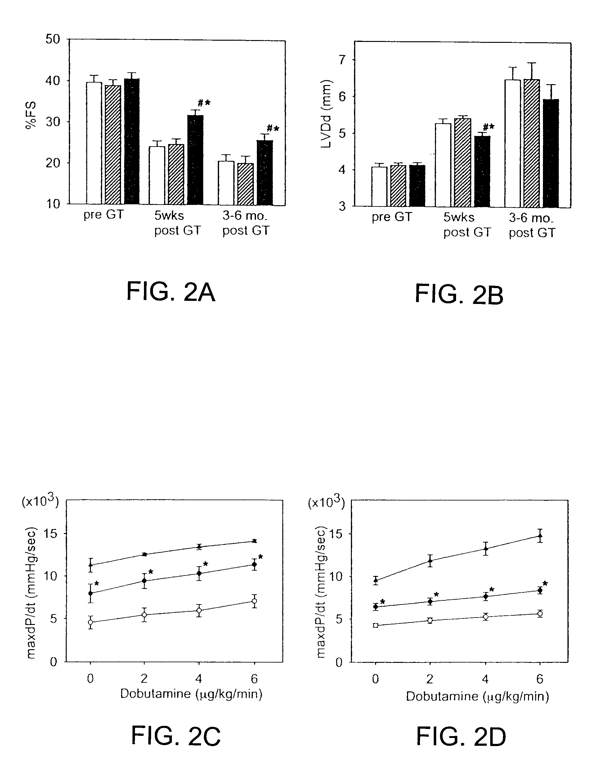
[0035]Transduction procedure using a reporter gene does not alter cardiac function. Normal hamsters (n=10) were studied by echocardiography (ECG) at 4 days post Ad.CMV LacZ administration. The percent fractional shortening (% FS) of the left ventricle was 43.88±5.45%, indicative of normal function and the left ventricle end-diastolic dimension (LVDd) was 4.38±0.22 mm, indicative of normal heart size. In a group of age-matched normal, untreated hamsters, the % FS was 46.83±2.43 and the LVDd was 4.12±0.3 mm (not significantly different). In the CM hamsters, some animals were examined by echocardiography both pre- and 6 days post-administration of Ad.CMV LacZ. The left ventricular function was depressed in control CM hamsters vs. wild-type hamsters as expected (% FS 27.77±3.17%, LVDd 5.44±0.25 mm) and somewhat further depressed 6 days later (% FS 23.59±4.93%, LVDd 5.60±0.37 mm, both p<0.05).


PUM

PropertyMeasurementUnit
temperatureaaaaaaaaaa
pressureaaaaaaaaaa

Abstract

Methods for the delivery of genes to improve cardiac function including the use of viral vectors, isolation of the heart from systemic circulation, and induction of hypothermia/cardiac arrest are described. The method results in high-level, long-term expression of reporter genes and enhanced cardiac function in hamster models of heart disease.

Description

CROSS-REFERENCES TO RELATED APPLICATIONS[0001]This application claims the benefit of priority of U.S. provisional application Ser. No. 60 / 231,821 filed Sep. 11, 2000, which is incorporated herein by reference in its entirety.GOVERNMENT INTEREST[0002]The invention was made with government support from the National Institutes of Health.BACKGROUND OF THE INVENTION[0003]Somatic gene transfer offers a myriad of possibilities for therapeutic usage in a number of diseases including congenital defects, as well as acquired forms of pathological abnormalities. There have been several critical limitations that have hindered the practical application of gene transfer in vivo. These include the duration of expression of transferred genes, the trade-off between tissue specificity and the efficiency of gene expression, and the adverse side effects of local inflammation provoked by vectors.[0004]In the field of cardiovascular medicine, gene therapy has been focused on vascular gene transfer, mainly...

Claims


Application Information

Patent Timeline
Patent Type & Authority Patents(United States)
IPC IPC(8): A01N43/04A01N63/00C12N15/00C12N5/00A61K38/17A61K38/46A61K48/00A61P9/00C12N15/12
CPCA61K38/1709A61K38/46A61K48/0008A61K48/0083A61K38/046A61K38/1787A61K38/177A61K31/417A61K31/4045A61K45/06C12N2750/14143A61K48/00C12N2710/10043A61K2300/00A61P9/00
Inventor CHIEN, KENNETH R.HOSHIJMA, MASAHIKOROSS, JR., JOHNIKEDA, YASUHIRO
Owner RGT UNIV OF CALIFORNIA
Why Eureka
Try Eureka