Morphogen-induced nerve regeneration and repair

a morphogen-induced nerve and regeneration technology, which is applied in the direction of animal cells, peptide/protein ingredients, impression caps, etc., can solve the problems of inability to repair the damage caused by these neuropathies, the neural pathways of mammals are particularly at risk, so as to reduce the risk of dying, significantly inhibit the effect of cellular metabolic dysfunction, and reduce the risk of death

Inactive Publication Date: 2009-07-07
STRYKER CORP
View PDF26 Cites 7 Cited by
  • Summary
  • Abstract
  • Description
  • Claims
  • Application Information

AI Technical Summary

Benefits of technology

[0023]In another aspect of the invention, the morphogens described herein are useful for providing neuroprotective effects to alleviate neural pathway damage associated with the body's immune / inflammatory response to an initial injury to nerve tissue. Such a response may follow trauma to nerve tissue, caused, for example, by an autoimmune dysfunction, neoplastic lesion, infection, chemical or mechanical trauma, disease, by interruption of blood flow to the neurons or glial cells, for example following ischemia or hypoxia, or by other trauma to the nerve or surrounding material. For example, the primary damage resulting from hypoxia or ischemia-reperfusion following occlusion of a neural blood supply, as in an embolic stroke, is believed to be immunologically associated. In addition, at least part of the damage associated with a number of primary brain tumors also appears to be immunologically related. Application of the morphogen directly to the cells to be treated, or providing the morphogen to the mammal systemically, for example, intravenously or indirectly by oral administration, may be used to alleviate and / or inhibit the immunologically related response to a neural injury. Alternatively, administration of an agent capable of stimulating morphogen expression and / or secretion in vivo, preferably at the site of injury, also may be used. Where the injury is to be induced, as during surgery or other aggressive clinical treatment, the morphogen or agent may be provided prior to induction of the injury to provide a neuroprotective effect to the nerve tissue at risk.
[0024]In still another aspect, the invention described herein provides methods for supporting the growth and maintenance of differentiated neurons, including inducing neurons to continue expressing their phenotype. It is anticipated that this activity will be particularly useful in the treatment of nerve tissue disorders where loss of function is caused by reduced or lost cellular metabolic function and cells become senescent or quiescent, such as is thought to occur in aging cells and to be manifested in Alzheimer's disease. Application of the morphogen directly to cells to be treated, or providing it systemically by parenteral or oral administration stimulates these cells to continue expressing their phenotype, significantly inhibiting and / or reversing the effects of the cellular metabolic dysfunction, thereby maintaining the neural pathway at risk. Alternatively, administration of an agent capable of stimulating endogenous morphogen expression and / or secretion in vivo may be used.

Problems solved by technology

Accordingly, the neural pathways of a mammal are particularly at risk if neurons are subjected to mechanical or chemical trauma or to neuropathic degeneration sufficient to put the neurons that define the pathway at risk of dying.
Currently no satisfactory method exists to repair the damage caused by these neuropathies, which include multiple sclerosis, amyotrophic lateral sclerosis (ALS), Huntington's chorea, Alzheimer's disease, Parkinson's disease (parkinsonism), and metabolically derived disorders, such as hepatic encephalopathy.
Currently, however, human fetal cell transplantation research is severely restricted.
Successful grafting appears to be limited, however, by a number of factors, including the length of the CNS axonal lesion to be bypassed, and the distance of the graft sites from the CNS neuronal cell bodies, with successful grafts occurring near the cell body.
Within the peripheral nervous system, this cellular regenerative property of neurons has limited ability to repair function to a damaged neural pathway.
Specifically, the new axons extend randomly, and are often misdirected, making contact with inappropriate targets that can cause abnormal function.
For example, if a motor nerve is damaged, regrowing axons may contact the wrong muscles, resulting in paralysis.
Efforts to repair peripheral nerve damage by surgical means has met with mixed results, particularly where damage extends over a significant distance.
Even where scar tissue formation has been reduced, as with the use of nerve guidance channels or other tubular prostheses, successful regeneration generally still is limited to nerve damage of less than 10 millimeters in distance.
In addition, the reparative ability of peripheral neurons is significantly inhibited where an injury or neuropathy affects the cell body itself or results in extensive degeneration of a distal axon.
Mammalian neural pathways also are at risk due to damage caused by neoplastic lesions.
Transformed cells of neural origin generally lose their ability to behave as normal differentiated cells and can destroy neural pathways by loss of function.
Metastatic tumors, which are a significant cause of neoplastic lesions in the brain and spinal cord, also similarly may damage neural pathways and induce neuronal cell death.
In addition, antibodies to N-CAM have been shown to disrupt functional recovery in injured nerves (Remsen (1990) Exp. Neurobiol. 110:268-273).

Method used


Image

  • Morphogen-induced nerve regeneration and repair
  • Morphogen-induced nerve regeneration and repair
  • Morphogen-induced nerve regeneration and repair

Examples


example 1

Identification of Morphogen-Expressing Tissue

[0087]Determining the tissue distribution of morphogens may be used to identify different morphogens expressed in a given tissue, as well as to identify new, related morphogens. Tissue distribution also may be used to identify useful morphogen-producing tissue for use in screening and identifying candidate morphogen-stimulating agents. The morphogens (or their mRNA transcripts) readily are identified in different tissues using standard methodologies and minor modifications thereof in tissues where expression may be low. For example, protein distribution may be determined using standard Western blot analysis or immunofluorescent techniques, and antibodies specific to the morphogen or morphogens of interest. Similarly, the distribution of morphogen transcripts may be determined using standard Northern hybridization protocols and transcript-specific probes.

[0088]Any probe capable of hybridizing specifically to a transcript, and distinguishin...

example 2

Morphogen Localization in the Nervous System

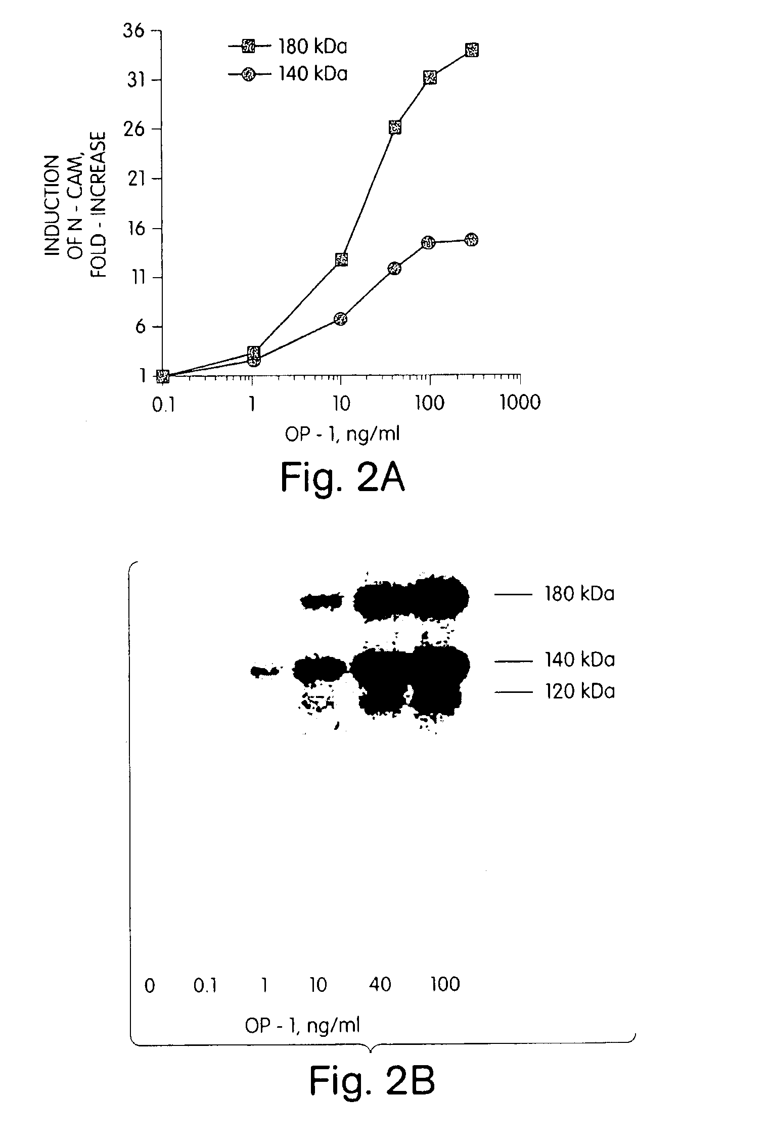
[0091]Morphogens have been identified in developing and adult rat brain and spinal cord tissue, as determined both by northern blot hybridization of morphogen-specific probes to mRNA extracts from developing and adult nerve tissue (see Example 1, above) and by immunolocalization studies. For example, northern blot analysis of developing rat tissue has identified significant OP-1 mRNA transcript expression in the CNS (U.S. Ser. No. 07 / 752,764, and Ozkaynak et al. (1991) Biochem. Biophys. Res. Comm., 179:11623 and Ozkaynak, et al. (1992) JBC, in press). GDF-1 mRNA appears to be expressed primarily in developing and adult nerve tissue, specifically in the brain, including the cerebellum and brain stem, spinal cord and peripheral nerves (Lee, S. (1991) PNAS 88: 4250-4254). BMP2B (also referred in the art as BMP4) and Vgr-1 transcripts also have been reported to be expressed in nerve tissue (Jones et al. (1991) Development 111:531-542), althoug...

example 3

Morphogen Enhancement of Neuronal Cell Survival

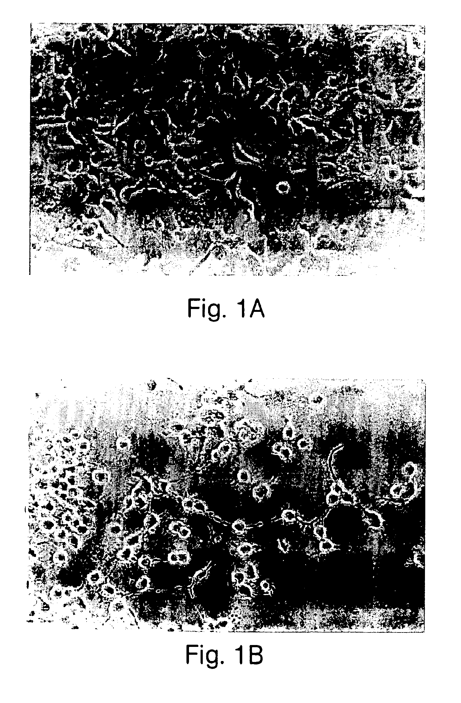
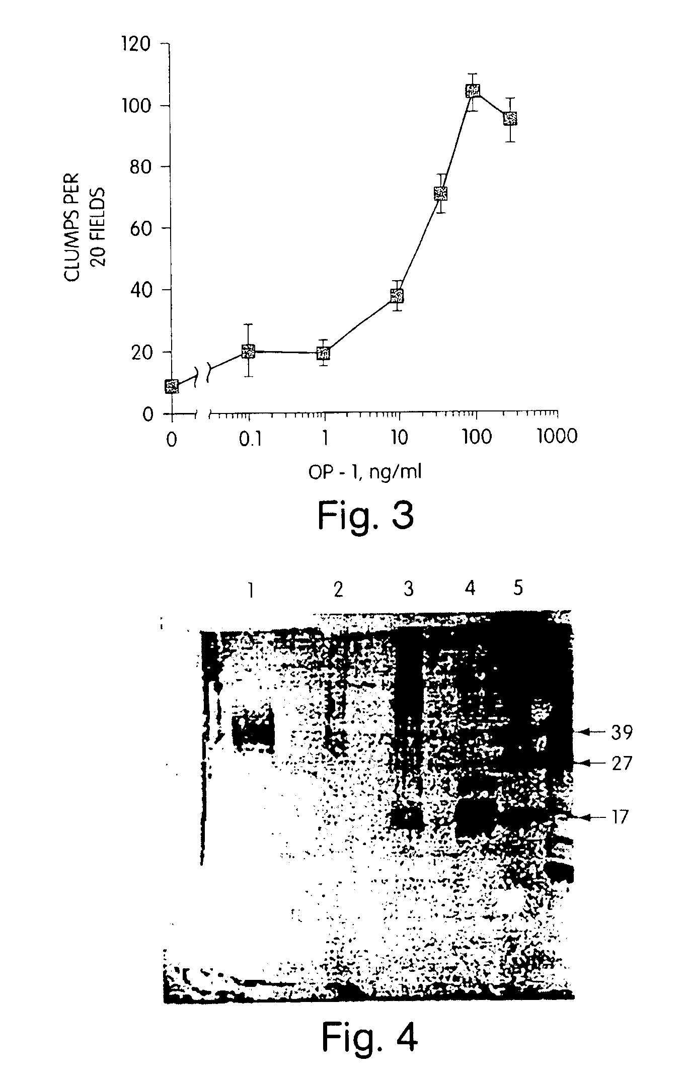
[0098]The morphogens described herein enhance cell survival, particularly of neuronal cells at risk of dying. For example, fully differentiated neurons are non-mitotic and die in vitro when cultured under standard mammalian cell culture conditions, using a chemically defined or low serum medium known in the art, (see, for example, Charness (1986) J. Biol. Chem. 26:3164-3169 and Freese et al. (1990) Brain Res. 521:254-264.) However, if a primary culture of non-mitotic neuronal cells is treated with a morphogen, the survival of these cells is enhanced significantly. For example, a primary culture of striatal basal ganglia isolated from the substantia nigra of adult rat brain was prepared using standard procedures, e.g., by dissociation by trituration with pasteur pipette of substania nigra tissue, using standard tissue culturing protocols, and grown in a low serum medium, e.g., containing 50% DMEM (Dulbecco's modified Eagle's medium), 50%...


PUM

PropertyMeasurementUnit
distanceaaaaaaaaaa
distanceaaaaaaaaaa
weightaaaaaaaaaa

Abstract

Disclosed are therapeutic treatment methods, compositions and devices for maintaining neural pathways in a mammal, including enhancing survival of neurons at risk of dying, inducing cellular repair of damaged neurons and neural pathways, and stimulating neurons to maintain their differentiated phenotype. In one embodiment, the invention provides means for stimulating CAM expression in neurons. The invention also provides means for evaluating the status of nerve tissue, including means for detecting and monitoring neuropathies in a mammal. The methods, devices and compositions include a morphogen or morphogen-stimulating agent provided to the mammal in a therapeutically effective concentration.

Description

CROSS REFERENCE TO RELATED APPLICATIONS[0001]This application is a continuation of U.S. Ser. No. 08 / 260,675, filed Jun. 16, 1994, now U.S. Pat No. 6,800,603, which is a file wrapper continuation of U.S. Ser. No. 08 / 126,100, filed Sep. 23, 1993, now abandoned, which is a file wrapper continuation of U.S. Ser. No. 07 / 922,813, filed Jul. 31, 1992, now abandoned, which is a continuation-in-part of both U.S. Ser. No. 07 / 752,764, now abandoned, and U.S. Ser. No. 07 / 753,059, now abandoned, both filed Aug. 30, 1991 and both continuations-in-part of U.S. Ser. No. 07 / 667,274, filed Mar. 11, 1991, now abandoned.BACKGROUND OF THE INVENTION[0002]The present invention relates to methods for enhancing the survival of neuronal cells in vivo and to methods, compositions and devices for maintaining neural pathways in vivo. More particularly, the invention provides methods for enhancing survival of neuronal cells at risk of dying, including methods for redifferentiating transformed cells of neural ori...

Claims


Application Information

Patent Timeline
Patent Type & Authority Patents(United States)
IPC IPC(8): A61K38/00C12N5/08G01N33/53C07K2/00G01N33/567C07K5/00C12N5/00C12N5/06A01N1/02A61F2/00A61K6/00A61K38/17A61K38/18A61L27/22A61L27/24C07K14/495C07K14/51C07K16/22
CPCA61K38/17A61K38/1703A61K38/1875A61L27/227A61L27/24A01N1/0226C07K14/51C07K16/22C07K14/495A61F2310/00365G01N2500/10A61K2300/00A61L2430/32
Inventor RUEGER, DAVID C.KUBERASAMPATH, THANGAVELOPPERMANN, HERMANNOZKAYNAK, ENGINPANG, ROY H. L.COHEN, CHARLES M.
Owner STRYKER CORP
Why Eureka
Try Eureka