

Antigenic modulation of cells
a cell and antigen technology, applied in the field of nonimmunogenic cellular compositions, can solve the problems of many organ transplant failures, large clinical problems, and significant transfusion reactions (to both major and minor red blood cell antigens), and achieve the effects of minimal modification of red blood cell stability, and increased red blood cell lysis
- Summary
- Abstract
- Description
- Claims
- Application Information
AI Technical Summary
Benefits of technology
Problems solved by technology
Method used
Image

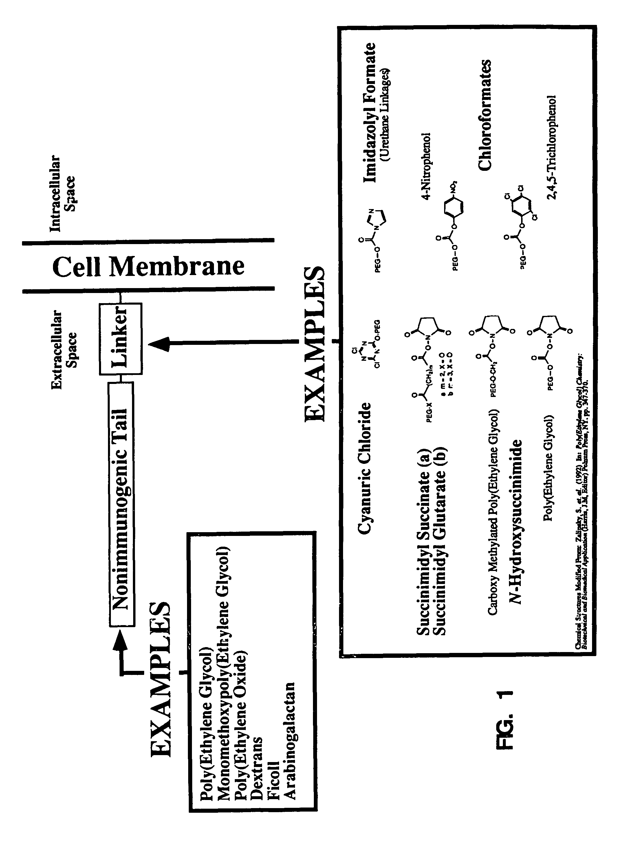
Examples
example i
Inhibition of Red Blood Cell Agglutination
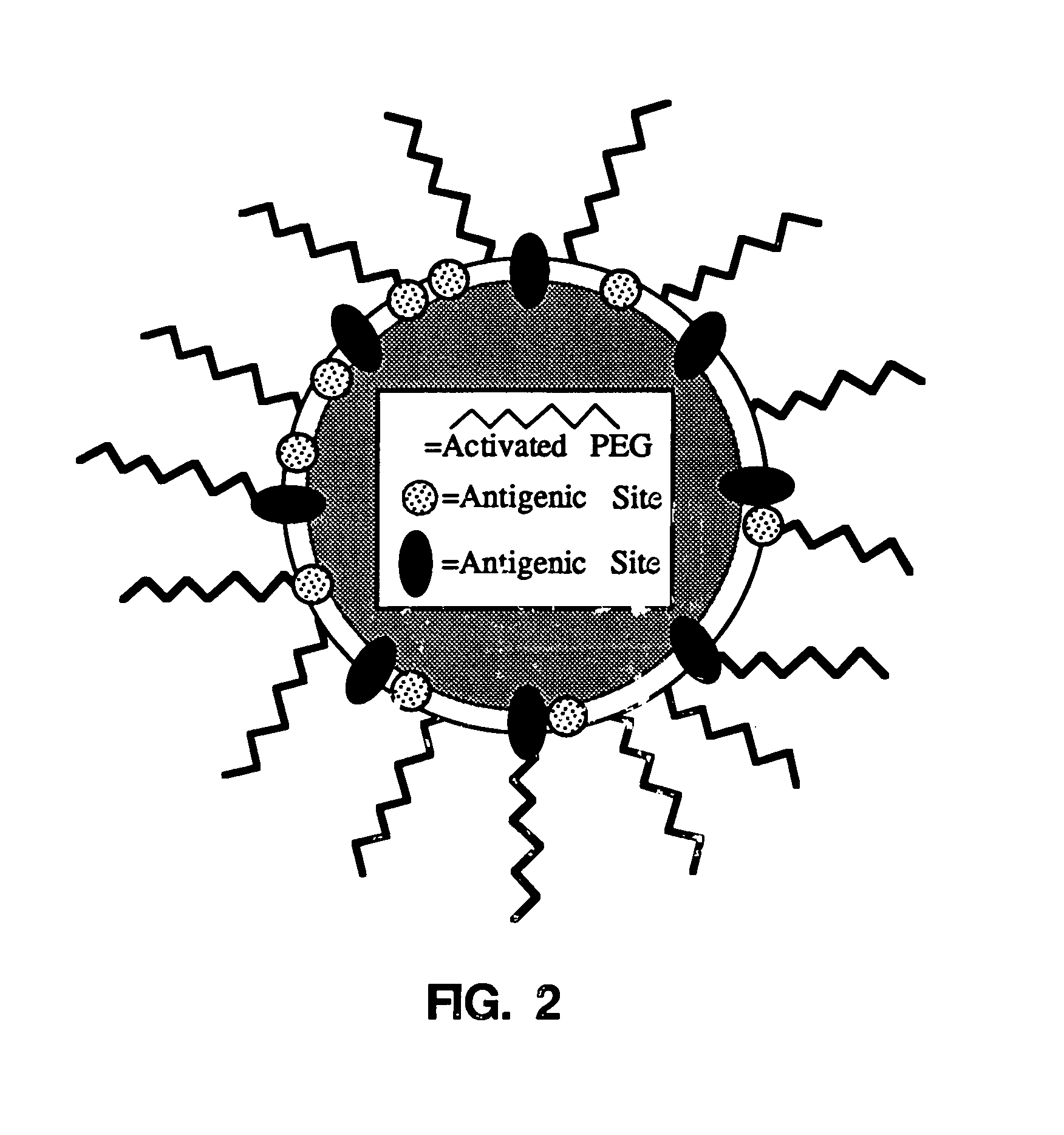
[0080]Normal red blood cells (erythrocytes) were washed 3× in isotonic saline. A red blood cell suspension of hematocrit about 12% is prepared in isotonic alkaline phosphate buffer (PBS; 50 mM K2HPO4 and 105 mM NaCl, pH about 9.2). Cyanuric chloride-activated methoxypolyethylene glycol (Sigma Chemical Co.) is added and the red cells are incubated for 30 minutes at 4° C. Cell derivatization can also be done under other pH and temperature conditions with comparable results to those presented. For example, red blood cells derivatized at pH 8.0 for 60 minutes at 22° C. demonstrated virtually identical characteristics to those derivatized at pH 9.2 for 30 minutes at 4° C. The extreme range of pH and temperature conditions make this procedure broadly applicable to a wide range of cells and tissues. The proposed mechanism of covalent reaction with external proteins and other membrane components is outlined below. Typical activated mPEG concentratio...
example ii
Effect on Red Blood Cell Stability
[0087]While mPEG-modification of red blood cells slightly increases red blood cell lysis, this lysis is less than 5% of the total red blood cell mass (FIG. 4). Furthermore, mPEG-attachment was found to have no effect on red blood cell osmotic fragility (FIG. 5). Red blood cell stability was minimally modified by the covalent attachment of mPEG. As shown in FIG. 4, red blood cell lysis was slightly increased by the attachment of mPEG. However, red blood cell lysis of the RBC during mPEG modification followed by 24 hours storage at 4° C. or after incubation at 37° C. was less than 5%. As shown in FIG. 5, osmotic fragility of the mPEG-treated red blood cells was also unaffected. Shown are the osmotic fragility profiles of control and mPEG-modified (3 and 6 mg / ml) red blood cells after 48 hours incubation at 37° C. Again, while a very minor increase in spontaneous lysis was observed, no significance differences in the osmotic lysis profiles were seen. E...
example iii
Inhibition of Antibody Binding
[0088]mPEG-modified red blood cells bind significantly less anti-A antibody (FIG. 6). As shown in FIG. 6, an ELISA assay of mPEG-treated human blood type A− red blood cells demonstrates significantly less antibody binding by mPEG-modified red blood cells. The control and mPEG red blood cells were mixed with an IgG anti-A antibody incubated for 30 minutes. The samples were extensively washed and a secondary antibody (anti-human IgG conjugated with alkaline phosphatase) was added to quantitate bound anti-Blood group A antibody.
PUM
| Property | Measurement | Unit |
|---|---|---|
| molecular weight | aaaaa | aaaaa |
| molecular weights | aaaaa | aaaaa |
| molecular weights | aaaaa | aaaaa |
Abstract
Description
Claims
Application Information