Use of adipose-tissue cell fractions for post-irradiation tissue regeneration

a technology of adiposetissue cells and adiposetissue cells, which is applied in the direction of biocide, plant growth regulators, biochemistry apparatus and processes, etc., can solve the problems of inability to repair damaged skin, so as to reduce tissue elasticity and improve skin regeneration. , the effect of increasing the production of collagen

Active Publication Date: 2012-05-08
INSTITUT DE RADIOPROTECTION & DE SURETE NUCLEAIRE
View PDF8 Cites 2 Cited by
  • Summary
  • Abstract
  • Description
  • Claims
  • Application Information

AI Technical Summary

Benefits of technology

[0027]Lesions and more particularly wounds and / or skin burns caused by irradiation, whether accidental or as a result of radiotherapy treatment, differ in nature from physiopathological trauma. The epidermis and keratinocytes are affected, tissue elasticity is reduced and the healing process is considerably slower.
[0028]The use of cells derived from the VSF of extramedullary adipose tissue acts by accelerating wound closure by acting at the level of keratinocyte differentiation and / or skin viscoelasticity, in particular by increasing the production of collagen. This has the advantage of giving a large quantity of tissues and cells and does not pose any particular ethical problems. It is moreover possible to envisage homologous or heterologous grafts and the possibility of treating several persons from a single individual. Implementation of the use according to the invention is made greatly easier by the source of the tissues used. It is indeed possible to maintain, multiply and even immortalise cells in vitro in a given medium. Freezing the cells can be envisaged in order to make cell pools available.
[0029]As lesions, and, more particularly, cutaneous lesions, are repaired much more quickly when used according to this invention, tissues are protected more quickly against external pathogenic agents.
[0030]The advantages in terms of costs are also considerable. Obtaining stem cells is easier because of their origin and the protocols are more straightforward and less expensive. As a matter of fact, many samples can be taken, notably during liposuction or dermolipectomy, those samples being usually destroyed. Sampling methods are not very invasive and as a result hospital and care time are much shorter. All these factors contribute to reducing the cost of the treatment described above.

Problems solved by technology

Disruption of these complex mechanisms has incapacitating consequences for the patients, in particular physical consequences or consequences on quality of life that can be as drastic as death.
Nevertheless, it has been shown that many pathological situations lead to a dysfunction in angiogenesis, loss of keratinocytes as well as impairment of skin regeneration.
However, these methods require cutting-edge technologies and are extremely expensive due to the availability, the obention and purification of raw materials.
They are also generally very dependent on potential contamination by micro-organisms and the relatively long period of time needed for the preparation of raw materials.
Several studies have shown that radiation can cause dry desquamation associated with atypical skin keratinisation or epiderm loss accompanied by ulceration.
These phenomena trigger a slowing down of the healing process.
In particular, local irradiation of the skin affects the healing process considerably.
Dilatation of the capillaries also causes acute erythema.
In particular, the fact that a drug is effective in improving the healing of mechanically caused wounds does not necessarily mean that this drug is effective in the treatment of lesions, notably cutaneous, linked to irradiation, because the healing of post-irradiation lesions involves much more complex mechanisms.
The use of stem cells is limited by their low number in most adult tissues and by the difficulty in extracting and purifying them.

Method used


Image

  • Use of adipose-tissue cell fractions for post-irradiation tissue regeneration
  • Use of adipose-tissue cell fractions for post-irradiation tissue regeneration
  • Use of adipose-tissue cell fractions for post-irradiation tissue regeneration

Examples


example 1

Obtaining and Preparing Adipose Tissue

[0050]An adipose tissue fragment is removed from subcutaneous adipose tissue taken from anaesthetised mice. After digestion of the extracellular matrix by proteolytic enzymes at 37° C. in collagenase for 45 minutes, to allow dissociation of tissue cells, various cell populations are selected by density difference according to the procedure described by Bjorntorp et al (6) or by the difference in antigen expression. The cell fraction that does not contain adipocytes corresponds to the vascular stroma fraction. Cells are labelled with CD34+, CD45− and CD31.

example 2

Effect of Cell Fractions According to the Invention on Healing

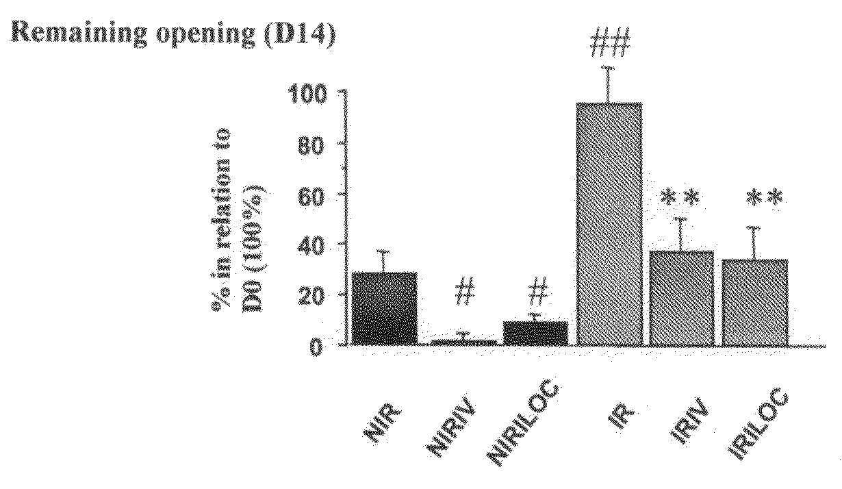
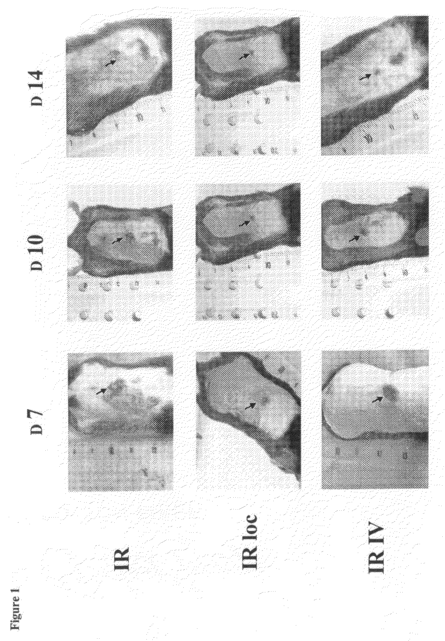
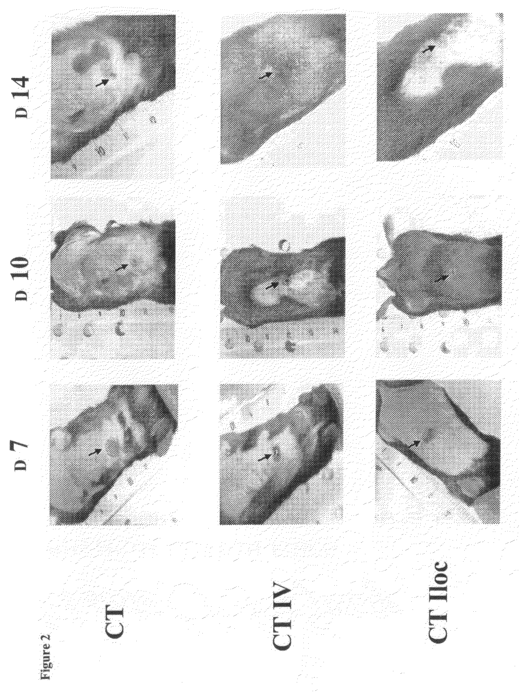
Localised irradiation with 20 Gy is carried out on the dorsal side of the mouse after shaving and anaesthetisia. Irradiation is carried out prior to application of a punch.

[0051]Wound Healing Model:

[0052]A 0.8 mm diameter punch is applied to the dorsal side of 8-week-old male mice (C57B16, Iffa Creddo) in order to produce a wound. Immediately after application of the punch, the cells obtained in Example 1 are injected locally (loc) or intravenously (iv). Animals receive 1×10⁶ cells and controls receive the solvent (saline phosphate buffer).

[0053]Each experiment group includes 5 individuals.

The experiments are repeated without preliminary irradiation of mice.

[0054]Technical Evaluation of Wound Healing:

The reduction in wound surface area is evaluated by direct observation at different times (D0, D4, D7, D10 and D14). Changes in the wounds of mice are monitored photographically. The results are reported in FIG. 1 (IR, IR Loc...


PUM

PropertyMeasurementUnit
diameteraaaaaaaaaa
timeaaaaaaaaaa
mechanicalaaaaaaaaaa

Abstract

The present invention concerns the use of cells derived from the cellular fraction of the vascular stroma of the extramedullary adipose tissue to promote the regeneration of tissue following lesions caused by irradiation. More specifically, the use according to the invention aims to prepare a drug for promoting regeneration of the skin, and in particular to repair the cutaneous wounds caused by irradiation.

Description

CROSS-REFERENCE TO RELATED APPLICATIONS[0001]The present application is a national phase entry under 35 U.S.C. §371 of International Application No. PCT / EP2007 / 055782, filed on Jun. 12, 2007, which claims priority from French Patent Application No. 0605190 filed Jun. 12, 2006, both of which are incorporated herein by reference.[0002]The present invention concerns the use of cells derived from the fraction of the vascular stroma of extramedullary adipose tissue to promote the regeneration of tissue following lesions caused by irradiation.BACKGROUND OF THE INVENTION[0003]The healing process involves a set of complex and perfectly orchestrated biological and molecular mechanisms, such as cell migration, cell proliferation or the depositing of extracellular matrix. An induced lesion constitutes the starting point for a cascade of events comprising interactions between local, regional and systemic growth factors, as well as participation at the cellular level of various elements such as ...

Claims


Application Information

Patent Timeline
Patent Type & Authority Patents(United States)
IPC IPC(8): A61K35/12C12N5/02A61K35/35
CPCA61K35/35A61L27/3834A61L27/60A61P1/00A61P11/00A61P15/00A61P17/00A61P17/02A61P19/00A61P35/00A61P39/00A61P43/00A61P9/00
Inventor TAMARAT, RADIABENDERITTER, MARC
Owner INSTITUT DE RADIOPROTECTION & DE SURETE NUCLEAIRE
Why Eureka
Try Eureka