Biocompatible scaffold for ligament or tendon repair
a biocompatible, ligament or tendon technology, applied in the field of biocompatible tissue implant devices, can solve the problems of surgical intervention, articular cartilage tissue injury will not heal spontaneously, and the cartilage and underlying bone of the joint will gradually be destroyed
- Summary
- Abstract
- Description
- Claims
- Application Information
AI Technical Summary
Benefits of technology
Problems solved by technology
Method used
Image
Examples
example 1
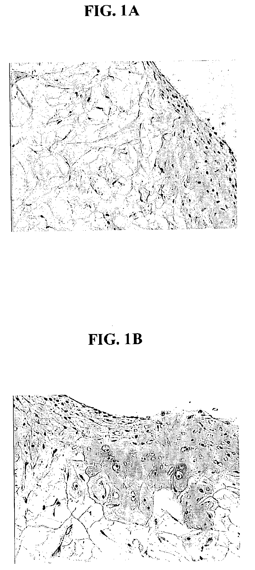
[0123]Healthy cartilage tissue from articulating joints was obtained from bovine shoulders. The cartilage tissue, which was substantially free of bone tissue, was minced using scalpel blades to obtain small tissue fragments in the presence of 0.2% collagenase. The size of the tissue fragments varied but on average should be approximately 1×1 mm in dimension. The minced tissue was then distributed uniformly on a 4×5 cm synthetic bioresorbable polycaprolactone / polyglycolic acid (PCL / PGA) scaffold. Ethylene oxide sterilized polymer scaffolds, were pre-soaked for 4 hours in Dulbecco's Modified Eagle's Medium prior to distribution of tissue fragments. The scaffold loaded with minced fragments was then placed in a 10 cm cell culture dish containing chondrocyte growth medium. The chondrocyte growth medium consisted of Dulbecco's modified eagles medium (DMEM-high glucose) supplemented with 20% fetal calf serum (FCS), 10 mM HEPES, 0.1 mM nonessential amino acids, 20 mg / ml of L-proline, 50 mg...
example 2
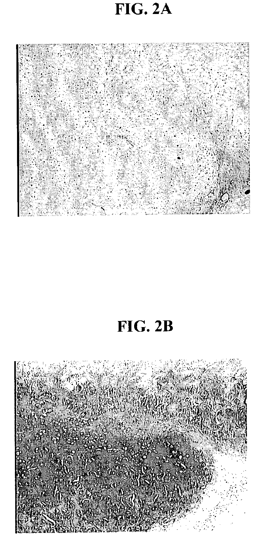
[0124]The bioresorbable scaffolds containing minced cartilage tissue and cells from Example 1 were also implanted into SCID mice. The objective was to evaluate the chondrocytic ingrowth of minced cartilaginous tissues into polymer scaffolds in vivo. Polymer scaffolds 5 mm in diameter, were subcutaneously implanted bilaterally in the lateral thoracic region of SCID mice. The implanted scaffold was permitted to support cell growth for four weeks. The subcutaneous implantation sites with their overlying skin were then excised and preserved in 10% buffered formalin fixative. Following fixation, each implantation site was processed for histology. Histological sections were stained with Hematoxylin and eosin, and Safranin-O. FIGS. 2 A and B show that abundant cells were distributed within the scaffold. The cells displayed chondrocyte-like morphology, as evidenced by the intense positive staining for Safranin O of the synthesized matrix.
example 3
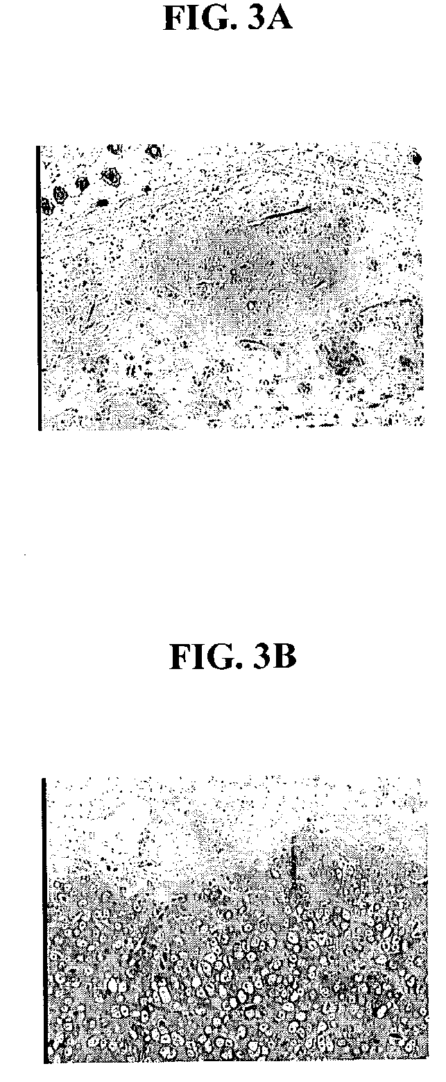
[0125]Minced cartilage tissue prepared according to the method described in Example 1 was distributed uniformly on a 4×5 cm synthetic bioresorbable polycaprolactone / polyglycolic acid (PCL / PGA) scaffold. Minced cartilage tissue fragments were adhered to the scaffolds with 1 mL of platelet rich plasma (PRP, Human). Sixty microliters (60 units) of thrombin were used to induce clot formation in the PRP. Control scaffolds loaded with minced cartilage fragments alone and scaffolds loaded with minced cartilage fragments adhered by PRP, were cultured in vitro for 1 week, and then implanted into SCID mice as described in the Example 2. FIG. 3A is a photomicrograph of a control scaffold loaded with minced tissue. FIG. 3B is a photomicrograph depicting a scaffold loaded with minced tissue and PRP. FIG. 3B demonstrates that PRP is beneficial in promoting the migration of the chondrocyte cells, and PRP is also beneficial in promoting the maintenance of the differentiated phenotype of the chondro...
PUM
 Abstract
Description
Claims
Application Information
 - R&D
- Intellectual Property
- Life Sciences
- Materials
- Tech Scout
- Unparalleled Data Quality
- Higher Quality Content
- 60% Fewer Hallucinations
Browse by: Latest US Patents, China's latest patents, Technical Efficacy Thesaurus, Application Domain, Technology Topic, Popular Technical Reports.
© 2025 PatSnap. All rights reserved.Legal|Privacy policy|Modern Slavery Act Transparency Statement|Sitemap|About US| Contact US: help@patsnap.com


