Preparation method of biological activity protein or polypeptides coating biology bracket

A bioactive protein and bio-scaffold technology, which is applied in the field of bio-scaffold preparation, can solve the problems of inflammation and allergic reaction clinical experiment results failure, etc.

Active Publication Date: 2009-03-25
LEPU MEDICAL TECH (BEIJING) CO LTD
View PDF6 Cites 19 Cited by
  • Summary
  • Abstract
  • Description
  • Claims
  • Application Information

AI Technical Summary

Problems solved by technology

However, these stents did not have good results in the clinical trial stage as shown in animal experiments, probably because they set up a layer of matrix layer on the stent body in order to

Method used


Image

  • Preparation method of biological activity protein or polypeptides coating biology bracket
  • Preparation method of biological activity protein or polypeptides coating biology bracket
  • Preparation method of biological activity protein or polypeptides coating biology bracket

Examples


Embodiment 1

[0049] Example 1 Preparation of Monoclonal Antibody CD34 Coated Biological Scaffold

[0050] After polishing, 316L stainless steel alloy or cobalt complex alloy material was used to engrave a vascular stent with a laser, and the monoclonal antibody CD34-coated biological stent was prepared according to the following steps:

[0051] 1. Cleaning of the surface of the bracket body:

[0052] (1) Grinding the surface of the bracket body with an abrasive belt, mechanically removing oxides on the surface, and adjusting the surface roughness;

[0053] (2) Use ultrasonic waves to clean with 75% medical ethanol solution, 99.5% acetone solution, and deionized water in sequence. The ultrasonic frequency is 100KHz, and the cleaning time is 10 minutes;

[0054] (3) Soak the stent body in a 30g / L, 80°C sodium hydroxide solution for 10 minutes to remove grease and scale;

[0055] (4) Place the cleaned stent body in a vacuum dryer, set the temperature at 40° C., and take it out after drying ...

Embodiment 2

[0062] Example 2 Preparation of monoclonal antibody CD34-coated bioscaffold with nanopores

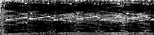
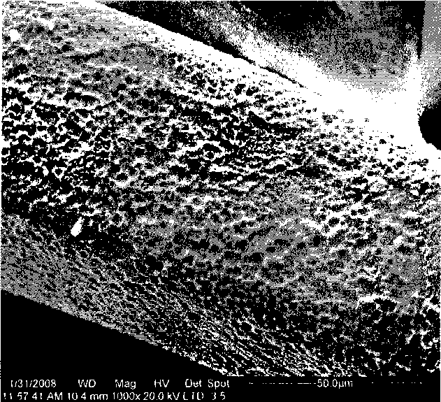
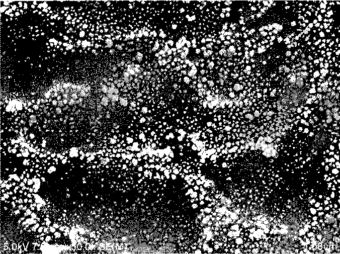
[0063] After polishing, 316L stainless steel alloy or cobalt complex alloy material is used to engrave a vascular stent with a laser; the surface of the stent is processed by chemical or physical methods, such as corrosion, anodic oxidation, micro-arc oxidation, micro-arc nitriding, etc. or a combination of these methods Holes are directly formed on the local surface of the raw material of the device body. The holes can be drug-loading grooves or pore structures, and the diameter of the holes is 200-300nm. Prepare the monoclonal antibody CD34-coated bioscaffold according to the following steps:

[0064] 1. Cleaning of the surface of the bracket body:

[0065] (1) Grinding the surface of the bracket body with an abrasive belt, mechanically removing oxides on the surface, and adjusting the surface roughness;

[0066] (2) Use ultrasonic waves to clean with 75% medical ethanol solution, ...

Embodiment 3

[0075] Example 3 Preparation of antibody-drug combined coating bioscaffold with nanopores

[0076] After polishing, 316L stainless steel alloy or cobalt complex alloy material is used to engrave a vascular stent with a laser; the surface of the stent is processed by chemical or physical methods, such as corrosion, anodic oxidation, micro-arc oxidation, micro-arc nitriding, etc. or a combination of these methods Holes are directly formed on the local surface of the raw material of the device body. The holes can be drug-loading grooves or pore structures, and the diameter of the holes is 200-300nm. Prepare the monoclonal antibody CD34-coated bioscaffold according to the following steps:

[0077] 1. Cleaning of the surface of the bracket body:

[0078] (1) Grinding the surface of the bracket body with an abrasive belt, mechanically removing oxides on the surface, and adjusting the surface roughness;

[0079] (2) Use ultrasonic waves to clean with 75% medical ethanol solution, 9...


PUM

PropertyMeasurementUnit
Diameteraaaaaaaaaa
Diameteraaaaaaaaaa

Abstract

The invention relates to a preparation method of a bioactive protein or polypeptide coat biological bracket. The method of anode polarization is adopted, a bracket body is taken as the anode, and is inserted into bioactive protein or polypeptide solution together with the cathode; under the action of the electric field of a power supply, the bioactive protein or polypeptide is ionized to have negative electrification to move towards the anode of the bracket and evenly coats on the surface of the bracket. The preparation method provided by the invention dose not need to additionally prepare a medicine loading coat, has no obvious interface, can envelop a bioactive protein or polypeptide coat on the whole bracket body or part surface of the bracket through the function of anode polarization; the bioactive protein or polypeptide has stable enveloping amount and is not easily eluted, has safe use, simple operation and low cost, and is suitable for large-scale production in industry.

Description

technical field [0001] The invention belongs to the field of biological scaffold preparation, and in particular relates to a method for preparing a bioactive protein or polypeptide-coated biological scaffold. Background technique [0002] Atherosclerosis causes the stenosis of the blood vessel lumen, which leads to the obstacle of blood supply and oxygen supply to the muscle and causes muscle ischemia and necrosis in the corresponding part. For half a century, arterial vascular diseases such as coronary heart disease have become one of the diseases that seriously threaten human health. [0003] In 1976, German scholar Andrea Gronzgo first proposed the idea of ​​installing stents in arteries. By the 1990s, coronary stents had been widely used in clinical treatment. Vascular stents are the main means of interventional therapy to prevent cardiovascular and peripheral vascular occlusive lesions. Keep the lumen open. Vascular stents can be divided into bare stents, drug-elutin...

Claims


Application Information

Patent Timeline
IPC IPC(8): A61L27/28A61L27/06A61L27/04A61L27/54A61L29/08A61L29/02A61L29/16A61L31/08A61L31/02A61L31/16A61F2/82
Inventor 陈永强余占江张正才蒲忠杰
Owner LEPU MEDICAL TECH (BEIJING) CO LTD
Why Eureka
Try Eureka