
Tissue engineering material-based culture method and applications of melanophore

A melanocyte and cell culture technology, applied in the field of biomedicine, can solve the problems of cell culture and biosafety, easily damaged tissue engineering epidermis, unable to meet transfer and transplantation and other problems, achieve good mechanical properties, maintain cell activity and shape good effect

Active Publication Date: 2010-10-13
HANGZHOU THIRD HOSPITAL +1
View PDF5 Cites 16 Cited by
  • Summary
  • Abstract
  • Description
  • Claims
  • Application Information

AI Technical Summary

Problems solved by technology

CN1786155A discloses selecting the membrane material or porous support of chitosan-collagen complex as load material, epidermal cells and melanocytes are inoculated on membrane surface or support, constructs the tissue engineering epidermis substitute with pigment, but this tissue engineering The epidermis is easily damaged during the later transfer and transplantation process
CN 1493367 and CellBiology International 2007, 31 (9): 985-990 have reported that collagen is selected as a material, loaded with keratinocytes, fibroblasts and melanocytes to construct artificial skin containing pigment, and expressed on nude mice, but The artificial skin will shrink during the preparation process, which will affect its subsequent use
CN 101605566A discloses the cultivation of proliferative melanocytes on polylactic acid film, but the preparation of polylactic acid film needs to use solvent casting method, which leaves hidden dangers for cell culture and biological safety
In addition, Sung-Jan Lin et al. (Biomaterials2005, 26 (12): 1413-1422) reported that chitosan was used as a load to inoculate melanocytes, and it was found that melanocytes clustered on the membrane, which affected cell viability. The prepared chitosan carrier cannot meet the requirements of transfer and transplantation

Method used


Image

  • Tissue engineering material-based culture method and applications of melanophore
  • Tissue engineering material-based culture method and applications of melanophore
  • Tissue engineering material-based culture method and applications of melanophore

Examples

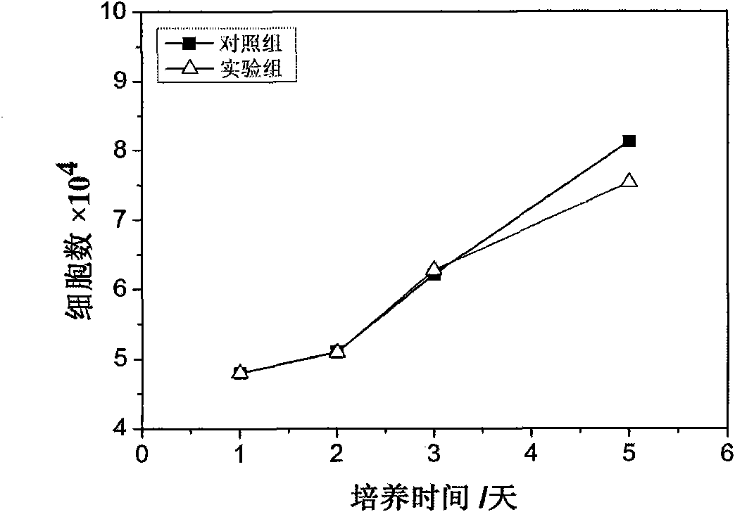
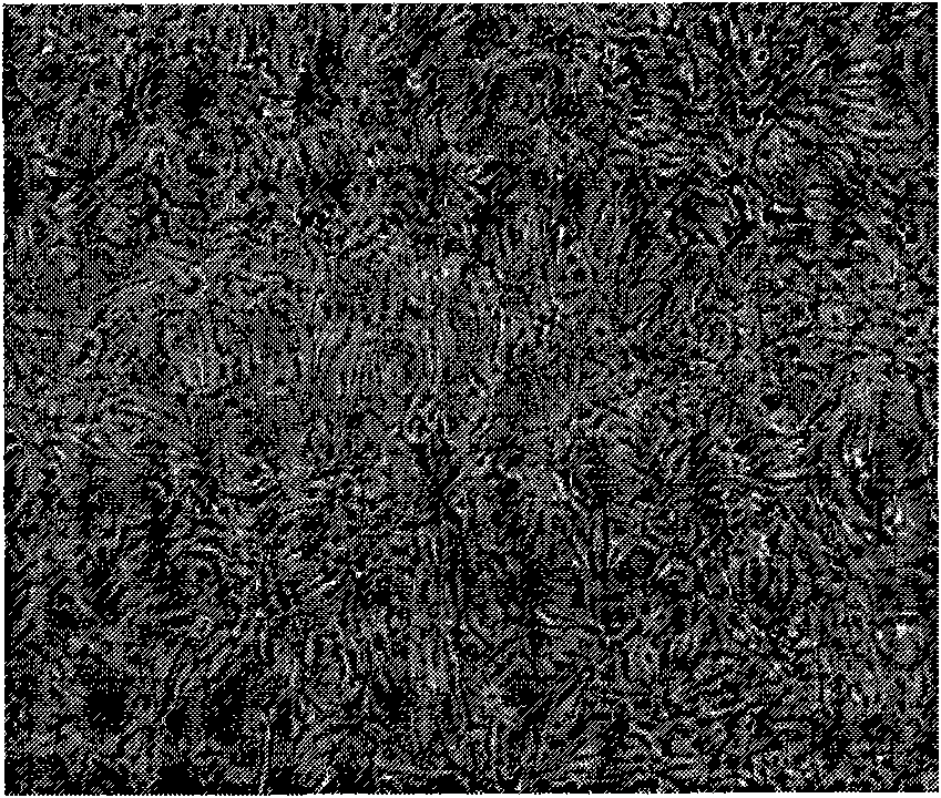
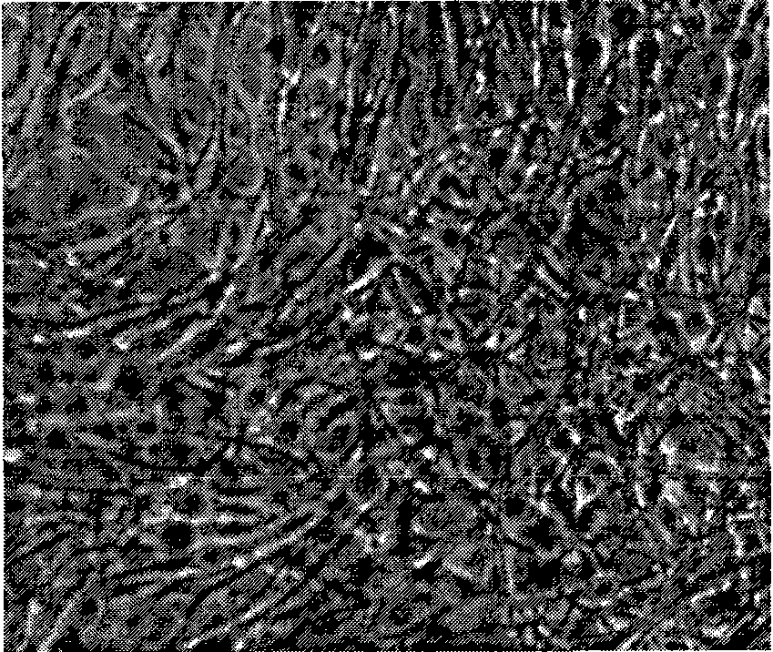

Embodiment 1

[0029] Embodiment 1. Preparation of chitosan-based carrier film material

[0030] a) dissolving chitosan with 1M acetic acid solution to make a chitosan solution with a concentration of 1.5%;

[0031] b) 2.5ml of chitosan solution was injected into the cell culture plate, the cell culture plate was placed in a blast oven, and dried at 50° C. for 6 hours;

[0032] c) Place the dried chitosan film in 0.5M Na 2 SO 4 Soak in the solution for 30 minutes, then rinse with plenty of pure water;

[0033] d) Soak the chitosan membrane with 0.1M NaOH solution for 30min to remove residual acetic acid on the membrane, and then wash it to pH=7.2-7.4 with a large amount of pure water;

[0034] e) placing the cell culture plate covered with the chitosan membrane in a blast oven, and drying at 37° C. for 24 hours;

[0035] f) obtaining a carrier membrane material that can be used for cell culture and transfer.

Embodiment 2

[0036] Embodiment 2. Preparation of chitosan-hyaluronic acid-based composite carrier membrane material

[0037] a) dissolving chitosan with 1M acetic acid solution to make a chitosan solution with a concentration of 1.5%;

[0038] b) Dissolving hyaluronic acid in purified water to prepare a 1% hyaluronic acid solution;

[0039] c) mixing chitosan and hyaluronic acid in a weight ratio of 20:1, stirring evenly to obtain a chitosan-hyaluronic acid mixed solution;

[0040] d) Inject 2.5ml of the mixed solution into the cell culture plate, place the cell culture plate in a blast oven, and dry at 50°C for 6 hours;

[0041] e) Place the dried chitosan-hyaluronic acid film in 0.5M Na 2 SO 4 Soak in the solution for 30 minutes, then rinse with plenty of pure water;

[0042] f) Soak the chitosan-hyaluronic acid film with 0.1M NaOH solution for 30 minutes to remove residual acetic acid on the film, and then wash it with a large amount of pure water to PH=7.2-7.4;

[0043] g) placing...

Embodiment 3

[0045] Embodiment 3. Preparation of chitosan-gelatin-based composite carrier membrane material

[0046] a) dissolving chitosan with 1M acetic acid solution to make a chitosan solution with a concentration of 1.5%;

[0047] b) dissolving the gelatin with 1M acetic acid solution to prepare a 1% gelatin solution;

[0048] c) mixing chitosan and gelatin in a weight ratio of 10:1, stirring evenly to obtain a chitosan-gelatin mixed solution;

[0049] d) Inject 2.5ml of the mixed solution into the cell culture plate, place the cell culture plate in a blast oven, and dry at 50°C for 6 hours;

[0050] e) Place the dried chitosan-gelatin film in 0.5M Na 2 SO 4 Soak in the solution for 30 minutes, then rinse with plenty of pure water;

[0051] f) Soak the chitosan-gelatin film with 0.1M NaOH solution for 30 minutes to remove residual acetic acid on the film, and then flush it to PH=7.2-7.4 with a large amount of pure water;

[0052] g) placing the cell culture plate covered with the...


PUM


Abstract

The invention discloses a tissue engineering material-based culture method and applications of melanophore. In the method of the invention, the tissue engineering technology is used to build carrier material with good biocompatibility and perform culture (co-culture) and transfer of melanophore or melanophore, fibroblast and keratinocyte and the method can be used in depigmentation diseases such as vitiligo and for the regulation of skin color. The invention relates to the preparation of the carrier material with biocompatibility and the building and transplanting of the cell-carrier composite material. The carrier material can be used to provide good carrier for cell culture, maintain cellular activity and solve the problem that the cell suspension is easy to lose in the cellular transplantation process; the inoculum density and culture time of cells can be regulated at the same time, the regulation to the skin colors of different individuals can be realized; and the carrier materialhas good performances such as damage resistance, tear resistance and easy transfer, has good function of promoting wound healing, and satisfies the use requirements of the large-area depigmentation disease and the regulation of skin color.

Description

technical field [0001] The invention belongs to the field of biomedicine, and in particular relates to a method for culturing melanocytes based on tissue engineering materials and its application. Background of the invention [0002] Vitiligo is an acquired skin depigmentation disease, manifested as localized or generalized depigmentation, characterized by the formation of white spots, and histology and immunocytochemistry showed that the epidermal melanocytes of the lesions disappeared. Vitiligo often brings a heavy psychological burden to patients, affects normal life such as work and study, and easily develops into a mental illness. [0003] Traditional treatment methods include oral medicine, ultraviolet radiation and surgical treatment. But drug therapy and ultraviolet light exposure have limited cure rates. Surgical treatment methods mainly include autologous epidermal transplantation, melanocyte suspension transplantation, etc. These treatment methods have been prov...

Claims


Application Information

Patent Timeline
IPC IPC(8): A61L27/60C12N5/071
Inventor 许爱娥王文俊董东许思原傅丽芳尉晓冬
Owner HANGZHOU THIRD HOSPITAL
Learn More