
Method for detecting tumor mutant gene in blood

A technology for mutated genes and tumors, applied in the field of capillary electrophoresis detection, to achieve the effect of ensuring new reproduction, ensuring accuracy, and less sample consumption

Inactive Publication Date: 2011-11-23
王荣
View PDF5 Cites 6 Cited by
  • Summary
  • Abstract
  • Description
  • Claims
  • Application Information

AI Technical Summary

Problems solved by technology

In the Chinese patent database from 1984 to 2010, the Chinese patent with the application number 200510026931.X "A PCR detection method and reagent system for tumor-related gene mutations" and the Chinese patent with the patent number 200910042599.4 "for the individualization of malignant tumors Gene chip and its application for drug-related gene mutation detection", Chinese patent "Method for detecting gene mutation" with application number 200810005523.X, and Chinese patent "Rapid detection of APC gene mutation" with application number 201010134207.X 200910201917.7 Chinese patent "A Primer for Detecting K-ras Gene Mutation and Its Application", the above-mentioned patent only discloses gene mutations using real-time quantitative fluorescent PCR, sequencing, high-resolution melting curve method, fluorescent double-loop probe technology, etc. The detection method detects tumor gene mutations in tissue or blood, but the method and application of capillary electrophoresis combined with SSCP and RFLP for detecting tumor mutation genes in blood have not been reported in the literature

Method used


Image

  • Method for detecting tumor mutant gene in blood
  • Method for detecting tumor mutant gene in blood
  • Method for detecting tumor mutant gene in blood

Examples


Embodiment 1

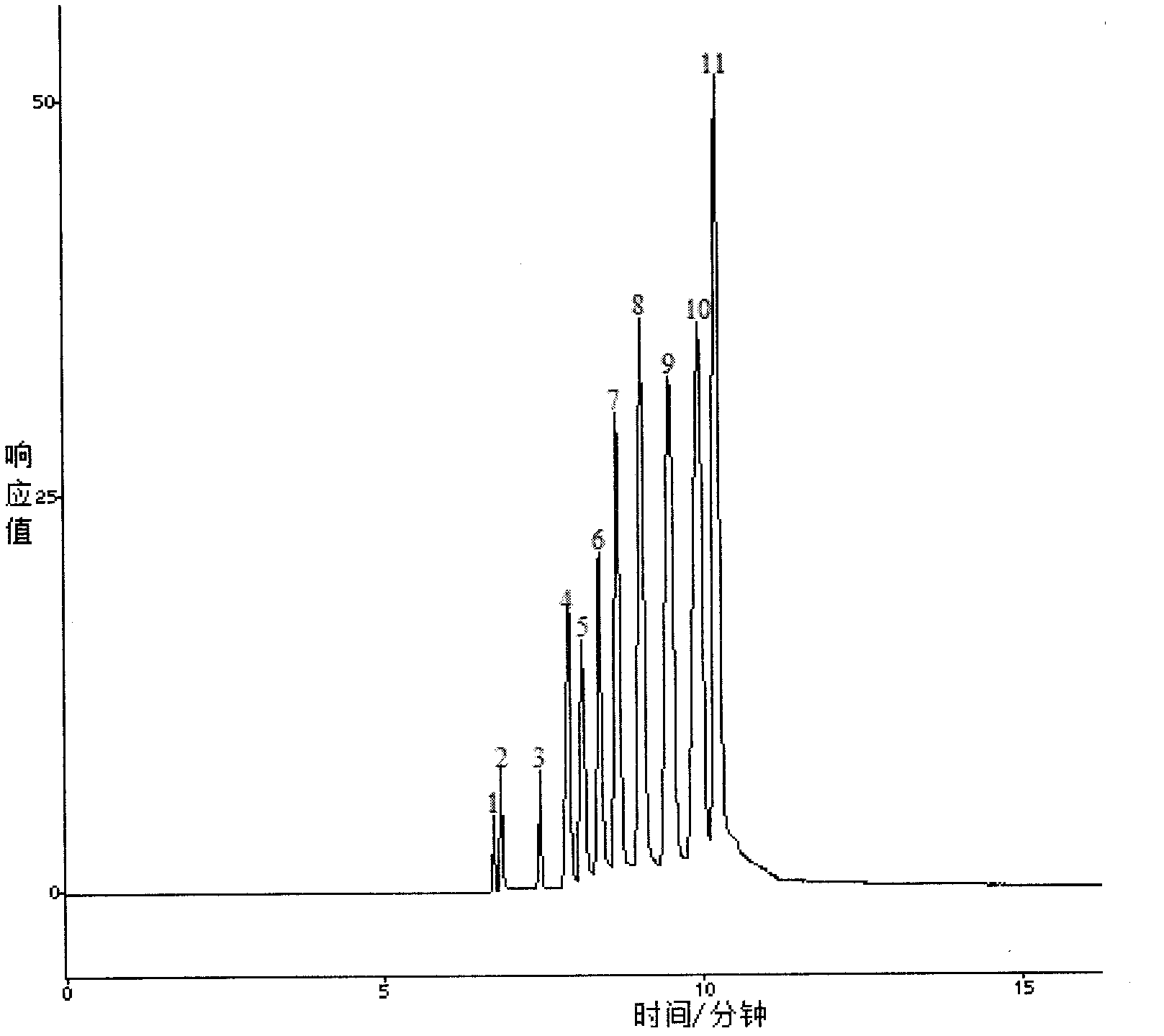
[0043] Example 1 A method for detecting tumor mutation genes in blood samples, which is used for the isolation of pUC19 DNA / MspI / HpaII DNA Marker.

[0044] (1) Preparation of polyethylene oxide sieving medium containing TBE:

[0045] Add polyethylene oxide into 1×TBE buffer solution to make the final solution concentration 3.0% and pH value 8.3, and use a DF-101S magnetic stirrer produced by Yuhua Instrument Co., Ltd., Gongyi City, Henan Province at 400 rpm Stir at a speed of 1 / min until the solution becomes clear, and the clear solution is filtered with a 0.45um water-based microporous membrane to obtain a polyethylene oxide sieving medium.

[0046] Among them: 1 × TBE buffer refers to adding 10.8 grams of Tris (Tris) produced by Shanghai Bioengineering Company, 5.5 grams of boric acid produced by Shanghai Shenggong Company, After stirring and dissolving 0.74 g of ethylenediaminetetraacetic acid disodium salt (EDTA) produced, add deionized water to dilute the solution to 1 l...

Embodiment 2

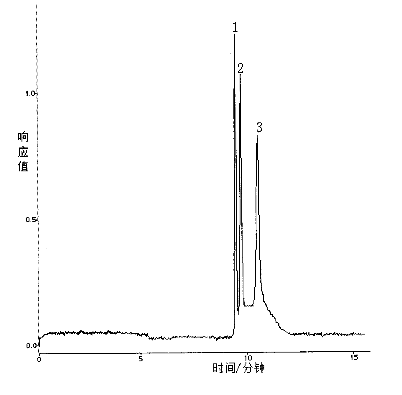
[0053] Embodiment 2 A method for detecting tumor mutation genes in blood, comprising the following steps:

[0054] (1) Preparation of polyethylene oxide sieving medium containing TBE:

[0055] Add polyethylene oxide into 1×TBE buffer solution to make the final solution concentration 2.5% and pH value 8.0, stir until the solution is clear, and filter the clear solution with a 0.22um water-based microporous membrane to obtain Polyethylene oxide sieving media.

[0056] Wherein the 1× TBE buffer solution is the same as in Example 1.

[0057] (2) Genomic DNA extraction from blood samples:

[0058] After adding Tris buffer Ⅰ to the blood sample to lyse the red blood cells, discard the supernatant, add Tris buffer Ⅱ to the pellet to suspend the cell mass, add proteinase K, and incubate in a water bath at 55°C for 4 hours; after the incubation, add phenol-chloroform to precipitate the protein , absorb the supernatant and add absolute ethanol to the supernatant, ice-bath for 1 h, an...

Embodiment 3

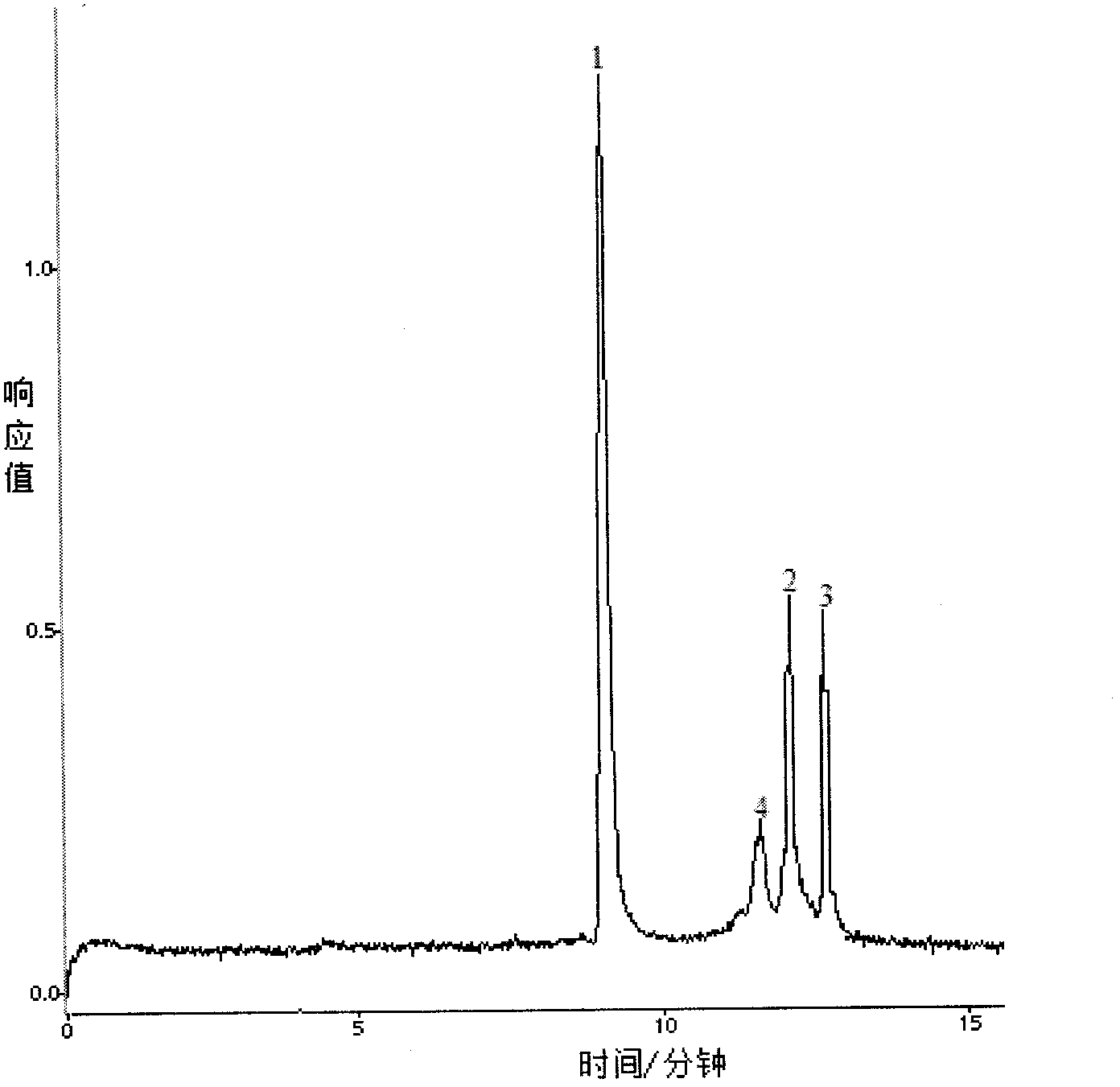
[0070] Embodiment 3 A method for detecting tumor mutation genes in blood, comprising the following steps:

[0071] (1) Preparation of polyethylene oxide sieving medium containing TBE:

[0072] Add polyethylene oxide into 1×TBE buffer solution to make the final solution concentration 2.5% and pH value 7.5, stir until the solution is clear, and filter the clear solution with a 0.22um water-based microporous membrane to obtain Polyethylene oxide sieving media.

[0073] Wherein the 1× TBE buffer solution is the same as in Example 1.

[0074] (2) Genomic DNA extraction from blood samples:

[0075] Add Tris buffer Ⅰ to the blood sample to lyse the red blood cells, discard the supernatant, add Tris buffer Ⅱ to the pellet to suspend the cell mass, add proteinase K, and incubate in a water bath at 56°C for 2 hours; after the incubation, add phenol-chloroform to precipitate the protein , absorb the supernatant and add absolute ethanol to the supernatant, ice-bath for 1 h, and centrif...


PUM


Abstract

The invention relates to a method for detecting a tumor mutant gene in blood. The method comprises the following steps of: (1) preparing a polyethylene oxide sieving medium containing TBE (Tetrabromoethane); (2) extracting a genome DNA (Deoxyribonucleic Acid) in a blood sample; (3) amplifying a gene target fragment needing to be detected: performing conventional PCR (Polymerase Chain Reaction) amplification by taking the genome DNA as an amplifying template to obtain a p53 gene PCR amplification sample and a k-ras gene PCR amplification sample; (4) treating the samples: performing denaturation or enzyme digestion on the p53 gene PCR amplification sample and the k-ras gene PCR amplification sample to obtain a denatured sample solution and an enzyme digestion solution respectively; and (5) detecting the samples: adding a 10*SYBR Green I fluorescence dye and the genome DNA into the denatured sample solution and the enzyme digestion solution respectively, uniformly mixing, adding deionized water till a system is 30-50 mu l, automatically filling the polyethylene oxide sieving medium by using a capillary electrophoresis apparatus pressure system and performing CE-SSCP (Capillary Electrophoresis-Single Strand Conformation Polymorphism) detection or CE-RFLP (Capillary Electrophoresis-Restriction Fragment Length Polymorphism) detection respectively. The method is easy to operate, and has high repeatability.

Description

technical field [0001] The invention relates to a capillary electrophoresis detection method in the field of analysis method application research, in particular to a method for detecting tumor mutation genes in blood. Background technique [0002] The detection of gene mutation plays a very important role in clinical disease diagnosis. At present, PCR-based polyacrylamide gel (PAGE) electrophoresis and various gene mutation detection techniques are often used to screen gene point mutations. These methods have certain advantages in meeting the needs of clinical rapid detection of gene mutations. limitation. Therefore, it is imperative to establish an accurate and rapid gene mutation detection method for clinical diagnosis. [0003] Capillary electrophoresis (CE) is a new separation and analysis technology combining electrophoresis and chromatography. It has the characteristics of high separation efficiency, fast analysis speed, less sample consumption and wide application r...

Claims


Application Information

Patent Timeline
Patent Type & Authority Applications(China)
IPC IPC(8): C12Q1/68G01N27/447
Inventor 王荣贾正平谢华谢希辉
Owner 王荣
Learn More