Culture method for retina progenitor cells and culture medium thereof
A retinal progenitor cell, cell culture technology, applied in animal cells, nervous system cells, vertebrate cells, etc., can solve problems such as plasma loss, pain caused by patients, physical diseases of patients, etc., to prevent premature aging or differentiation, Increase the number of growth and reproduction, break through the effect of small cell yield
- Summary
- Abstract
- Description
- Claims
- Application Information
AI Technical Summary
Problems solved by technology
Method used
Image
Examples
Embodiment 1
[0040] Embodiment 1: culture method of the present invention carries out retinal progenitor cell culture
[0041] Sphere-forming culture of retinal progenitor cells: After the tissue is sterilized, take the retinal tissue, digest it with enzymes, centrifuge at 1000r for 3 minutes, take the precipitate, and use DMEM / F12 basal medium (add N-2 supplement, the concentration is 1:100, Epidermal growth factor EGF, the concentration is 10ng / ml, basic growth factor bFGF, the concentration is 10ng / ml, L-glutamine, the concentration is 1:100) mix and count the sediment, according to 1-2×10 5 / cm 2 Inoculate in low-adsorption T25 culture flasks at 37°C, 5% CO 2 Cultured in a constant temperature incubator, the next day the cells formed into spheres.
[0042] Preparation of sodium alginate gel: Use deionized water as the solvent of sodium alginate, first weigh the sodium alginate powder, slowly add the powder into a centrifuge tube containing a small amount of solvent (20ml), and add th...
Embodiment 2
[0045] Embodiment 2: MTT assay cell viability and proliferation rate
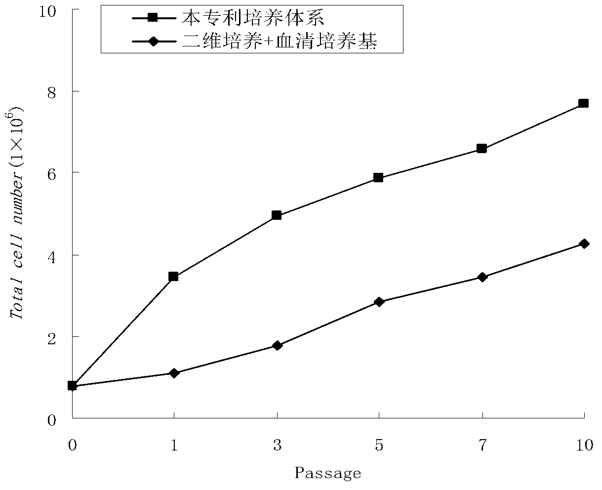
[0046] Referring to the experimental method described in Example 1, the retinal progenitor cells in the logarithmic growth phase of the third generation were taken, and a single cell suspension was prepared, and 750 cells / well were inoculated in the culture medium prepared in Example 1 and covered with sodium alginate gel. The MTT test was carried out every day within 6 days in a 96-well culture plate. At the same time, a traditional culture method was established as a control, that is, retinal progenitor cells were cultured in a suspension or adherent manner, and the medium was 10%-20% serum medium.
[0047] Add 10μl MTT (1g / L) to each well of cells and continue to culture for 4h. The culture medium was removed by centrifugation, the cells were lysed by adding dimethyl sulfoxide, and the OD value was measured at a wavelength of 570 nm. The wells without MTT were used as the background, and the OD values ...
Embodiment 3
[0049] Example 3: Cell cycle detection by flow cytometry
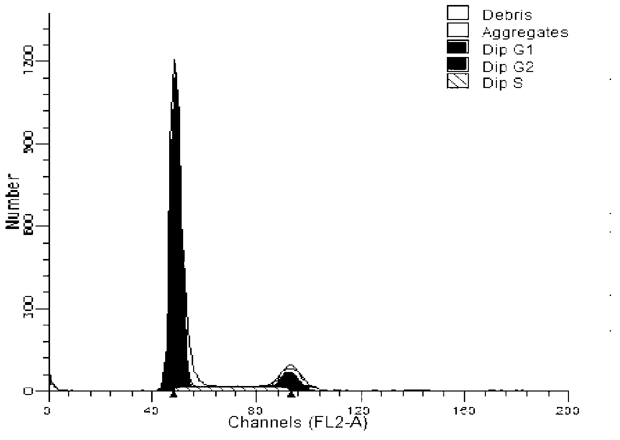
[0050] Take 1×10 retinal progenitor cells cultured by the method described in Example 1 6 One, adding propidium iodide PI dye, staining for 20min, adding PBS and centrifuging to wash, and flow cytometry to detect the cycle of the cells. Cell cycle detection uses PI (propidium iodide) staining to detect the cell cycle, which can be combined with DNA and RNA in cells. After RNA is digested with RNA inhibitors, the fluorescence intensity of PI bound to DNA detected by flow cytometry It directly reflects the amount of DNA in the cell.
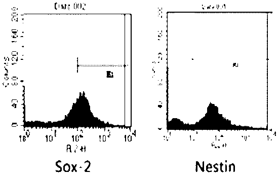
[0051] figure 2 and image 3 The cells tested are the 10th passage cells, from figure 2 It can be seen that the cells cultivated by the method of this patent are still in a vigorous proliferative phase, and there is no sign of aging; image 3 The results of flow cytometry identification proved that the retinal progenitor cells cultured at the 10th passage in vitro expressed the retin...
PUM
 Abstract
Description
Claims
Application Information
 - R&D
- Intellectual Property
- Life Sciences
- Materials
- Tech Scout
- Unparalleled Data Quality
- Higher Quality Content
- 60% Fewer Hallucinations
Browse by: Latest US Patents, China's latest patents, Technical Efficacy Thesaurus, Application Domain, Technology Topic, Popular Technical Reports.
© 2025 PatSnap. All rights reserved.Legal|Privacy policy|Modern Slavery Act Transparency Statement|Sitemap|About US| Contact US: help@patsnap.com


