
Adenovirus multi-fluorescent quantitative PCR (polymerase chain reaction) detection kit and using method thereof

A technology of multiplex fluorescence quantification and detection kit, applied in the field of molecular biology, can solve the problems of instability, pollution in PCR process, easy formation of aerosol by plasmids, etc., and achieve the effects of convenient preparation, stable cost and low cost.

Inactive Publication Date: 2013-06-19
北京海斯凯生物科技有限公司
View PDF0 Cites 17 Cited by
  • Summary
  • Abstract
  • Description
  • Claims
  • Application Information

AI Technical Summary

Problems solved by technology

[0008] Whether it is a competitive internal standard or a non-competitive internal standard, there are mainly three forms: 1) Plasmid or DNA, RNA fragments: This form of internal standard is easy to prepare, but it is often unstable, and the plasmid is easy to form aerosol, It is easy to cause pollution in the PCR process, especially because it is a DNA fragment, which often does not participate in the sample extraction process and is only added in the process of fluorescent quantitative PCR, so it can only monitor the PCR process, but not the quality of sample extraction.
The current papers on adenovirus fluorescent quantitative PCR detection reported in the literature mainly use plasmids as internal reference substances or quality controls; As an internal reference, it can monitor the release of virus particles and the efficiency of nucleic acid extraction, making up for the lack of plasmid DNA or naked DNA, RNA fragments as internal reference or quality control products, but due to the difficulty in the preparation and transportation of conventional virus particles, and for some Virus particles (HBV and HCV) of patient specimens also have ethical issues, so they are less used; 3) virus-like particles: certain viruses such as MS2 bacteriophage, filamentous phage, Lamda phage and tobacco mosaic virus (TMV) The shell can withstand the action of RNase and DNase, so exogenous target sequences can be wrapped into the shell of these viruses, which can protect them from the degradation of RNase and DNase in the environment

Method used


Image

  • Adenovirus multi-fluorescent quantitative PCR (polymerase chain reaction) detection kit and using method thereof
  • Adenovirus multi-fluorescent quantitative PCR (polymerase chain reaction) detection kit and using method thereof
  • Adenovirus multi-fluorescent quantitative PCR (polymerase chain reaction) detection kit and using method thereof

Examples


Embodiment 1

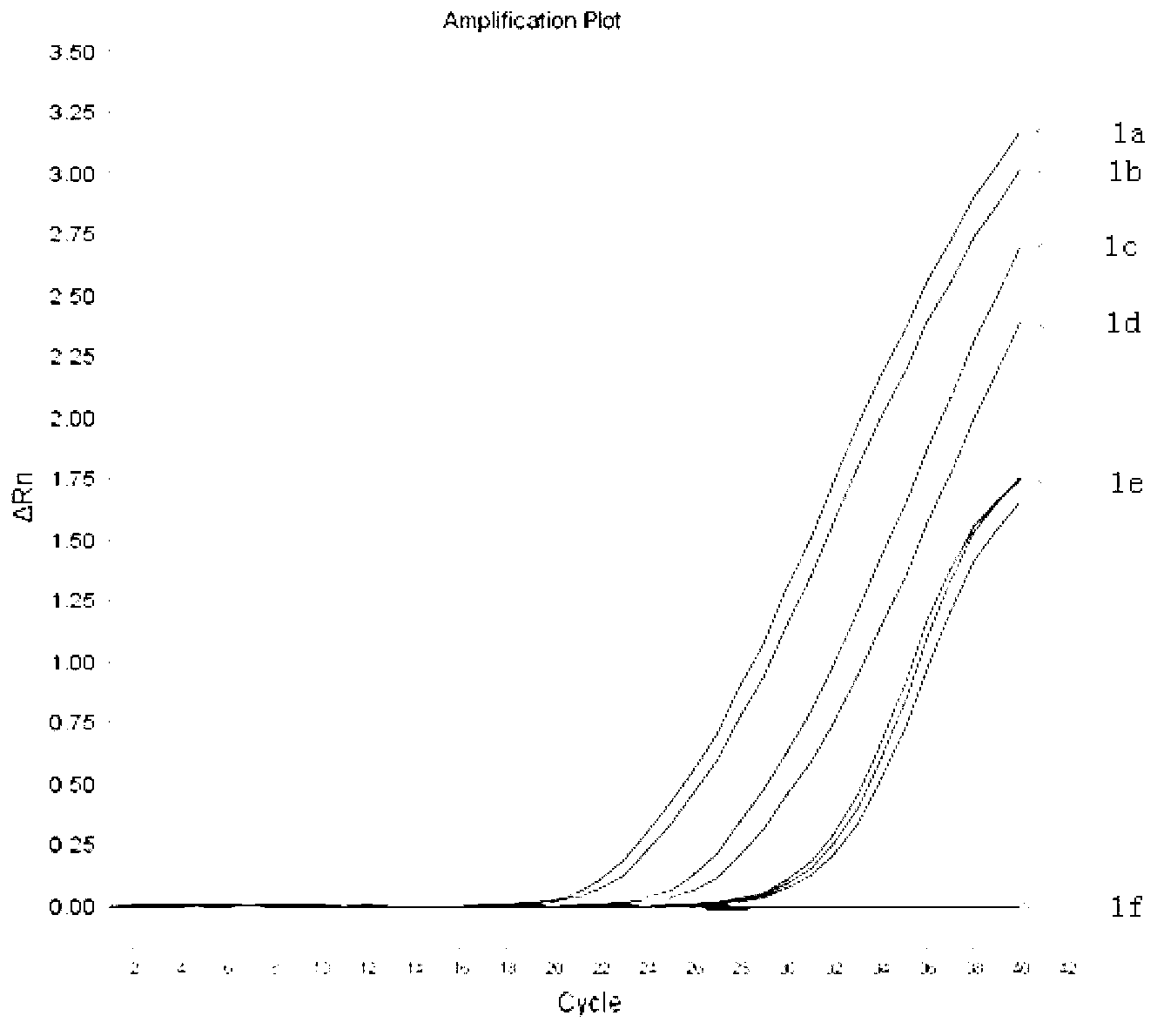
[0072] Example 1: Detection of Adenovirus Type 7 from Oral Swabs

[0073] 1. Preparation of various reagents:

[0074] Preparation of lysate: Accurately weigh 30g guanidine isothiocyanate, dissolve in 25ml 0.1M Tris-HCl (pH6.4), then add 8.8ml 0.5M EDTA (pH 8.0), 13.2ml ddH 2 O, mix well and set aside.

[0075] Preparation of nucleic acid binding solution: Accurately weigh 3g of amorphous silica, dissolve in 25ml of ddH 2 O, mix well and let stand at room temperature for 24 hours; remove 22ml of supernatant, add 22ml of ddH 2 Mix well with O, and let stand at room temperature for 5 hours; remove 22ml of the supernatant, mix the remaining suspension thoroughly, add 7μl of concentrated hydrochloric acid to adjust the pH to 2, and the final concentration is 1g / ml. Put the above suspension into a small glass bottle, autoclave it, and store it at room temperature in the dark.

[0076] Washing solution preparation: Prepare 70% ethanol with DEPC-treated deionized water as a washi...

Embodiment 2

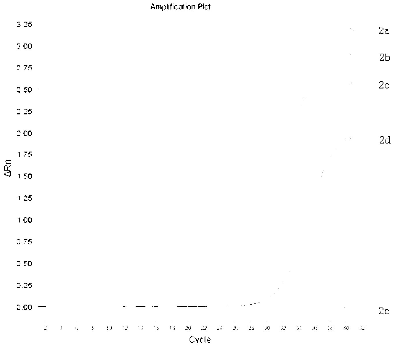
[0098] Example 2: Detection of adenovirus type 40 from feces

[0099] 1. Viral nucleic acid extraction:

[0100] Take the adenovirus type 40 positive water sample stool suspension verified by conventional PCR sequencing stored in the laboratory, centrifuge at 5000×g for 1 min, aspirate 100 μl of the supernatant for HAdV DNA extraction, and set up a negative and positive quality control at the same time, add to the above test tube Add 10 μl of internal reference substance, add 200 μl of lysis solution and 10 μl of nucleic acid binding solution, shake for 15 s, centrifuge at 6000×g for 1 minute, carefully discard all the liquid, save the precipitate, wash the precipitate once with 500 μl of lysis solution, and wash with 400 μl of washing solution The precipitate was washed twice with 1 ml of cold acetone once, and finally eluted with 30 μl of DEPC water preheated to 70°C.

[0101] 2. PCR sample loading and fluorescent quantitative PCR detection are the same as in Example 1

[...

Embodiment 3

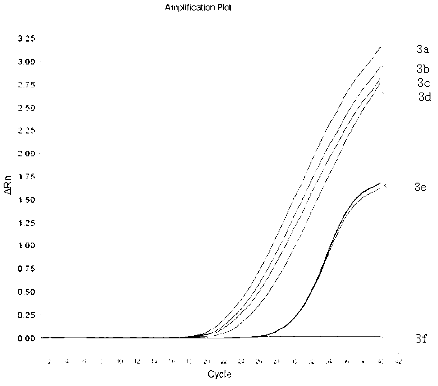
[0104] Example 3: Detection of Adenovirus Type 3 from Serum

[0105] 1. Viral nucleic acid extraction

[0106] Take 1ml of HBV, HIV, and HCV negative serum, add 10μl of adenovirus type 3 virus culture cultured in vitro, mix well, take 100μl for HAdV DNA extraction, and set up a negative and positive quality control at the same time, add 10μl of internal Add 200μl lysis solution and 10μl nucleic acid binding solution to the reference sample, shake for 15s, centrifuge at 6000×g for 1 minute, carefully discard all the liquid, save the precipitate, wash the precipitate once with 500μl lysis solution and twice with 400μl washing solution and 1 ml of cold acetone to wash the precipitate once, and finally eluted with 30 μl of DEPC water preheated to 70°C.

[0107] 2. PCR sample loading and fluorescent quantitative PCR detection are the same as in Example 1

[0108] 3. The test results are as follows: image 3 Shown:

[0109] The amplification of the internal reference substance o...


PUM

PropertyMeasurementUnit
Pre-denaturedaaaaaaaaaa

Abstract

The invention discloses an adenovirus multi-fluorescent quantitative PCR (polymerase chain reaction) detection kit, comprising PCR liquid, a primer probe combination, a positive quality control sample, and a negative quality control sample. The adenovirus multi-fluorescent quantitative PCR detection kit also comprises liquid for nucleic acid extraction and an inner reference substance for nucleic acid extraction, wherein the inner reference substance is inactivated T4 bacteriophage; the primer probe combination comprises a primer probe (general: SEQ ID NO4-6; subtype SEQ ID NO7-9) for adenovirus nucleic acid and a primer probe (SEQ ID NO1-3) for the inner reference substance; the 5' terminals of the SEQ ID NO3, 6, 9 are connected to a fluorescent report group; and the 3' terminals are connected to a fluorescent quenching group. A using method of the kit is also disclosed by the invention. Viral nucleic acid extraction is carried out by the liquid for nucleic acid extraction before the fluorescent quantitative PCR detection; and the inner reference substance is added in the extraction process of the viral nucleic acid. The kit and the using method thereof disclosed by the invention have the characteristics of being convenient to use, and accurate in detection result; all adenoviruses can be detected; and B and E subgroups can be parted.

Description

technical field [0001] The invention relates to the field of molecular biology, in particular to an adenovirus multiplex fluorescent quantitative PCR detection kit and a use method thereof. Background technique [0002] Adenovirus (adenovirus) is a particle with a diameter of 70-90nm without an envelope, which is composed of 252 capsomers arranged in an icosahedron. The capsid contains 240 hexads, 12 pentads and 12 cilia, in addition to other small proteins such as VI, VIII, IX, IIIa and IVa2. The hexaplex is the main protein that forms the 20 triangular faces of the virus capsid. The top of the 12 is a complex composed of 5 pentad subunits and 3 ciliary proteins. Protruding, the tips of the cilia form the scolex. The pentad and the scolex region of the cilium can bind to the virus receptor on the cell surface and play a very important role in the process of virus infection of cells. [0003] Since the discovery and successful isolation of adenovirus in the 1950s, more th...

Claims


Application Information

Patent Timeline
IPC IPC(8): C12Q1/70C12Q1/68G01N21/64
Inventor 辛忠涛牛宇辰姜曰晓张英民
Owner 北京海斯凯生物科技有限公司
Learn More