

Collagen/chitosan micro-nano fiber composite hemostatic membrane material and preparation method thereof
A fiber composite and chitosan technology, applied in medical science, absorbent pads, bandages, etc., can solve the problems of insufficient mechanical properties of collagen-based hemostatic agents, failure to meet market requirements, insufficient purity of type I collagen, etc., and achieve good biological Effects of degradability, promotion of wound healing, good biocompatibility
- Summary
- Abstract
- Description
- Claims
- Application Information
AI Technical Summary
Problems solved by technology
Method used
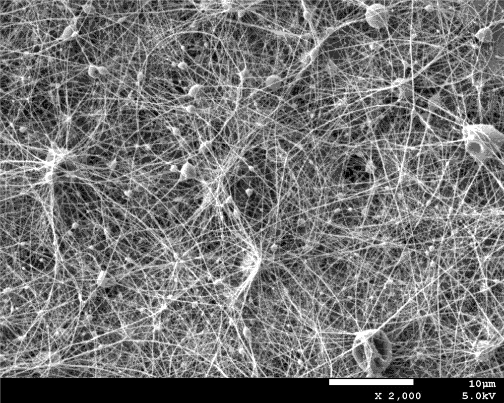
Image

Examples
Embodiment 1
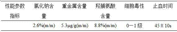
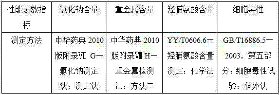
[0029](1) Type I collagen extraction: Take 10 parts by weight of medical biological skin, cut it into small pieces of 0.5cm×0.5cm, wash it with ultrapure water for 5 times, and then soak it in 0.05mol / L Tris, 1mol / L L NaCl, in a Tris-NaCl buffer solution with a pH of 7.5, place it in an ultrasonic cleaner with a frequency of 20kHz, and act for 2 hours; pour off the buffer solution, rinse the skin twice with distilled water; add 30 times of 0.5M acetic acid solution, soaked for 2 hours, crushed and homogenized with a homogenizer at a constant temperature of 4°C, and then transferred to a reaction kettle, adding 0.2 parts by weight of pepsin; under ultrasonic conditions with a frequency of 30kHz, slowly stirred at 4°C Enzyme hydrolysis for 36 hours, and act for 1 hour every 3 hours; after the reaction is completed, stop stirring, filter the reaction solution, adjust the pH of the filtrate to 7.0, add ammonium sulfate powder with a final concentration of 1.5mol / L, and let it stand...
Embodiment 2
[0036] (1) Type I collagen extraction: Take 10 parts by weight of medical biological skin, cut it into small pieces of 0.5cm×0.5cm, wash it with ultrapure water for 5 times, and then soak it in 0.05mol / L Tris, 1mol / L L NaCl, in a Tris-NaCl buffer solution with a pH of 7.5, place it in an ultrasonic cleaner with a frequency of 30 kHz, and act for 2 hours; pour off the buffer solution, rinse the skin twice with distilled water; add 40 times of 0.5M acetic acid Solution, soaked for 2 hours, crushed and homogenized with a homogenizer at a constant temperature of 4°C, then transferred to a reaction kettle, added 0.2 parts by weight of pepsin, and slowly stirred the enzyme at 4°C under ultrasonic conditions with a frequency of 30kHz Solution for 24 hours, every 3 hours for 1 hour. After the reaction is completed, stop stirring, filter the reaction solution with suction, adjust the pH of the filtrate to 7.5, add ammonium sulfate powder with a final concentration of 1.5mol / L, and let ...
Embodiment 3
[0043] (1) Type I collagen extraction: Take 10 parts by weight of medical biological skin, cut it into small pieces of 0.5cm×0.5cm, wash it with ultrapure water for 5 times, and then soak it in 0.05mol / L Tris, 1mol / L L NaCl, in a Tris-NaCl buffer solution with a pH of 7.5, place it in an ultrasonic cleaner with a frequency of 30 kHz, and act for 2 hours; pour off the buffer solution, rinse the skin twice with distilled water; add 50 times of 0.5M acetic acid solution, soaked for 2 hours, crushed and homogenized with a homogenizer at a constant temperature of 4°C, and then transferred to a reaction kettle, adding 0.2 parts by weight of pepsin; under the condition of ultrasonic waves with a frequency of 20kHz, the enzyme was slowly stirred at 4°C Decompose for 36 hours, and act for 1 hour every 3 hours; after the reaction is completed, stop stirring, filter the reaction solution with suction, adjust the pH of the filtrate to 7.3, add ammonium sulfate powder with a final concentra...
PUM
| Property | Measurement | Unit |
|---|---|---|
| shear viscosity | aaaaa | aaaaa |
Abstract
Description
Claims
Application Information