Biological repairing material for abdominal wall defect and preparation method of biological repairing material

A bio-repair, abdominal wall technology, applied in the field of biomedical engineering, can solve the problems of insufficient remodeling of the host's new autologous tissue, weak abdominal wall at the defect, poor mechanical strength of biological patches, etc., and achieve good biocompatibility and biomechanics. performance effect

Inactive Publication Date: 2015-02-18
SHANGHAI NINTH PEOPLES HOSPITAL SHANGHAI JIAO TONG UNIV SCHOOL OF MEDICINE
View PDF4 Cites 21 Cited by
  • Summary
  • Abstract
  • Description
  • Claims
  • Application Information

AI Technical Summary

Problems solved by technology

However, the acellular matrix biological patch also has some problems: 1. The mechanical strength of the biological patch is poor, and the degradation rate is too fast when implanted in the human body, and the host's new autologous tissue is not fully remodeled, resulting in weak abdominal wall and hernia at the defect site relapse
2. The surface of biological patch materials is dense, and it is difficult for host cells to migrate into the material, but only adhere to the surface of the material, resulting in a greatly reduced repair effect
However, its hydrophilicity and biocompatibility are slightly poor, and it is difficult for cells to migrate into the material

Method used


Image

  • Biological repairing material for abdominal wall defect and preparation method of biological repairing material
  • Biological repairing material for abdominal wall defect and preparation method of biological repairing material
  • Biological repairing material for abdominal wall defect and preparation method of biological repairing material

Examples


Embodiment 1

[0032] Embodiment 1 preparation test example

[0033] (1) Pre-treatment: Donor pigs are taken from local slaughterhouses, aged 25-30 weeks, weighing 30-40kg, male or female. After euthanasia with anesthesia, a median incision from the xiphoid process to the lower abdomen was selected, and the subcutaneous tissue and superficial fascia were free to expose the rectus abdominis tissue. Muscle, rinse repeatedly with deionized water at 0-8°C or buffered saline solution (PBS) with a pH value of about 7.4 to remove blood clots on the surface, remove surface fat tissue and fascia tissue under sterile conditions, and cut into 2×2cm 2 For large and small flakes, continue to rinse with deionized water or buffered saline solution (PBS) with a pH value of about 7.4.

[0034] (2) Decellularization treatment: In this part, freeze-thaw method, alkyl glycoside and nuclease solution are combined for decellularization treatment. The steps are as follows. The above-mentioned rectus abdominis ti...

Embodiment 2

[0037] Example 2 In vitro test

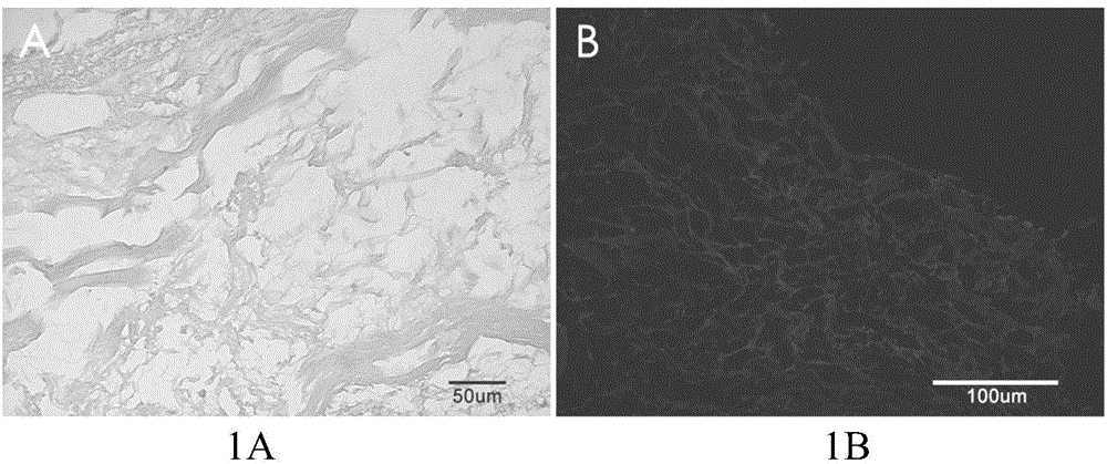
[0038] (1) Determination of decellularization degree:

[0039] Hematoxylin-eosin staining (HE staining) of skeletal muscle acellular matrix material embedded in paraffin (attached figure 1 A) and 4,6-diamidino-2-phenylindole fluorescent staining (DAPI staining) (attached figure 1 As shown in B), it is confirmed that there is no obvious cell nucleus residue inside the material, and the protein collagen fibers are arranged in an irregular shape.

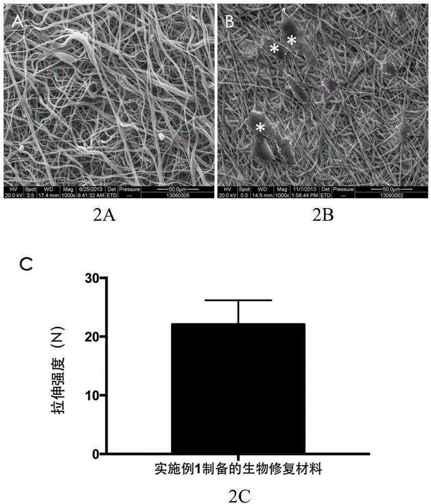
[0040] (2) Determination of fiber structure and mechanical properties:

[0041] The skeletal muscle acellular matrix / polycaprolactone blended electrospun fiber membrane prepared in Example 1 was detected by scanning electron microscopy (SEM), which showed that the fiber structure of the material was arranged in a cord-like disorder, and the fiber diameter was several Between hundreds of nanometers and several microns (with figure 2 shown in A).

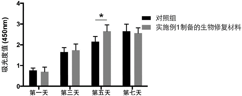
[0042] Rat fibroblasts were planted on the skeletal...

Embodiment 3

[0046] Example 3 In vivo experiments verify the application of composite nanofibrous membranes in abdominal wall defects

[0047] In order to check the specific effect of the polycaprolactone / skeletal muscle acellular matrix fiber nanofilm prepared in Example 1, we used it to repair the rat full-thickness abdominal wall defect model, and the detailed steps were as follows: SD female rats (12 Only) given 10% chloral hydrate intraperitoneal injection anesthesia (0.4ml / 100g), routine abdominal skin preparation, sterilized drape. A longitudinal incision was made in the center of the abdomen, about 5 cm long, and the subcutaneous tissue was cut open. The subcutaneous tissue was bluntly separated to both sides along the incision. A full-thickness rat abdominal wall defect model of 2×2 cm2 was repaired with the fibrous nanomembrane scaffold prepared in Example 1, sutured with 4-0 silk interrupted sutures, and the skin and subcutaneous tissue were sutured with 3-0 silk interrupted sut...


PUM

PropertyMeasurementUnit
concentrationaaaaaaaaaa

Abstract

The invention relates to the technical field of biological medical engineering, and discloses a biological repairing material for an abdominal wall defect and a preparation method of the biological repairing material. The method comprises the following steps: (1) performing pre-treatment on the muscular tissue of rectus abdominis of a dead animal; (2) performing cell extraction treatment to prepare a skeletal muscle acellular matrix biological thin sheet; (3) dissolving the skeletal muscle acellular matrix biological thin sheet into pepsase for digestion; (4) performing electrostatic spinning on skeletal muscle acellular matrix microparticles obtained by digestion and polycaprolactone to obtain a polycaprolactone / skeletal muscle acellular matrix blended electrostatic spinning fiber film which is the biological repairing material disclosed by the invention. According to the method, a skeletal muscle acellular matrix from abdominal wall tissue and polycaprolactone are combined by an electrostatic spinning technology; the preparation materials can be degraded, and products from degradation are harmless to a human body; the prepared biological repairing material can well simulate constituents and the structure of an extracellular matrix and is good in biocompatibility and biomechanical property.

Description

technical field [0001] The invention relates to the technical field of biomedical engineering, in particular to a biological repair material for abdominal wall defect and a preparation method thereof. Background technique [0002] Abdominal wall defect is a common disease in clinical surgery today. Abdominal wall hernia, severe abdominal wall trauma, and extended resection of abdominal wall tumors can all cause the formation of abdominal wall defect. Among them, inguinal hernia and incisional hernia are the most common abdominal wall defects. The incidence rate of inguinal hernia in European and American countries is 1-5‰. ‰, the number of adult inguinal hernia surgery cases to be carried out in my country reaches 3-4 million cases every year. The recurrence rate of abdominal incisional hernia is about 2-20% in abdominal surgery, and even as high as 50% when the defect is too large or accompanied by infection. The classic treatment method is direct suture repair, and the po...

Claims


Application Information

Patent Timeline
Patent Type & Authority Applications(China)
IPC IPC(8): A61L27/18A61L27/36A61L27/50D04H1/728
Inventor 顾岩杨志宋致成杨建军聂鑫刘正尼王恵春
Owner SHANGHAI NINTH PEOPLES HOSPITAL SHANGHAI JIAO TONG UNIV SCHOOL OF MEDICINE
Why Eureka
Try Eureka