Method for preparing folic acid functional nanofibers for target capture of cancer cells

A nanofiber, targeted capture technology, applied in the direction of fiber type, fiber processing, conjugated synthetic polymer rayon, etc., can solve the problems of complex operation, high cost, cumbersome steps, etc., and achieve the effect of improving the capture ability

Inactive Publication Date: 2015-07-22
DONGHUA UNIV
View PDF3 Cites 10 Cited by
  • Summary
  • Abstract
  • Description
  • Claims
  • Application Information

AI Technical Summary

Problems solved by technology

The method is complicated to operate, and the steps of self-assembly are cumbersome and expensive
[0008] The surface of polyethyleneimine (PEI) contains a large number of amino groups, which is easy to modify and low in cost. The results of searching

Method used


Image

  • Method for preparing folic acid functional nanofibers for target capture of cancer cells
  • Method for preparing folic acid functional nanofibers for target capture of cancer cells
  • Method for preparing folic acid functional nanofibers for target capture of cancer cells

Examples


Embodiment 1

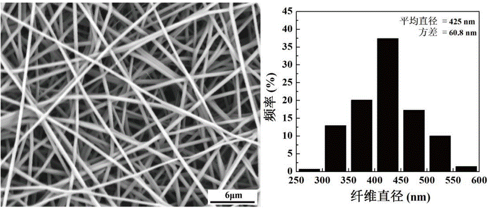
[0066] Prepare a 12wt% PVA solution with water as a solvent, and at the same time stir slowly with a glass rod for about 30 minutes until the PVA powder is completely swollen. Then, put a rotor in the beaker, place it on a magnetic stirrer, heat and stir in an 80°C water bath for 3 hours, and let it stand to cool. Then, PEI was added at a mass ratio of PVA:PEI=3:1, water was used as a solvent, dissolved by ultrasonic vibration and magnetically stirred overnight to make it uniformly dispersed, and configured as a PVA / PEI blended solution. Electrospinning experiments were carried out at room temperature and humidity less than 50%. The electrospinning device consists of a high-voltage power supply (which can provide a DC voltage of 0-40 kV), a syringe pump, a syringe, a stainless steel needle with an inner diameter of 1.0 mm, and a grounded collecting aluminum plate. The technological parameters in the spinning process are set as follows: the voltage is 18.6kv, the flow rate is ...

Embodiment 2

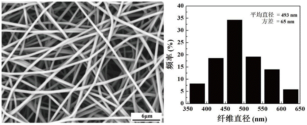
[0068] Adopt glutaraldehyde vapor crosslinking method to carry out crosslinking treatment to the PVI / PEI nanofiber that implements 1 to obtain, and its specific operation process is, the fiber membrane is covered on the petri dish that fills 20ml 25% glutaraldehyde solution, two Put them together in a desiccator, vacuum cross-linking for 18h. Rinse the cross-linked fiber membrane in water, and then take it off. At this time, it can be found that the fiber membrane is insoluble in water, that is, the modification treatment is completed. Then wash it with deionized water for 3-5 times, and put it into a vacuum drying oven for 12-24h. SEM results showed that after cross-linking with glutaraldehyde vapor, the fiber morphology was still good, and the average diameter of the cross-linked nanofibers was 493nm, which was 70nm different from the average diameter of 425nm of the PVA / PEI nanofibers without hydrophobic treatment. Left and right, it shows that on the glutaraldehyde cross-...

Embodiment 3

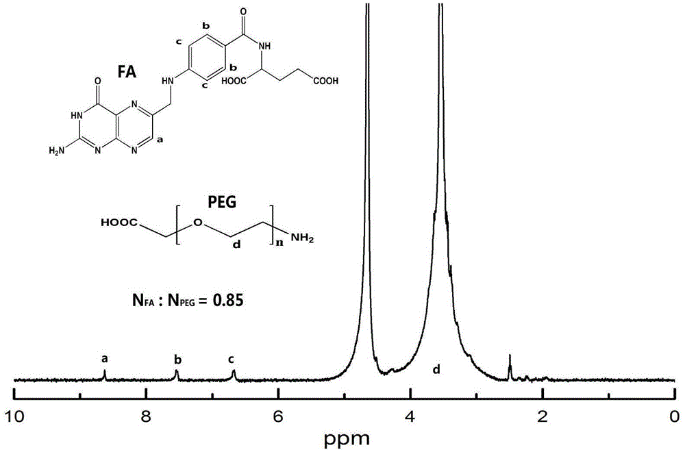
[0070] Preparation of FA-PEG-COOH. Known NH 2 -M of PEG-COOH W The molecular weight of FA is 441.4; the molecular weight of EDC·HCl is 191.7; the molecular weight of NHS is 115.09. Weigh NH 2 - PEG-COOH 79.42 mg, dissolved in 5 ml of DMSO. Take FA:EDC·HCl:NHS:NH 2 - PEG-COOH molar ratio = 2.5:2:2:1 Calculate the required mass of FA to be 43.82mg, which is dissolved in 5ml DMSO. The required EDC·HCl and NHS were 15.22 mg and 9.14 mg, respectively, dissolved in 1 ml of DMSO. The carboxyl groups in FA were activated by EDC·HCl and NHS, and the reaction time was 3 h. FA after activation of the carboxyl group for 3 h, added to NH 2 -PEG-COOH, mixed on a magnetic stirrer for 3 days.

[0071] After the reaction, the reaction product was transferred to a dialysis bag with a molecular weight cut off of 1000, and dialyzed in distilled water for 3 days (6x2L). Then freeze-drying is carried out to obtain the FA-PEG-COOH reaction product. figure 1 shown 1 The H NMR spectrum indi...


PUM


Abstract

The invention relates to a method for preparing folic acid functional nanofibers for target capture of cancer cells. The method comprises the following steps: (1) preparing a PVA/PEI nanofiber membrane by electrostatic spinning, and carrying out vacuum drying; (2) after drying, using glutaraldehyde for crosslinking; (3) activating carboxyl of folic acid FA through EDC.HCl and NHS, and reacting the carboxyl-activated FA with polyethylene glycol PEG with carboxyl at one end and amidogen at the other end to obtain FA-PEG-COOH; (4) activating carboxyl of FA-PEG-COOH, adding FA-PEG-COOH into the PVA/PEI nanofiber membrane, and carrying out shaking reaction to obtain the folic acid functional nanofibers; adding triethylamine and acetic anhydride for acetylation treatment, washing and drying. The prepared nanofibers have good biocompatibility and structural stability, is short in time to capture the cancer cells and strong in specificity, and has a wide application prospect.

Description

technical field [0001] The invention belongs to the field of preparation of nanomaterials for targeting and capturing cancer cells, in particular to a method for preparing folic acid functionalized nanofibers for targeting and capturing cancer cells. Background technique [0002] Circulating tumor cells (CTCs) are tumor cells that survive in the blood circulation system during tumor metastasis, and the generation of these cells is considered to be a necessary prerequisite for tumor metastasis. Malignant tumor is one of the major diseases with the highest mortality rate in my country, and more than 90% of the deaths of tumor patients are caused by tumor metastasis. Invasion and metastasis are one of the most prominent features of malignant tumors. Tumor cells detach from the primary tumor spontaneously or due to diagnostic and therapeutic operations, undergo epithelial-mesenchymal transition (EMT), and thus have flow characteristics. When they enter the peripheral blood, they...

Claims


Application Information

Patent Timeline
IPC IPC(8): D06M15/53D06M13/123D01F8/10D01F8/16D01D5/00D04H1/4382D04H1/728D06M101/24D06M101/30
Inventor 史向阳范章余赵毅丽
Owner DONGHUA UNIV
Why Eureka
Try Eureka