
Sequential enzyme surface co-display system and use thereof

A surface co-display and co-display technology, applied in the field of biotechnology and analysis, can solve the problems of reduced detection efficiency, loss of enzyme activity, low efficiency, etc., to solve the complex extraction process, low stability, and good stability.

Inactive Publication Date: 2016-06-01
QINGDAO INST OF BIOENERGY & BIOPROCESS TECH CHINESE ACADEMY OF SCI
View PDF3 Cites 9 Cited by
  • Summary
  • Abstract
  • Description
  • Claims
  • Application Information

AI Technical Summary

Problems solved by technology

However, excessive chemical cross-linking will lose the activity of the enzyme and reduce the efficiency of detection
Through molecular level manipulation, it is possible to effectively retain the activity of the enzyme when it is immobilized and control the spatial position of the enzyme. However, the current research only stays on a single enzyme molecule, and the improvement effect on the sequential enzyme sensor is negligible.
The development of a multi-enzyme co-display system is expected to improve the stability of sequential enzymes. For example, Zhou et al. took the maltose sensor as the research object and proposed a fusion protein solution. Molecular manipulation was carried out at the gene level, and the GOD-connecting peptide- The fusion gene of GA expresses the fusion protein in methylotrophic yeast, realizes the ratio control of the two enzymes, and significantly improves the performance of the maltose sensor. The disadvantage is that the construction method of the fusion protein is complicated and inefficient.
The above co-display systems are all based on the cohesin-dockerin interaction to display multiple enzymes on the surface of yeast and other cells at a ratio of 1:1. However, the development of co-display systems for sequential enzymes at other ratios has not been reported.
In addition, the above studies are mainly used in the production of ethanol, and there are few studies on bioanalysis. Therefore, it is of great significance to develop sequential enzyme sensors using the sequential enzyme scale co-display system.

Method used


Image

  • Sequential enzyme surface co-display system and use thereof
  • Sequential enzyme surface co-display system and use thereof
  • Sequential enzyme surface co-display system and use thereof

Examples


Embodiment 1

[0046] Acquisition of cell surface display sequence enzyme expression vector:

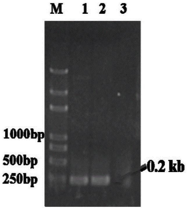
[0047] 1. The gene fragment docc encoding the dockerin protein comes from the strain Clostridium cellulolyticumH10; doct comes from the strain ClostridiumthermocellumATCC27405, wherein the length of the docc gene fragment is 213bp, and the length of the doct gene fragment is 204bp. According to the gene sequences of the two, primers are used from the respective genomic DNAs with high-fidelity polymerases Amplify the gene of interest separately. The target gene PCR reaction system is as follows: 0.25μL ExTaq, 5μL 10×ExTaqbuffer, 4μL 2.5mMdNTP, 1μL genomic DNA, 1μL DocC-F and DocT-F, 1μL DocC-R and DocT-R, 37.75μL ultrapure water. PCR reaction conditions: 94°C for 5min; 94°C for 30s, 58°C for 30s, 72°C for 20s, 30 cycles; 72°C for 5min; after gene amplification, use 1% agarose gel to detect the size and purity of the target gene (see figure 1 ), and then ligated to the pMD-19Tsimple vector (TaKaRa, ...

Embodiment 2

[0059] Acquisition of cells displaying sequence enzymes on the cell surface:

[0060] 1. Expression of fusion protein. The recombinant vectors obtained in Example 1 were respectively transformed into Escherichia coli Escherichiacoli BL21 (DE3), inoculated in fresh LB liquid medium containing kanamycin (30 μg / ml) after enzyme digestion and identification, and cultured with shaking until the absorbance (OD600 ) to 0.6, add 0.5mM, 0.2mM, and 1mM IPTG (isopropyl-β-D-thiogalactopyranoside) respectively, culture at 25°C for 20h, and induce the expression of GA-DocC and DocT-GDH respectively , INP-CohC / CohT and INP-CohC / CohT / CohC fusion proteins.

[0061] Escherichia coli E.coliBL21 (DE3) was cultured in LB medium, and the composition of LB medium was: 5g / L yeast extract, 10g / L peptone, 10g / L NaCl.

[0062] 2. Dispense the bacteria liquid into 50mL centrifuge tubes and centrifuge at 6000rpm for 5min, discard the supernatant, wash twice with PBS buffer of pH 7.4 after collecting all...

Embodiment 3

[0066] Determination of single-enzyme assembler activity of GA-DocC and DocT-GDH:

[0067] 1. Enzyme activity assay of GA-DocC single enzyme assembly. Take 2.5ml GA-DocC crude enzyme solution and mix with 20μl INP-CohC / CohT / CohC bacterial solution. Incubate at 25°C and 180rpm for 2h, centrifuge at 6000rpm for 3min to remove the supernatant, wash twice with the same buffer solution, and measure GA enzyme activity. With 10mM maltose as substrate, react at 70°C for 15min. After the reaction was completed, the glucose concentration in the product was measured with a glucose kit. Finally, the enzymatic activity of GA-DocC single-enzyme assembly was calculated. The GA activity unit U is defined as the amount of enzyme required to release 1 μmol of glucose per minute under certain reaction conditions. The enzyme activity of whole cells displayed by GA at a ratio of 2:1 is 0.82U / OD 600 whole cells.

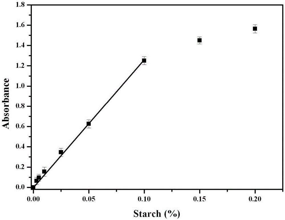
[0068] 2. NADH standard curve preparation. NADH is composed of NAD + It is re...


PUM


Abstract

The invention relates to the technical field of biotechnology and analysis technology and concretely relates to a sequential enzyme proportional bacterial surface co-display system and a use thereof. The sequential enzyme surface co-display system comprises a gene sequence (ga) for coding a target protein glucoamylase, a gene sequence (gdh-m) of a glucose dehydrogenase mutant, a gene sequence (coh-dock) of a cohesion-dockerin protein and a gene sequence inaPb-N of an ice nucleating protein N end structural domain for transmembrane positioning and transport. The bacterial surface display system is used for starch detection. The starch detection method has high sensitivity and simple processes. The whole cell catalyst can be used in the fields of biosensing, foods, medicine and health, and biological energy sources.

Description

technical field [0001] The invention relates to the fields of biotechnology and analysis technology, in particular to a sequential enzyme surface co-display system and its application. Background technique [0002] Sequential enzyme sensor refers to the sequential and coordinated completion of a series of catalytic reactions by a variety of related enzymes. The substrate is catalyzed by two steps of enzymes to generate final products and by-products. The by-products generally have chemical or physical activity and can be converted into signals to be detected. The concentration of the substrate can be determined according to the detection signals. For example, starch or maltose is hydrolyzed by glucoamylase (GA) to produce glucose, which is then oxidized by glucose oxidase (GOD) to produce gluconolactone and hydrogen peroxide, which can be oxidized on the electrode to generate current to realize its detection. The preparation of sequential enzyme sensors is generally to immo...

Claims


Application Information

Patent Timeline
IPC IPC(8): C12N1/21C12N15/70C12N11/18C12N11/16C12Q1/40C12Q1/32C12R1/19
Inventor 刘爱骅梁波
Owner QINGDAO INST OF BIOENERGY & BIOPROCESS TECH CHINESE ACADEMY OF SCI
Learn More