Method for detecting thrombin protein concentration

A technology of thrombin and protein, which is applied in the fields of biomedicine and protein detection, can solve the problems of inability to control the range of detection objects, difficulty in meeting detection requirements, and limitation of practical applications, etc., to achieve the expansion of dynamic detection range, wide application range, detection sensitive effect

Inactive Publication Date: 2017-12-22
INST OF CHEM CHINESE ACAD OF SCI
View PDF1 Cites 6 Cited by
  • Summary
  • Abstract
  • Description
  • Claims
  • Application Information

AI Technical Summary

Problems solved by technology

[0004] At present, the detection methods of thrombin protein (Thrombin) based on nucleic acid aptamers mainly include fluorescence resonance energy transfer, electrochemical method, surface plasmon resonance technology and absorbance method, etc., but these methods have high technical requirements and low sensitivity, which are difficult to meet the current detection requirements. requirements
Moreover, based on the detection of these methods, the range of detection objects cannot be well regulated, thus limiting the practical application of these methods

Method used


Image

  • Method for detecting thrombin protein concentration
  • Method for detecting thrombin protein concentration
  • Method for detecting thrombin protein concentration

Examples


Embodiment 1

[0046] Embodiment 1, the preparation of aldehyde glass substrate

[0047] Put the small glass slide in acetone solution for ultrasonic cleaning, then wash it with ultrapure water, and then use 1M NaOH solution for 10 minutes to remove the oil on the surface, then wash it with deionized water and absolute ethanol, and extinguish it at 250°C. Bacteria treatment for 3h. Put the above-mentioned processed small slides into newly prepared piranha solution (piranha solution, volume ratio, 98% concentrated H 2 SO 4 / 30%H 2 o 2 =7:3) treated at 90°C for 30 minutes to hydroxylate the glass surface. The hydroxylated slides were then placed in a toluene solution containing 1% APTES and incubated at room temperature for 2 hours to aminate the surface of the slides. The aminated glass slide was incubated in Tris-HCl buffer containing glutaraldehyde (1% v / v) for 3 hours at room temperature, and then washed twice with Tris-HCl buffer to obtain a modified glass substrate.

Embodiment 2

[0048] Hybridization and immobilization of embodiment 2, DNA

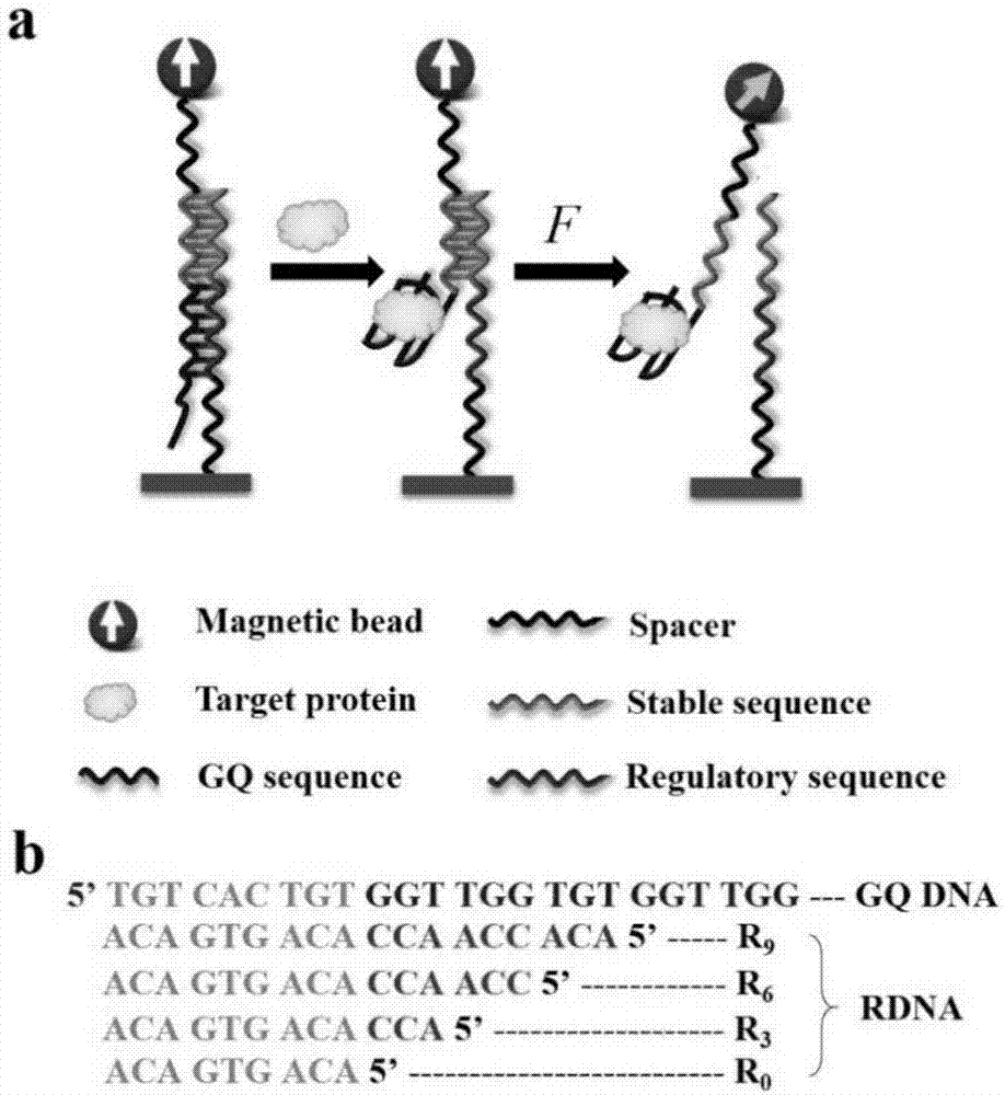
[0049] Mix 50uL 3uM thrombin nucleic acid aptamer (GQ DNA) with 50uL 3uM complementary DNA sequence R i (i=1, 2, 3...15) were mixed in equal volumes, reacted at 90° C. for 5 minutes, and naturally cooled to room temperature to obtain hybridized double-stranded DNA. 20 uL of the hybridized double-stranded DNA solution was drip-coated on the surface of the glass substrate modified in Example 1 of the present invention, and incubated at 4° C. for 10 h. Then use Tris-HCl buffer solution to wash away unbonded double-stranded DNA molecules on the surface, and then incubate with a reference buffer solution containing BSA with a mass volume fraction of 1% for 30 minutes, and then connect to magnetic beads M-280. The process is as follows: figure 1 shown.

Embodiment 3

[0050] Embodiment 3, the measurement of force spectrum

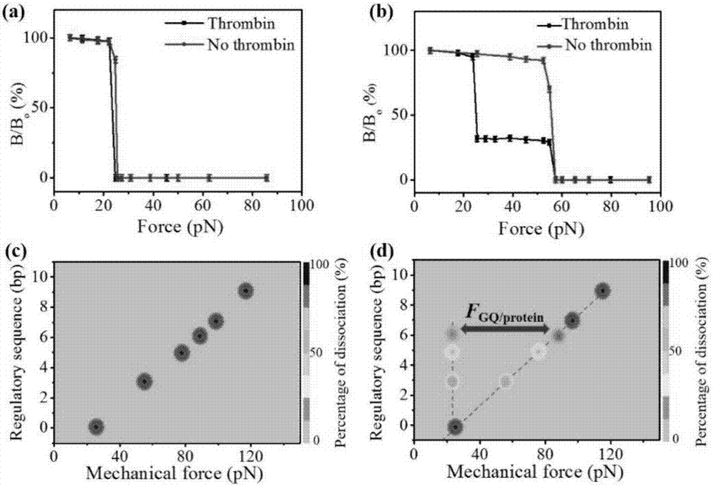
[0051] Add 20uL of 300nM thrombin protein to the above-mentioned glass substrate connected with DNA, and the buffer solution used for dissolving thrombin protein is Tris-HCl buffer (20mM Tris-HCl, 140mM NaCl, 5mM KCl, 1mM CaCl 2 ,1mMMgCl 2 , pH=7.4), under the condition of 37°C, incubate the thrombin protein with the double-stranded DNA for 30min, and wash away the unreacted thrombin protein with standard buffer solution. The above treated sample to be tested was magnetized under a magnet (~0.8T) for 2 minutes, and then centrifuged at 1000 rpm for 5 minutes to remove physical adsorption. The magnetic signal is then detected on an ultra-low field atomic magnetometer. Gradually increase the magnitude of the centrifugal force, and the centrifugation time is still 5 minutes each time, and record the magnetic signal intensity corresponding to each centrifugal force. Due to the non-covalent bond between the double-stranded ...


PUM

PropertyMeasurementUnit
strengthaaaaaaaaaa

Abstract

The invention discloses a method for detecting a thrombin protein concentration. The method for detecting the thrombin protein concentration comprises the following steps of modifying a substrate; connecting DNA with the modified substrate; connecting and magnetizing magnetic beads; measuring an interface point of a complementary DNA chain; detecting the thrombin protein through sequence regulation; detecting the thrombin protein through mechanical force regulation. According to the method disclosed by the invention, one end of a nucleic acid aptamer is modified by the magnetic beads; by means of measuring a low-intensity magnetic field generated by the magnetic beads, the size of self residual magnetism of the magnetic beads is obtained; by means of magnetic signal strength change, a concentration of a target detection object is obtained at the same time; not only is a range of the target detection object dynamically regulated by a complementary sequence of the nucleic acid aptamer, but also a detection limit of the target object is widened and flexibility of the target object is reduced by the mechanical force change.

Description

technical field [0001] The invention relates to a method for detecting thrombin protein concentration, which belongs to the field of protein detection in the field of biomedicine. Background technique [0002] Thrombin is a serine protease that plays an important regulatory role in the blood coagulation system and plays a very important role in physiology. It participates in some physiological and pathological responses of the human body, such as tissue repair, inflammation, platelet activation and wound healing. Thrombin has dual properties of procoagulation and anticoagulation in the coagulation system, and changes in its content in the blood can cause abnormal coagulation. At the same time, it is an important measure of blood coagulation mechanism, and it is also a diagnostic marker for the diagnosis of certain diseases such as cartilage synovial inflammation. Therefore, the detection of thrombin content in the body is of great significance to the diagnosis, treatment, ...

Claims


Application Information

Patent Timeline
Patent Type & Authority Applications(China)
IPC IPC(8): C12Q1/56G01N27/72
CPCC12Q1/56G01N27/72
Inventor 姚立鲁攀
Owner INST OF CHEM CHINESE ACAD OF SCI
Why Eureka
Try Eureka