Biochip for detecting myocardial infarction marker, and detection method applying same

A biochip and marker technology, applied in chemical instruments and methods, laboratory containers, instruments, etc., can solve the problem that the equipment cannot be miniaturized and POCT, reduce detection precision, detection sensitivity and specific interference, etc. problems, to avoid missed detection and false negatives, shorten the diagnosis time, and increase the rescue time.

Inactive Publication Date: 2018-12-21
微粒云科技(北京)有限公司
View PDF3 Cites 9 Cited by
  • Summary
  • Abstract
  • Description
  • Claims
  • Application Information

AI Technical Summary

Problems solved by technology

[0008] The first-generation detection technology competition method (radioimmunoassay, RIA): There are many factors affected, and the RIA sample requires a large amount. Because extraction is usually required before detection, it not only increases the operation steps, but also because the recovery rate of extraction is usually Only 80% to 90%, the coefficient of variation (CV) is relatively large, which reduces the precision of detection;
[0009] The second-generation detection method non-competitive method (radioimmunoassay, RIA): immunoassay using double-antibody sandwich, no extraction, high antibody specificity, its sensitivity, precision and specificity are better than competitive methods, but time-consuming Still long and difficult to apply to automation;
[0010] The third-generation detection method enzyme immunoassay, stationary phase immunochromatography: the double antibody plus core method immunoassay solid phase chroma

Method used


Image

  • Biochip for detecting myocardial infarction marker, and detection method applying same
  • Biochip for detecting myocardial infarction marker, and detection method applying same
  • Biochip for detecting myocardial infarction marker, and detection method applying same

Examples


Embodiment 1

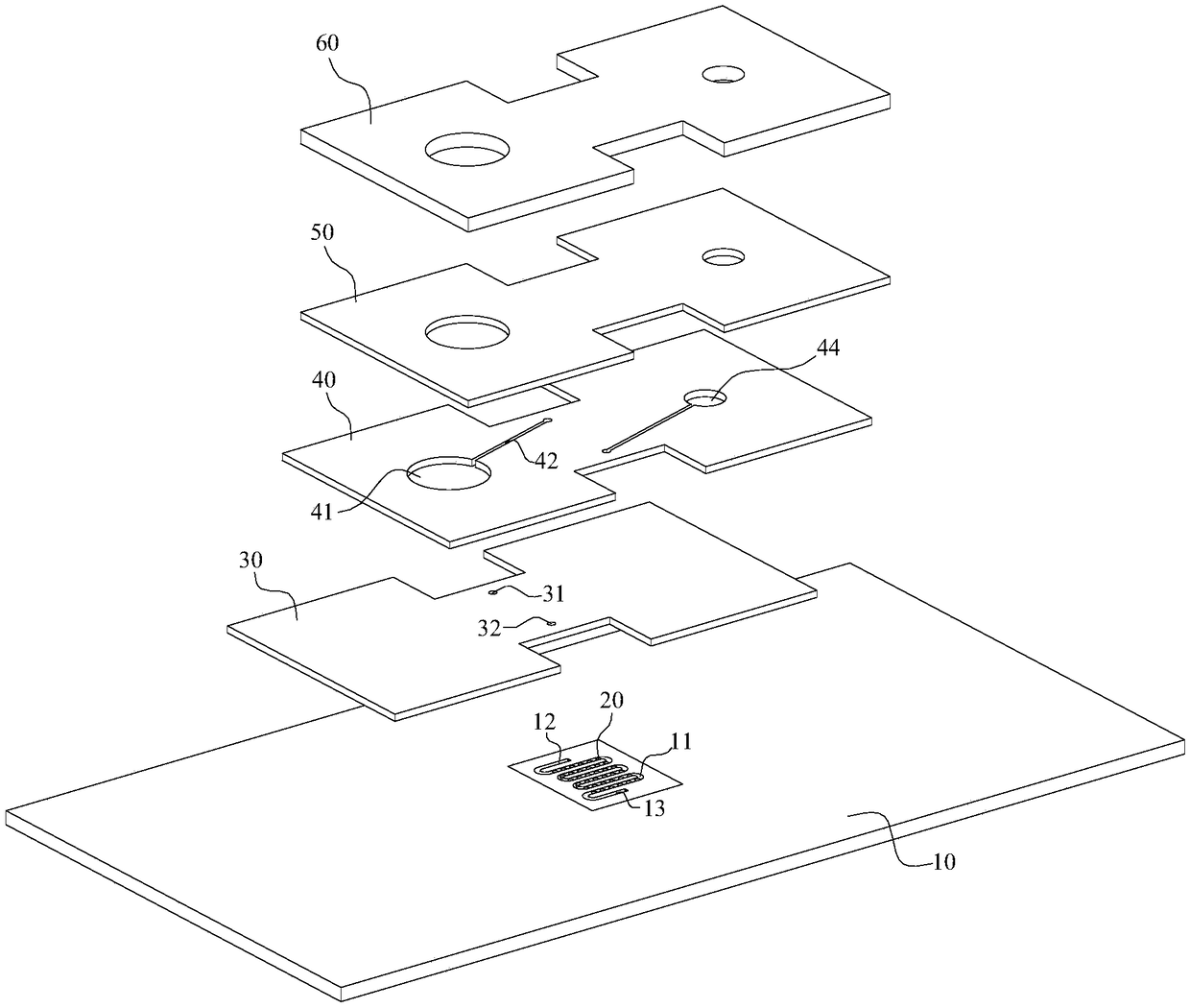
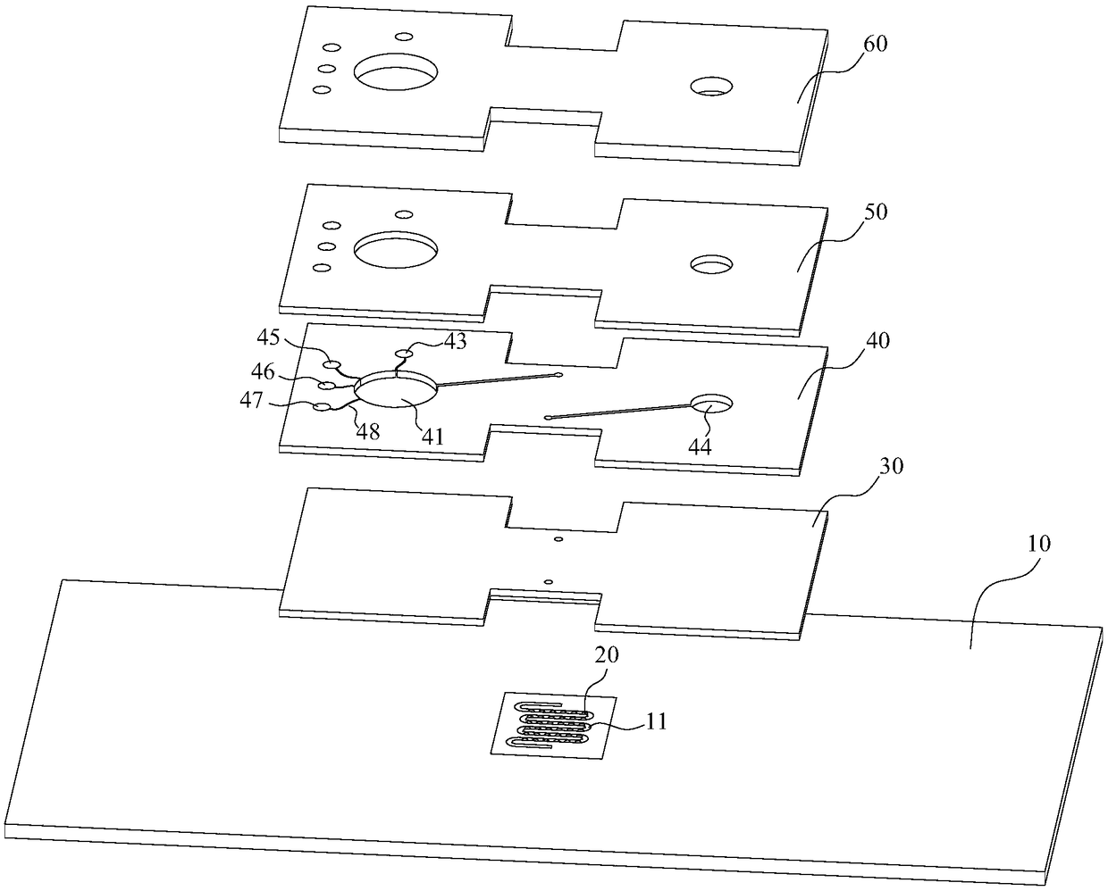
[0091] The present invention is a biochip for detection of myocardial infarction markers, the biosensor is pre-coated with myocardial infarction markers (CTI / CK-MB / MYO) spotting monoclonal antibody, and then the first layer 30, the second layer 40. The microfluidic channel sealing layer 50 and the whole blood filter layer 60 are pasted on the top of the microfluidic channel 11 of the PCB board 10 in sequence, thereby sealing the microfluidic channel 11, so that the reaction can be carried out under airtight conditions, achieving micro-full laboratory Effect. The first layer 30 is a biosensor airtight layer, containing the sampling hole 31 and the waste liquid hole 32; The pump guides the sample to be tested to flow in one direction from the mixing area 41 to the detection area (where the biosensor is located) and the waste liquid area 44; the microfluidic channel sealing layer 50 is the third layer to ensure that the sample flows in a closed pipeline, At the same time, the wh...

Embodiment 2

[0094] The preparation method of the biochip for the detection of three myocardial infarction markers (CTI / CK-MB / MYO) of the present invention:

[0095] Using the coupling method of superparamagnetic nano-magnetic particles, the superparamagnetic nano-magnetic particles include mixed magnetic beads with a diameter of 30nm and a diameter of 50nm. The reaction mechanism of the nano-magnetic particles is covalent bonding. The concentration of sodium acetate is reduced to 30nm and 50nm black colloidal superparamagnetic supersensitive magnetic particle mixed suspension, and the antibody / avidin with amino group is coupled. Quantitative detection reagent preparation steps are as follows,

[0096] 1. Preparation of 30nm+50nm superparamagnetic ultrasensitive magnetic particles:

[0097] (1) Preparation of 30nm magnetic beads: Take 0.75ml~2.25ml 4% Fe 3 o 4 Add the solution to 300ml of ultrapure water to make the final concentration of the iron oxide solution 0.01% to 0.03%, heat it ...

Embodiment 3

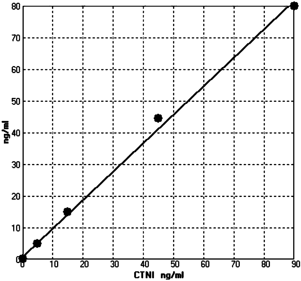
[0173] The specific steps of the three markers of myocardial infarction (CTI / CK-MB / MYO) detection of the present invention:

[0174] (1) Take the enterprise quality control product and biochip quantitative detection kit out of the refrigerator, and equilibrate to room temperature;

[0175] (2) Turn on the chip detector, perform system self-test and preheat for 5 minutes;

[0176] (3) Tear open the required aluminum foil bag containing the chip and take out the biochip, and mark the type and concentration of the quality control substance tested in the ID information position.

[0177] (4) Adding samples: Use a 100ul micropipette to respectively pipette 25ul of the internal control products of the three markers of myocardial infarction into the corresponding chip injection holes, and insert the chip into the detector;

[0178] (5) Detection: Select the corresponding myocardial infarction marker detection module of the detector to start the detection, and the device will automat...


PUM

PropertyMeasurementUnit
Diameteraaaaaaaaaa
Diameteraaaaaaaaaa

Abstract

The invention relates to a biochip for detecting a myocardial infarction marker, and belongs to the technical field of POCT detection. The biochip comprises biosensors, which are a wafer platform anda spotted antibody coated on the wafer platform, wherein the spotted antibody can form an immune complex with a magnetic bead conjugated antibody and the marker protein, and complex aggregation of magnetic beads will be formed on the biosensor coated with the spotted antibody when the blood sample contains the myocardial infarction marker protein. In the technical solution, the magnetic beads havea large specific surface area, and more antibodies are combined to improve the sensitivity of the detection; at the same time, the biomolecules and proteins are not magnetic, the magnetic beads not bound to the sensor cannot excite the magnetic field to generate magnetic resistance signals, and the specificity is high than the traditional chemiluminescence method. The invention also provides a detection method for applying the biochip, which can realize micro-measurement and micro-detection, and shortens the diagnosis time by controlling the detection time of 15 minutes to 20 minutes.

Description

technical field [0001] The invention relates to a biochip, which is mainly used for detecting the detection of myocardial infarction markers in blood samples. The invention also provides a detection method using the biochip, which belongs to the technical field of POCT detection. Background technique [0002] Cardiovascular disease is a serious disease that endangers human health and life, and has become the number one killer of human health in the 21st century. According to the "China Cardiovascular Disease Report 2015" and "China Cardiovascular Disease Report 2016" compiled by the National Center for Cardiovascular Diseases, the number of patients with cardiovascular diseases in my country is still growing rapidly. According to statistics, the number of patients with cardiovascular disease was 290 million in 2014, and this number has climbed to 290 million by 2015. According to the report estimates, about 3.5 million people die of cardiovascular disease in my country ever...

Claims


Application Information

Patent Timeline
IPC IPC(8): G01N35/00B01L3/00
CPCB01L3/5027G01N35/00
Inventor 张永勇
Owner 微粒云科技(北京)有限公司
Why Eureka
Try Eureka