Method for preparing artificial muscle tendons by using biological three-dimensional printing and electrospinning technology

An electrospinning technology and three-dimensional printing technology are applied in the field of artificial muscle preparation by using biological three-dimensional printing and electrospinning technology, which can solve the problems of difficult to achieve rapid repair of damaged tissue, affecting the normal function of tendon, low colonization efficiency, etc. The effect of tendon repair and normal function recovery, and the production method is simple and convenient

Inactive Publication Date: 2019-05-03
SHANGHAI NINTH PEOPLES HOSPITAL AFFILIATED TO SHANGHAI JIAO TONG UNIV SCHOOL OF MEDICINE
View PDF7 Cites 7 Cited by
  • Summary
  • Abstract
  • Description
  • Claims
  • Application Information

AI Technical Summary

Problems solved by technology

[0003] At present, tendon scaffolds prepared by tissue engineering methods mainly include fiber bundles directly as scaffolds, braided scaffolds, knitted scaffolds, and electrospun nanofiber scaffolds. It is beneficial to production and application. In addition, the tendon scaffold prepared by the above method has low adhesion and colonization efficiency of seed cells during repair, and it is difficult to achieve the effect of rapid repair of damaged tissue. Moreover, the obtained tendon scaffold is easy to adhere to the surrounding tissue in vivo. , affect the normal function of the tendon repair, electrospinning technology is a good method to prepare nanofibers, nanofibers can be well bionic extracellular matrix structure, so it has a wide range of applications in the field of tissue engineerin

Method used


Image

  • Method for preparing artificial muscle tendons by using biological three-dimensional printing and electrospinning technology
  • Method for preparing artificial muscle tendons by using biological three-dimensional printing and electrospinning technology
  • Method for preparing artificial muscle tendons by using biological three-dimensional printing and electrospinning technology

Examples


preparation example Construction

[0033] S2, the preparation of nanofiber film;

[0034] S3, the preparation of cell coating;

[0035] S4. Treatment of the nanofibrous membrane-cell coating.

[0036] Specifically, for the cultivation of seed cells in the S1 step, tendon stem cells are cultured in an incubator with DMEM medium containing 10% fetal bovine serum, bone marrow stem cells and adipose stem cells are cultured with 10% fetal bovine serum, connective tissue growth factor CTGF25ng / ml, ascorbic acid 25uM α-MEM medium cultured in the incubator.

[0037] Specifically, the incubator used in the S1 step contains 5% CO 2 , the temperature in the incubator is 37°C.

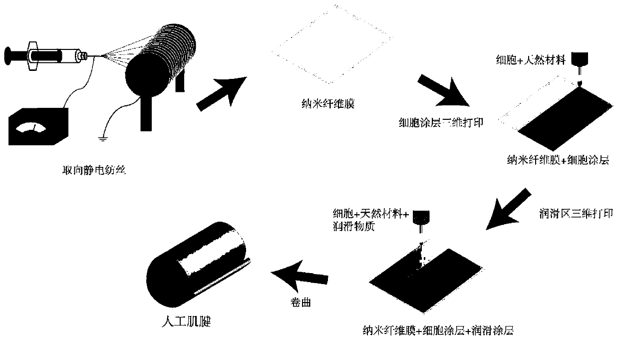
[0038] Specifically, the preparation of the nanofiber membrane in the step S2 includes the following steps:

[0039] S21. Preparation of materials: Measure an appropriate amount of biopolymer material and electrospinning solvent, and then put natural materials into the electrospinning solvent for mixing. The mass fraction of biopolymer materia...

Embodiment 1

[0050] Polycaprolactone, a polymer material, is dissolved in hexafluoroisopropanol solvent, with a total mass fraction of 12%. Adjust the electrospinning process parameters: turn on the orientation, the flow rate of the propulsion pump is 1.5mL / h, the external high voltage is 10kv, the receiving distance is 10cm, and the nanofiber film is obtained by electrospinning;

[0051] Mix tendon stem cells with 10g of sodium alginate gel and add them into the cylinder of the 3D printer. Set it to 0 / 90°, print on the surface of the obtained nanofiber membrane, start the printing program, and print a single-layer rectangular bracket with a length of 3 cm and a width of 1 cm;

[0052] Mix tendon stem cells, cytokine BMP7, and 10 g of sodium alginate gel into the barrel of the three-dimensional printer. The printing temperature is set at 37 ° C, the diameter of the print head is 200 μm, the layer height is 200 μm, and the air pressure of the barrel is 800KPa. The printing path is set to 0...

Embodiment 2

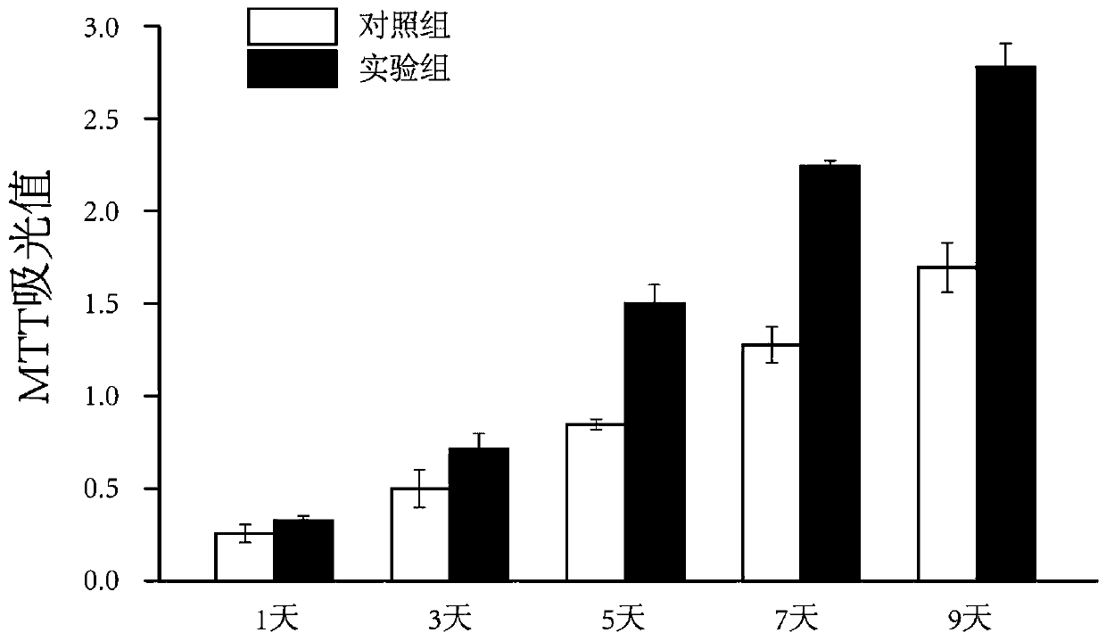
[0055] The biocompatibility of the tendon scaffold prepared by three-dimensional bioprinting technology combined with electrospinning technology was characterized. The obtained nanofiber membrane was curled and placed in an incubator for culture. A single layer of nanofiber membrane was planted with tendon stem cells on the surface as a control. For comparison, after 1, 3, 5, 7 and 9 days of culture, the tenocyte viability in the tendon scaffold was detected by MTT method. The tenocytes on the fibrous membrane had good proliferation behavior, and the cell viability was better, and the proliferation behavior was more significant than that of the control group.


PUM

PropertyMeasurementUnit
Lengthaaaaaaaaaa
Diameteraaaaaaaaaa
Lengthaaaaaaaaaa

Abstract

The present invention belongs to the technical field of biological three-dimensional printing and particularly discloses a method for preparing artificial muscle tendons by using a biological three-dimensional printing and an electrospinning technology. The preparation steps are as follows: S1, seed cell culturing; S2, nano fiber membrane preparing; S3, cell coating layer preparing; and S4, nano fiber membrane/cell coating layer treating. The biological three-dimensional printing and the electrospinning technology are combined, the electrospinning method is used to obtain directional nano fiber membranes, the biological three-dimensional printing technology is used for printing on the nano fiber membranes to obtain lubricating cell coating layers to rapidly realize adhesion and colonization of seed cells and obtain the high polymer material artificial muscle tendons containing the cells and having good lubricating performance, the manufacture method is simple and convenient, better simulates tissue morphology, cell composition and lubricating performance of the natural muscle tendon tissues, and is conducive to muscle tendon repair and normal function recovery.

Description

technical field [0001] The invention belongs to the technical field of biological three-dimensional printing and electrospinning, and in particular relates to a method for preparing artificial tendon by using biological three-dimensional printing and electrostatic spinning technology. Background technique [0002] Tendon is composed of dense connective tissue. Tendon injury is a common sports injury, and it is also a common type of clinical soft tissue injury. The current treatment methods have certain limitations or deficiencies. Tissue engineering technology provides a more comprehensive method for clinical tendon repair. An ideal and physiological method to repair tendon defects with tissue engineering technology, that is, a small amount of seed cells are cultured in vitro, and after expansion, they are combined with biodegradable scaffolds to form a complex, and the cells proliferate and differentiate after implanting them in the defect site , Secrete matrix, repair dama...

Claims


Application Information

Patent Timeline
IPC IPC(8): A61L27/44A61L27/38A61L27/52D04H1/728
Inventor 乔之光唐佳昕戴尅戎孙彬彬王友连梅菲
Owner SHANGHAI NINTH PEOPLES HOSPITAL AFFILIATED TO SHANGHAI JIAO TONG UNIV SCHOOL OF MEDICINE
Why Eureka
Try Eureka