Preparation method and imaging method of quantum dots for clinical skeletal in vivo imaging

A technology of in vivo imaging and quantum dots, applied in preparations for in vivo experiments, nanotechnology for materials and surface science, chemical instruments and methods, etc. supply and metabolism status, bone tissue sensitivity and resolution are not as good as CT and X-ray, and cannot dynamically observe bone tissue metabolism, etc., to achieve deep tissue penetration ability, good biocompatibility, fidelity and resolution increase Effect

Active Publication Date: 2021-10-22
SUZHOU UNIV
View PDF2 Cites 0 Cited by
  • Summary
  • Abstract
  • Description
  • Claims
  • Application Information

AI Technical Summary

Problems solved by technology

Although modern imaging technologies, such as CT (Computed Tomography), MRI (Magnetic Resonance Imaging) and ECT (Emission Computed Tomography), including SPECT (single-photon ECT) and PET (positron ECT), have shown great advantages in disease diagnosis. , especially for skeletal system imaging, CT still needs to rely on high-energy X-ray assistance; MRI imaging has no ionizing radiation, but its sensitivity and resolution to bone tissue are not as good as CT and X-ray, and may be harmful to patients with cardiac pacemakers. Some metal foreign bodies cannot be the first choice for MRI examination, and factors such as high price limit its application in bone imaging; as for ECT, it can not only perform plane imaging and tomographic imaging, but also static imaging and dynamic imaging. It is often used clinically in the examination of tumors in bone and other parts, especially in the detection of bone metastatic tumors. However, the basic principle of imaging is still to inject radioactive isotopes into the human body. Differences in radioactivity concentration are formed between the inside and outside of organs or between diseased parts and normal tissues. These detected differences are processed by computer and then imaged, so there are still certain restrictions in clinical application.
[0003] The above imaging methods still have the following defects and deficiencies: 1. Clinical bone imaging is mainly based on the penetrability, fluorescence effect and photosensitive effect of X-rays, which restricts the human body from being used for a long time; 2. The ionizing radiation that X-rays have not only The patient's health poses a certain impact, and it also brings potential harm to medical staff, especially for pregnant women and infants, it is restricted, and it brings a lot of concerns to doctors and patients in clinical use; 3. X-ray imaging is the second 3D static imaging cannot dynamically observe bone tissue metabolism, and cannot more specifically reflect the blood supply and metabolic status of bones and surrounding tissues; 4. Can not clearly image irregular human bones (such as sternum, ribs, scapula, etc.) , ultimately need to use high-energy CT to identify
In summary, there is currently no safe and reliable bone imaging technology

Method used


Image

  • Preparation method and imaging method of quantum dots for clinical skeletal in vivo imaging
  • Preparation method and imaging method of quantum dots for clinical skeletal in vivo imaging
  • Preparation method and imaging method of quantum dots for clinical skeletal in vivo imaging

Examples


Embodiment 1

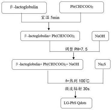
[0026] Preparation of quantum dots for clinical bone imaging in vivo, the specific process is as follows:

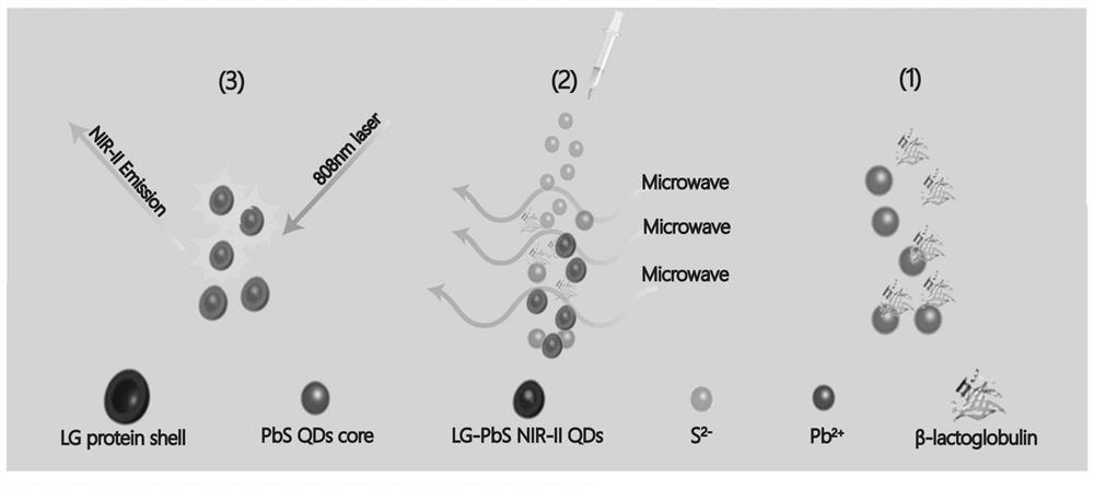
[0027] Take 500 μL of β-lactoglobulin (β-LG) at a concentration of 50 mg / mL and 500 μL of Pb(CH 3 COO) 2 Mix and stir for 5 min at room temperature; then titrate the pH value of the mixed solution to 7.5 with 1M NaOH; 2 S was quickly injected into the mixed solution with a pH value of 7.5, heated to 100 °C, and maintained under microwave irradiation for 30 s to obtain the final product PbS QDs, and the final product was stored at 4 °C. The preparation method flow is as follows figure 1 As shown, the synthesis schematic is shown as figure 2 shown.

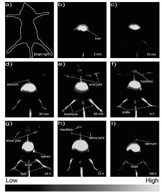
[0028] The method for using the quantum dots prepared above for clinical bone imaging in vivo is as follows:

[0029] In this example, mice were selected as the experimental research object. After the mice were anesthetized, 300 μL of LG-PbS QDs were injected through the tail vein of the mice. The original solution of LG...


PUM


Abstract

The invention discloses a preparation method of quantum dots for clinical bone imaging in vivo, specifically as follows: take an appropriate amount of β-lactoglobulin aqueous solution (β-lactoglobulin, LG) and Pb(CH 3 COO) 2 Mix the aqueous solution, mix and stir for 5min at room temperature; then titrate the pH value of the mixed solution to 7.5 with 1M NaOH; 2 S was quickly injected into the solution with a pH value of 7.5, heated to 100 °C, and maintained under microwave irradiation for 30 s to obtain the final product LG‑PbS QDs. The imaging method of the quantum dots prepared by the aforementioned method is as follows: a certain amount of LG‑PbS QDs is injected into the subject, an 808nm diode laser and a 850‑1000nm short-pass filter are used as the excitation source, and an InGaAs CCD camera is used to collect NIR‑II Fluorescence image, the emission signal is filtered by a 1100nm long-pass filter; the signals are recorded at different time points, and NIR‑II Movie imaging under 808nm laser excitation is performed at the same time. The quantum dot preparation and imaging method provided by the invention is energy-saving and environment-friendly, and realizes non-invasive and radiation-free animal bone imaging.

Description

technical field [0001] The invention relates to a preparation method and an imaging method of quantum dots used for clinical skeletal in vivo imaging, and belongs to the technical field of skeletal system imaging. Background technique [0002] Since Roentgen in Germany discovered X-rays in 1895 and X-rays have been used clinically in 1896, X-ray imaging is still the most routine diagnostic method for bone diseases. Although modern imaging technologies, such as CT (Computed Tomography), MRI (Magnetic Resonance Imaging) and ECT (Emission Computed Tomography), including SPECT (single-photon ECT) and PET (positron ECT), have shown great advantages in disease diagnosis. , especially for skeletal system imaging, CT still needs to rely on high-energy X-ray assistance; MRI imaging has no ionizing radiation, but its sensitivity and resolution to bone tissue are not as good as CT and X-ray, and may be harmful to patients with cardiac pacemakers. Some metal foreign bodies cannot be th...

Claims


Application Information

Patent Timeline
Patent Type & Authority Patents(China)
IPC IPC(8): C09K11/66C09K11/02B82Y20/00B82Y30/00B82Y40/00A61K49/00
Inventor 车艳军陈俊郭江博罗宗平
Owner SUZHOU UNIV
Why Eureka
Try Eureka