Preparation method of gelatin microspheres and application thereof

A technology of gelatin and microspheres, applied in the field of interventional medicine, can solve the problems of limiting biological applications, lack of cell adhesion sites, and reducing the biological activity of loaded cells, and achieve the effects of cell safety, good cell loading, and simple preparation process

Active Publication Date: 2019-09-06
SUN YAT SEN UNIV
View PDF6 Cites 7 Cited by
  • Summary
  • Abstract
  • Description
  • Claims
  • Application Information

AI Technical Summary

Problems solved by technology

However, these carriers lack cell adhesion sites, greatly limiting cell proliferation, migration, and tissue formation.
Cross-linking using cytotoxic ...

Method used


Image

  • Preparation method of gelatin microspheres and application thereof
  • Preparation method of gelatin microspheres and application thereof
  • Preparation method of gelatin microspheres and application thereof

Examples


Embodiment 1

[0033] The preparation of embodiment 1 gelatin microsphere

[0034] 1. Fabrication of microfluidic devices

[0035] Prepare a PTFE tube with a length of about 35cm (inner diameter 0.3mm, outer diameter 0.6mm), insert a 26G needle into one end of the PTFE tube, insert a 31G needle into the PTFE tube in parallel at a distance of 20cm from the needle, and keep the needle tip in the middle of the tube; use AB Fix the needle and PTFE tube at the joint with rubber seal. After the device is completed, first check the connectivity and tightness of the device. Use a 5mL syringe to absorb deionized water, insert a 31G needle, and manually push the syringe. If liquid flows out of the PTFE tube, then Internal connectivity is good. Connect the syringes to the 20G needle at the same time, block the outlet of the PTFE tube, and squeeze the two syringes manually at the same time. If no liquid leaks out, the device is well sealed. Dry the prepared device for later use.

[0036] 2. Preparati...

Embodiment 2

[0044] Embodiment 2 Fibroblast adhesion and proliferation characterization on the surface of gelatin microspheres

[0045] To test the adhesion and proliferation of fibroblasts on the surface of gelatin microspheres, the specific process is:

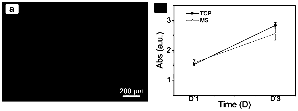
[0046] Spread gelatin microspheres on the bottom of a 48-well plate (non-adhesive plate), and NIH-3T3 cells (mouse embryonic fibroblast cell line) in a 5×10 4The cells / well density was inoculated, and each group had three parallel samples, and the control group was TCP (adhesive type). On days 1 and 3, use CCK-8 solution to detect cell viability;

[0047] The staining result is as figure 2 shown, where figure 2 a is the result of cell death and life staining on the surface of the microspheres 2 days after inoculation, the results show that the cells grow well on the surface of the microspheres, and there are no dead cells in the field of view. Within 3 days, the proliferation rate of the MS group was good, such as figure 2 b, sho...

Embodiment 3

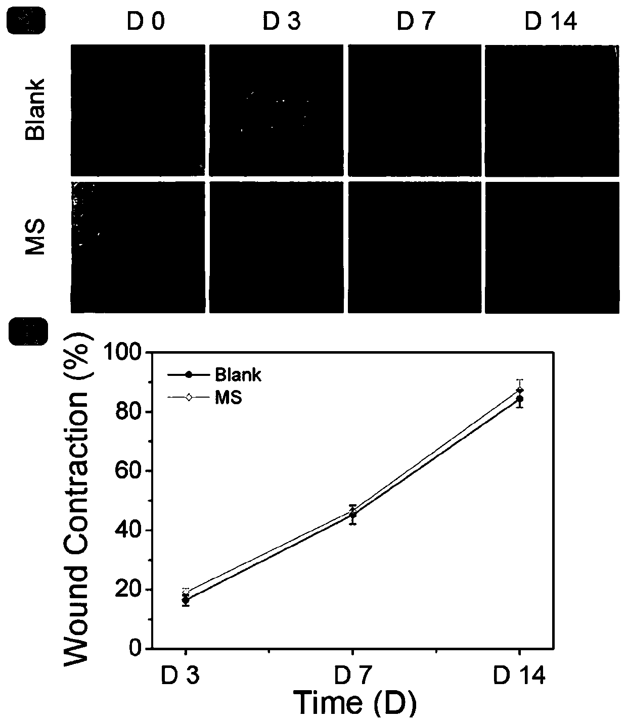
[0048] Example 3 Testing the effect of MS on full-thickness skin excision wounds

[0049] 1. Modeling of Type I Diabetic Rats (TIDM)

[0050] Fifteen SD rats (8 weeks old, weighing 200-250 g, male) were used to induce TIDM to model, and fasted for 24 hours before model making. Weigh each rat's body weight before modeling, and take tail vein blood to measure the fasting blood glucose value of each rat;

[0051] 1% STZ (streptozotocin) solution was injected intraperitoneally at a dose of 50 mg / kg. After the operation was completed, the rats were housed in separate cages. After modeling, blood was collected from the tail vein to measure the blood glucose of the rats. If the blood glucose value before induction was <8.9mmol / L, and the random blood glucose of rats was ≥16.7mmol / L for three consecutive days after induction, the model was considered successful. If the induced TIDM model fails, feed the rats with a normal diet and monitor the blood sugar level. After the blood sug...


PUM

PropertyMeasurementUnit
The inside diameter ofaaaaaaaaaa
Outer diameteraaaaaaaaaa
The average particle sizeaaaaaaaaaa

Abstract

The invention discloses a preparation method of gelatin microspheres and an application thereof. The preparation process comprises the following steps: S1. a gelatin aqueous solution is used as the internal phase; an oil phase is an external phase, and the flow rates of the inner and outer phases are respectively controlled by a microfluidic device to be 1 to 10 [mu]L/min and 20 to 200 [mu]L/min;and the external phase is the collected phase, and the micro-droplets are collected; and S2. the collected micro-droplets are gelatinized at 4 to 12 DEG C, the oil phase on the surface is removed, anda crosslinking reaction is carried out with a genipin solution for 1 h, and after the reaction is completed, the gelatin microspheres are obtained by separation. The gelatin microsphere of the invention not only has a simple preparation process, is safe to the cells, is non-toxic, and has certain mechanical strength, can load the cells well, does not affect the activity of the cells, and can be applied as a cell carrier; the gelatin microsphere has a certain promoting effect on the healing of diabetic wounds and the formation of wound blood vessels, also has a good degradation effect, and canbe used as an accelerator drug for wound healing and blood vessel formation.

Description

technical field [0001] The invention relates to the technical field of interventional medicine, and more specifically, to a preparation method and application of gelatin microspheres. Background technique [0002] Transplantation of mesenchymal stem cells (MSCs) has emerged as a promising therapeutic strategy. MSCs can be stably expanded ex vivo while maintaining the ability to differentiate into epidermal and dermal lineage cells, and even skin appendages such as hair follicles, sweat glands, and microvessels can be rapidly regenerated in MSC-treated wounds. Injecting the cells directly into the repair site minimizes the invasiveness of the procedure. However, in this way, the degree of integration of cells into host tissues is low, and the cell survival rate is low, resulting in a low success rate of clinical transplantation. [0003] To address the above issues, a potentially attractive strategy is to suspend stem cells in hydrogels, which is expected to improve the sur...

Claims


Application Information

Patent Timeline
IPC IPC(8): A61K9/16A61K47/42A61K35/545A61K35/28A61P17/02A61P3/10
CPCA61K9/1658A61K9/0014A61K35/545A61K35/28A61P17/02A61P3/10
Inventor 李燕史明张浩宋婷高芸芬
Owner SUN YAT SEN UNIV
Why Eureka
Try Eureka