
Method for preparing cell-free medical implant material

An implant material and cell-free technology, which is applied in the field of preparation of medical implant materials, can solve the problems of ECM three-dimensional structure damage, harsh and excessive decellularization process, and achieve mild and targeted action mode and good tissue repair function Effect

Pending Publication Date: 2019-11-05
上海白衣缘生物工程有限公司
View PDF10 Cites 6 Cited by
  • Summary
  • Abstract
  • Description
  • Claims
  • Application Information

AI Technical Summary

Problems solved by technology

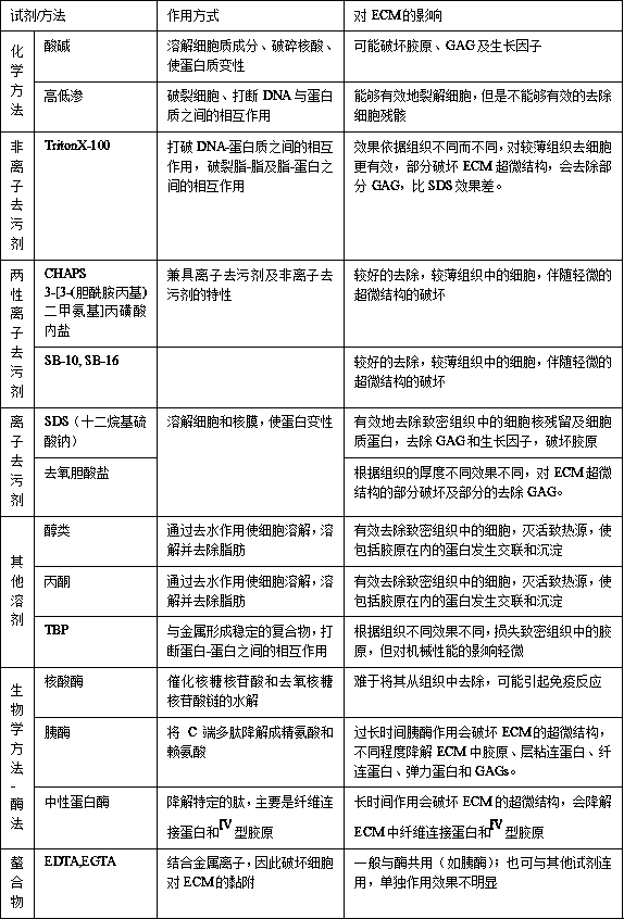
[0039] At the same time, another technical problem that accompanies it is: in order to ensure the high safety of biological patches, usually all manufacturers require complete decellularization in actual operation, so the decellularization process is very harsh and even a bit excessive. This will lead to a greater loss of effective active ingredients in the ECM, as well as damage to the three-dimensional structure of the ECM; thereby weakening the mechanical properties of the decellularized material
[0040] At present, in the practical application of some biological materials, it may be precisely because of the acid, alkali or protease used in the process of decellularization. This treatment method will damage the three-dimensional structure of the ECM, which will lead to the mechanical properties of the biological materials. There is a relatively obvious decline in the upper part; so that the mechanical strength required for tissue repair in some specific parts cannot be reached; for example, the hernia mesh, because the normal abdominal wall load force is about 19N, if the mechanical strength of the mesh is too small, there will be tearing. Therefore, in the process of preparing such decellularized biological patches, additional attention needs to be paid to the mechanical properties of such medical biomaterials after decellularization; currently there are additional cross-linking methods or other physical methods, such as adding Single-layer sheets are placed horizontally or vertically, and multi-layer cross sections are overlapped to enhance mechanical properties

Method used


Image

  • Method for preparing cell-free medical implant material

Examples


Embodiment 1

[0096] Example 1 (Commercial pork SIS+0.25%Saponin)

[0097] The preparation of the acellular medical material of porcine small intestine submucosa, the specific steps are as follows:

[0098] 1) Material selection: Fresh small intestines of commercial pork pigs are selected in slaughterhouses as raw materials for decellularization

[0099] 2) Pretreatment: use physical scraping method to remove the mucous membrane layer, muscular layer, serosa layer and lymph nodes of the pig small intestine, separate the submucosa, soak in 0.5% acetic acid solution for 30 minutes, the ratio of pig small intestine to acetic acid solution is 1 :5, then soaked in water to obtain the decellularized raw material, i.e. small intestinal submucosa, hereinafter referred to as SIS material

[0100] 3) Disinfection: use a mixed solution containing 1.0% peracetic acid and 15% ethanol, the ratio of the SIS material to the mixed aqueous solution is 1:10, and immerse at room temperature for 100 minutes un...

Embodiment 2

[0105] Embodiment 2 (commodity pig SIS+0.25%SDS)

[0106] In this example, the decellularized raw material is the small intestine tissue of commercial pork pigs. In the preparation method, the steps of pretreatment of the pig small intestine tissue, disinfection, degreasing, decellularization, DNA and α-Gal antigen removal, freeze-drying, and sterilization are completely complete. Same as Example 1; the difference is only in the choice of decellularization reagent, in this example, 0.25% SDS was used to replace the 0.25% saponin solution in Example 1.

Embodiment 3

[0107] Example 3: Optical observation and active ingredient detection of acellular medical materials.

[0108] Method: fixed in formalin, embedded in paraffin, cut the decellularized material in the example into thin slices, dewaxed with xylene, dehydrated with alcohol, stained with hematoxylin-eosin, observed the residual cells and Matrix fiber structure.

[0109] Results: In all the decellularized medical materials obtained in the two examples, no cells and their fragments were observed; the collagen fibers were continuous and varied in thickness, but there was no obvious fracture.

[0110] For the cell-free medical implant materials prepared in 1-2 examples, the content of the important active ingredient hyaluronic acid (HA) was detected using a commercial ELISA kit; Various decellularized materials were processed, and the test results are as follows:

[0111] group Sources and processing methods of decellularized materials Hyaluronic acid HA (ug / mg) Exa...


PUM


Abstract

The invention belongs to the field of medical repair materials and relates to a method for preparing a cell-free medical implant material. In the method for preparing the cell-free materials, neitherchemically synthesized detergents nor protease products are used to remove cells; but a plant-derived nonionic surfactant is used as a decellularizing agent, thus avoiding the damage of chemical detergents, protease preparations and the like to an ECM three-dimensional structure and the loss of effective active ingredients in ECM. The cell-free medical material prepared by the method can not onlybetter maintain the three-dimensional structure of ECM, but also allow more effective active ingredients to be remained in ECM and endowed with good mechanical properties. Thereby, the ability to induce chemotaxis and differentiation of host cells is stronger, and the effect of promoting tissue repair and regeneration will be better.

Description

technical field [0001] The invention relates to a method for preparing a medical implant material, in particular to a method for preparing a cell-free medical implant material. Background technique [0002] Clinical implantable medical biomaterials are changing from traditional non-absorbable materials to new biomaterials that are natural, degradable, and capable of actively inducing tissue regeneration; based on the principles of tissue engineering, animal tissues are used as raw materials to remove tissue The cells in the tissue retain a relatively complete extracellular matrix (Extracellular Matrix, ECM), which is used as a raw material to prepare decellularized medical materials for tissue repair and regeneration, which is a major development direction of surgical medicine in the future. [0003] Decellularized ECM usually has a three-dimensional structure and contains various macromolecular substances such as collagen, elastin, glycosaminoglycans (including hyaluronic a...

Claims


Application Information

Patent Timeline
Patent Type & Authority Applications(China)
IPC IPC(8): A61L27/36A61L27/50
CPCA61L27/3633A61L27/3687A61L27/50
Inventor 韩韦红葛翠兰钱锵张国强
Owner 上海白衣缘生物工程有限公司
Learn More