Biological patch for tympanic membrane repair and preparation method thereof

A biological patch and patch technology, applied in tissue regeneration, medical science, prosthesis, etc., can solve the problems of ECM structure damage, slow degradation, weak induction ability, etc., to achieve the promotion of the formation of new blood vessels and basement membrane, Mild mode of action, promotes repairing effect

Pending Publication Date: 2019-11-15
上海白衣缘生物工程有限公司
View PDF7 Cites 4 Cited by
  • Summary
  • Abstract
  • Description
  • Claims
  • Application Information

AI Technical Summary

Problems solved by technology

[0026] The object of the present invention is to aim at the existing tympanic membrane biopatch derived from dermis, which has the disadvantages of complex process, slow degradation, and more elastin, less active ingredients, and weak inductive ability; Materials, the decellularization process is harsh, resulting in serious loss of active ingredients in the patch, damage to the ECM structure, and weak ability to induce cell growth, which cannot effectively meet the actual clinical needs; for some shortcomings and defects of the aforementioned otology patch; The inventor focused on the selection of patch raw materials, the selection of decel

Method used


Image

  • Biological patch for tympanic membrane repair and preparation method thereof

Examples


Embodiment 1

[0088] Example 1 (decellularization reagent 0.25% Saponin)

[0089] For the repair of the eardrum, the preparation of the decellularized patch from the porcine small intestine submucosa, the specific steps are as follows:

[0090] 1) Collection, cleaning, and pretreatment: Pretreatment: select commercial pork pigs from slaughterhouses, and clean their fresh small intestine tissue; use physical scraping to remove the mucous membrane layer, muscular layer, serosa layer, Lymph nodes were separated from the submucosa, soaked in 0.5% acetic acid solution for 30 minutes, the ratio of porcine small intestine to acetic acid solution was 1:5, and then soaked in purified water for 3 times to obtain the raw material of the biological patch, namely the small intestinal submucosa, as follows Referred to as SIS material;

[0091] 2) Disinfection: use a mixed aqueous solution containing 1.0% peracetic acid and 15% ethanol, the ratio of the SIS material to the mixed aqueous solution is 1:10,...

Embodiment 2

[0096] Example 2 (decellularization reagent 0.25% SDS)

[0097] The steps of tissue material selection, pretreatment, disinfection, degreasing, cell removal, DNA removal and α-Gal antigen removal, freeze-drying, and sterilization are exactly the same as in Example 1; the difference is only in the selection of the cell-removing reagent in the fourth step. In this example, 0.25% SDS was used to replace the Quil-A solution in Example 1.

Embodiment 3

[0098] Example 3: Thickness detection, optical observation and active ingredient detection of decellularized patches for tympanic membrane repair.

[0099] The tympanic membrane biological patch is composed of three layers of SIS decellularized, and the measured thickness is 0.16mm.

[0100] Optical microscope observation:

[0101] Methods: Formalin-fixed, paraffin-embedded, the patch in the example was cut into thin slices, dewaxed with xylene, dehydrated with alcohol, stained with hematoxylin-eosin, and the residual cells and matrix fiber structure were observed under a microscope.

[0102] Results: In all the decellularized patches in the two examples, no cells and their fragments were observed; the collagen fibers were continuous and varied in thickness, but there was no obvious fracture.

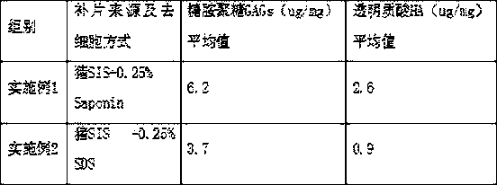
[0103] For the tympanic membrane biopatch samples prepared in the two examples, commercial ELISA kits were used to detect the content of important active ingredients glycosaminoglycans...


PUM


Abstract

The invention belongs to the field of otology repair materials, and relates to a biological patch for tympanic membrane repair and a preparation method thereof. The otology repair patch of the invention overcomes the shortcomings of the prior patch due to an improper decellularizing process and a complicated process, and causes serious loss of effective active ingredients in the ECM. The biological patch adopts the plant-derived saponin as a decellularizing reagent, which avoids the residue and toxicity of a chemical reagent, and the mild decellularizing reagent is highly targeted and mainly binds to the cell membrane lipid; the decellularizing reagent has little damage on an ECM stereoscopic three-dimensional structure and the active components thereof; and more biologically active components such as glycosaminoglycan (including hyaluronic acid) can be retained. The patch of the invention has a better ECM scaffold structure and more active components, and has stronger ability to induce cell chemotaxis and proliferation, is beneficial to faster and better repair of the tympanic membrane, and also has a certain anti-infective effect; at the same time, the method of the invention issimple and convenient to operate, and has no damage to the ECM.

Description

technical field [0001] The invention relates to the field of tympanic membrane repair materials in otology, in particular to a decellularized patch for tympanic membrane repair and a preparation method thereof. Background technique [0002] The adult tympanic membrane is about 9 mm long, 8 mm wide, and 0.1 mm thick; although the tympanic membrane is very thin, its anatomical structure has three layers (tense parts): the outer layer is the epithelial layer, which is continuous with the skin of the external auditory canal, and is essentially a Skin epithelial cells, the main components are collagen and skin cells; the middle layer is composed of radial and circular cilia, which has certain elasticity and tension, and is also collectively called the fibrous layer (radial on the outside and wheel-shaped on the inside); the inner layer is the mucous membrane The tympanic layer is connected with the tympanic membrane and is formed by its continuation. Type VI collagen forms the st...

Claims


Application Information

Patent Timeline
IPC IPC(8): A61L27/36A61L27/58
CPCA61L27/3604A61L27/3629A61L27/3645A61L27/3687A61L27/58A61L2430/14
Inventor 韩韦红葛翠兰钱锵张国强
Owner 上海白衣缘生物工程有限公司
Why Eureka
Try Eureka