

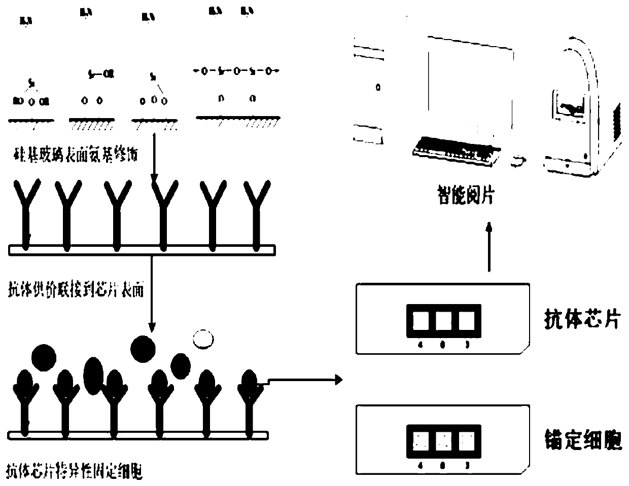
Antibody chip for blood cell separation counting and preparation method thereof
A technology of antibody chips and blood cells, which is applied in the direction of analyzing materials, measuring devices, instruments, etc., can solve the problems of expensive instruments, cumbersome operations, and long time consumption
- Summary
- Abstract
- Description
- Claims
- Application Information
AI Technical Summary
Problems solved by technology
Method used
Image

Examples
Embodiment 1
[0071] Example 1 Preparation of Antibody Chip for Separation and Counting of Blood Cells
[0072] S1. Hydroxylation;
[0073] (1) prepare 1wt% analytical sodium hydroxide;
[0074] (2) Place the glass slide in sodium hydroxide solution and soak for 1 hour;
[0075] (3) Transfer to an ultrasonic cleaning machine for ultrasonication for 15 minutes, wash with purified water three times, and then dry in an oven at 100° C. for 1 hour.
[0076] S2. Amination;
[0077] (1) prepare 5% silane coupling agent KH550 ethanol aqueous solution;
[0078] (2) Soak the glass slide in 5% silane coupling agent KH550 ethanol aqueous solution for 2 hours;
[0079] (3) Wash twice with purified water, then twice with 95% (v / v) ethanol, and then dry at 110° C. for 15 minutes.
[0080] S3. Formylation;
[0081] (1) prepare 2wt% glutaraldehyde aqueous solution;
[0082] (2) Soak all slides in wt 2% glutaraldehyde aqueous solution for 1 hour.
[0083] (3) Take out the slides, place them in purifi...
Embodiment 2
[0101] Example 2 Preparation of Immunocytochemical Staining Analysis and Counting Kit
[0102] Immunocytochemical Staining Analysis and Counting Kit contains antibody chip, diluent, hydrogen peroxide solution, staining solution and counterstaining solution for blood cell separation and counting.
[0103] The antibody chip used for blood cell separation and counting was prepared according to Example 1.
[0104] Diluents may be prepared by dissolving the diluent tablet in an aqueous solution. Diluent tablets are commercially available, purchased from Shanghai Baokang Biological High-tech Co., Ltd., and the main components are sodium chloride, potassium chloride, potassium dihydrogen phosphate, disodium hydrogen phosphate, citric acid, Tween 20, etc., with One tablet can be dissolved in 40ml aqueous solution to make phosphate buffer solution with pH 7.0-7.4. Its specifications are 10 pieces / pack, packed in sealed aluminum foil bags, and kept in an airtight place with shading. ...
Embodiment 3
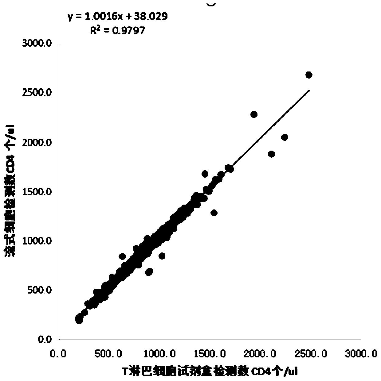
[0108] Example 3 Detection of Immunocytochemical Staining Analysis and Counting Kit
[0109] Applicable instruments:
[0110] Optical microscope or automatic counting microscope system with 10x objective lens.
[0111] Sample request:
[0112] Collection of venous blood samples: EDTA should be used as an anticoagulant for the samples (1.0-1.5mg EDTA: 1ml whole blood). All specimens must be inoculated within 6 hours after blood collection.
[0113] It is strictly forbidden to store in the refrigerator.
[0114] Please refuse to accept the sample when it is found that the blood sample has hemolysis, coagulation, plasma ratio imbalance, storage at extreme temperature, and no valid serial number.
[0115] Detection method:
[0116] 1. Solution preparation
[0117] 1) Prepare phosphate buffer solution: add 1 pack of phosphate buffer solution to 400ml of pure water, and stir evenly.
[0118] Note: The phosphate buffer solution is freshly prepared when testing, and it can be w...
PUM
 Abstract
Description
Claims
Application Information