Fluorescent probe targeting CKIP-1 and used for detecting osteoporosis and application of fluorescent probe in in-vivo detection

A technology for CKIP-1, osteoporosis, applied in the direction of analysis, application, diagnostic recording/measurement using fluorescence emission, which can solve the lack of biomarkers or indicators, and the inability to monitor bone density or osteoporosis status in living animals and other problems, to achieve the effect of fast detection speed, high sensitivity and simple operation method

Active Publication Date: 2021-01-29
ACADEMY OF MILITARY MEDICAL SCI
View PDF3 Cites 2 Cited by
  • Summary
  • Abstract
  • Description
  • Claims
  • Application Information

AI Technical Summary

Problems solved by technology

In the presence of Cathepsin K, the probe changes from its originally quenched form to fluorescent, which makes it sensitive to changes in bone resorption. These probes can only monitor bone formation or bone resorption activity, ...

Method used


Image

  • Fluorescent probe targeting CKIP-1 and used for detecting osteoporosis and application of fluorescent probe in in-vivo detection
  • Fluorescent probe targeting CKIP-1 and used for detecting osteoporosis and application of fluorescent probe in in-vivo detection
  • Fluorescent probe targeting CKIP-1 and used for detecting osteoporosis and application of fluorescent probe in in-vivo detection

Examples


Embodiment 1

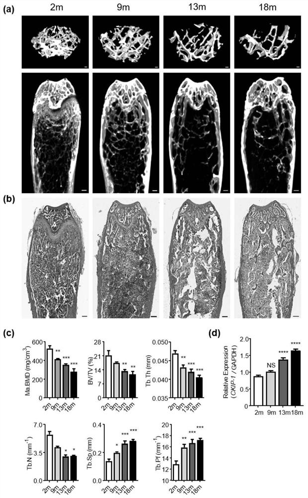
[0051] Example 1, Correlation between CKIP-1mRNA expression and osteoporosis

[0052] 1. To construct an aging osteoporosis model, use C57BL / 6J male mice (n=5 / group) of different ages (9m, 13m and 18m), and use 2m mice as a control group (n=5).

[0053] 2. To construct a postmenopausal osteoporosis model, C57BL / 6J female mice (20) were sham-operated (sham) or ovariectomized (OVX) at the age of 4 months, and raised under the same feeding conditions for 8 weeks, specifically The surgical operation is as follows: After the mice were weighed and anesthetized successfully, the ovaries were removed and placed in a dirt tray. After complete hemostasis was confirmed, the tissue was also brought into the abdominal cavity. Subsequently, the same operation was performed at the corresponding position on the contralateral side, and the right ovary of the mouse was excised, and finally disinfected with iodophor. Sham-operated group: After exposing the bilateral ovaries, no resection was p...

Embodiment 2

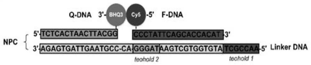
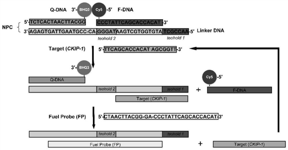
[0074] Example 2, preparation and detection of near-infrared fluorescent probes based on CKIP-1

[0075] 1. Mix 50μL Tris-HCl buffer (pH7.4, 10mM MgCl2), 20μL Linker DNA strand (100μM), 20μL Q-DNA strand (100μM) and 10μL F-DNA strand (100μM) in a PCR tube, and incubate at 95°C 10 minutes, and then lower the temperature to 37°C with a gradient of 0.1°C / s. The formed Linker / F / Q double chain structure was determined by UV spectrophotometer. Then the probe is further mixed with the fuel chain, the mixing molar ratio is 1:5, and 3000 (Thermo Fisher Scientific, USA) was incubated at room temperature for 30 min to complete the preparation of the probe system.

[0076] When the probe is used for live cell detection, the preparation system and steps are as follows: Lipofectamine TM 3000 Reagent 6μl and Opti-MEM TM Mix 100 μl of culture medium (solution A), probe (5 μg / μl) 8 μl, P3000 TM Reagent 16 μl and Opti-MEM TM Mix 100 μl of medium (solution B), incubate at room temperatur...

Embodiment 3

[0089] Example 3. Fluorescence imaging of CKIP-1-based near-infrared fluorescent probes in living cells and in vivo fluorescence imaging of mice

[0090] 1. Inoculate the mouse macrophage precursor cell line RAW264.7 cells in a confocal culture dish, add 10% fetal bovine serum, penicillin (100 μg / mL) and streptomycin (100 μg / mL) in DMEM medium , after culturing at 37°C under 5% carbon dioxide for 24 hours, replace the complete medium with the probe-containing culture solution and immediately place the culture dish in a live cell imaging system to monitor the changes of the probe in the cells for 22 hours. 1.5h after adding the probe, the cells began to show near-infrared fluorescence, reached the peak fluorescence signal at 6h, and then gradually decreased, see Figure 8 (a)-(b). In contrast, there was no activation probe in MC3T3-E1 cells with very low background CKIP-1 expression, see Figure 8 (c), indicating its high selectivity.

[0091] 2. After injecting the near-inf...


PUM


Abstract

The invention discloses a biomarker for detecting osteoporosis as well as a preparation method and application of an activatable near-infrared fluorescent probe of the biomarker. The biomarker provided by the invention has negative correlation with various primary osteoporosis. The activatable near-infrared probe of the biomarker comprises a fluorescent strand, a quenching strand and a fuel strand. A specific preparation method comprises the following steps: mixing a Cy5-labeled DNA fluorescent strand (FDA), a BHQ3-labeled DNA quenching strand (QDA) and a hybridized connection DNA strand (Linker DNA) according to a proper ratio, and conducting reacting to obtain the probe. The probe of the invention has the advantages of simple preparation, sensitive detection, good selectivity and the like. The probe can be used as a new index to monitor bone mass change, provides a new candidate means for clinical diagnosis of osteoporosis, and provides a new approach for personalized treatment and prognosis monitoring of osteoporosis.

Description

technical field [0001] The invention relates to a biomarker CKIP-1 for detecting osteoporosis and a preparation method and application thereof of an activatable near-infrared fluorescent probe. Background technique [0002] Osteoporosis (OP) is a degenerative skeletal disease characterized by decreased bone mass and deterioration of bone microarchitecture, which ultimately leads to skeletal fragility and increased fracture risk. It occurs due to disturbances in bone remodeling, which in turn is primarily regulated by bone formation and bone resorption. Osteoporosis includes two types: primary (type I: postmenopausal, type II: elderly) and secondary (eg, congenital disease, drugs, and weight loss / discontinuation). Due to its high morbidity and mortality, it has become a major public health problem worldwide. It is estimated that by 2020, the number of osteoporosis patients in the United States will increase from about 10 million to more than 14 million; in China, the preval...

Claims


Application Information

Patent Timeline
IPC IPC(8): A61B5/00
CPCA61B5/0071A61B5/4509A61B2503/40
Inventor 张令强崔宇杨曦
Owner ACADEMY OF MILITARY MEDICAL SCI
Why Eureka
Try Eureka