3D cell microsphere paraffin sectioning method
A technology of paraffin sectioning and microspheres, applied in sampling, measuring devices, instruments, etc., can solve the problems of insufficient wax soaking and difficult sectioning, etc.
- Summary
- Abstract
- Description
- Claims
- Application Information
AI Technical Summary
Problems solved by technology
Method used
Image
Examples
Embodiment 1
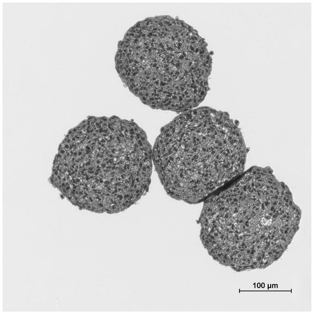
[0050] Embodiment 1 HE stained human hepatic stellate cell (LX-2) cell microsphere paraffin section
[0051] In this embodiment, paraffin sections of LX-2 cell microspheres were prepared according to the method of the present invention, and the sections were stained with HE. The specific steps are as follows:
[0052] 1. Sample fixation: Collect the cultured 3D hepatocyte microspheres into a centrifuge tube. After the cell spheres are sedimented or centrifuged at low speed to the bottom of the centrifuge tube, remove the supernatant medium and add an appropriate amount of 10% neutral formalin , fixed at room temperature for 30 minutes;
[0053] 2. Pre-staining: add water-soluble eosin dye solution and dye for 2 minutes;
[0054] 3. Dehydration: Remove the eosin dye solution and add gradient ethanol or its solution for gradient dehydration, followed by ① 75% ethanol for 5 minutes, ② 90% ethanol for 5 minutes, ③ 95% ethanol for 3 minutes, ④ absolute ethanol for 3 consecutive ti...
Embodiment 2
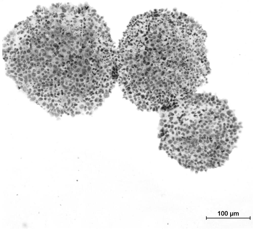
[0075] Example 2 Paraffin sections of HepG2 and LX-2 cells co-cultured with immunohistochemical staining
[0076] This embodiment mainly uses the method of the present invention to prepare paraffin sections of HepG2 and LX-2 cell co-cultured cell microspheres, wherein the steps from sample fixation to rehydration are the same as in Example 1, and immunohistochemical staining is used for the sections. The specific dyeing steps are as follows:
[0077] 1. Antigen retrieval: put the rack with slices into PBS (0.01M, pH 7.2-7.4) for cleaning, and microwave Tris / EDTA antigen retrieval solution (10mM Tris, 1mM EDTA, 0.05% Tween-20, pH 9.0 ) to 100°C, put the slide rack with slices into the boiling Tris / EDTA repair solution, heat on medium heat for 20 minutes, take out the container and place it in ice water for rapid cooling for about 10 minutes.
[0078] 2. Permeabilization: Incubate the sample in 1x TBS (pH 7.6) containing 0.025% Triton X-100 for 10 minutes, wash with PBS 3 times...
Embodiment 3
[0101] Example 3 Immunofluorescent staining of HepG2 and LX-2 cells co-cultured cell microsphere paraffin section
[0102] In this example, paraffin sections of co-cultured cell microspheres of HepG2 and LX-2 cells were prepared according to the method of the present invention, wherein the steps from sample fixation to rehydration were the same as in Example 1, and the sections were stained with immunofluorescence, and the details of the staining Proceed as follows:
[0103] 1. Antigen retrieval: put the rack with slices into PBS (0.01M, pH 7.2-7.4) for cleaning, and microwave Tris / EDTA antigen retrieval solution (10mM Tris, 1mM EDTA, 0.05% Tween-20, pH 9.0 ) to 100°C, put the slide rack with slices into the boiling Tris / EDTA repair solution, heat on medium heat for 20 minutes, take out the container and place it in ice water for rapid cooling for about 10 minutes.
[0104] 2. Permeabilization: Incubate the sample in 1x TBS (pH 7.6) containing 0.025% Triton X-100 for 10 minut...
PUM
| Property | Measurement | Unit |
|---|---|---|
| thickness | aaaaa | aaaaa |
| melting point | aaaaa | aaaaa |
| diameter | aaaaa | aaaaa |
Abstract
Description
Claims
Application Information
 - R&D
- Intellectual Property
- Life Sciences
- Materials
- Tech Scout
- Unparalleled Data Quality
- Higher Quality Content
- 60% Fewer Hallucinations
Browse by: Latest US Patents, China's latest patents, Technical Efficacy Thesaurus, Application Domain, Technology Topic, Popular Technical Reports.
© 2025 PatSnap. All rights reserved.Legal|Privacy policy|Modern Slavery Act Transparency Statement|Sitemap|About US| Contact US: help@patsnap.com


