Bionic natural tendon-bone gradient interface patch material and preparation method thereof
A tendon and patch technology, applied in the field of tendon patch materials, can solve problems such as the preparation method of tissue engineering tendon-bone interface patch materials with orientation structure and mineralization components that have not been reported, so as to reduce the healing of scars, Excellent elasticity and good tissue compatibility
- Summary
- Abstract
- Description
- Claims
- Application Information
AI Technical Summary
Problems solved by technology
Method used
Image
Examples
Embodiment 1
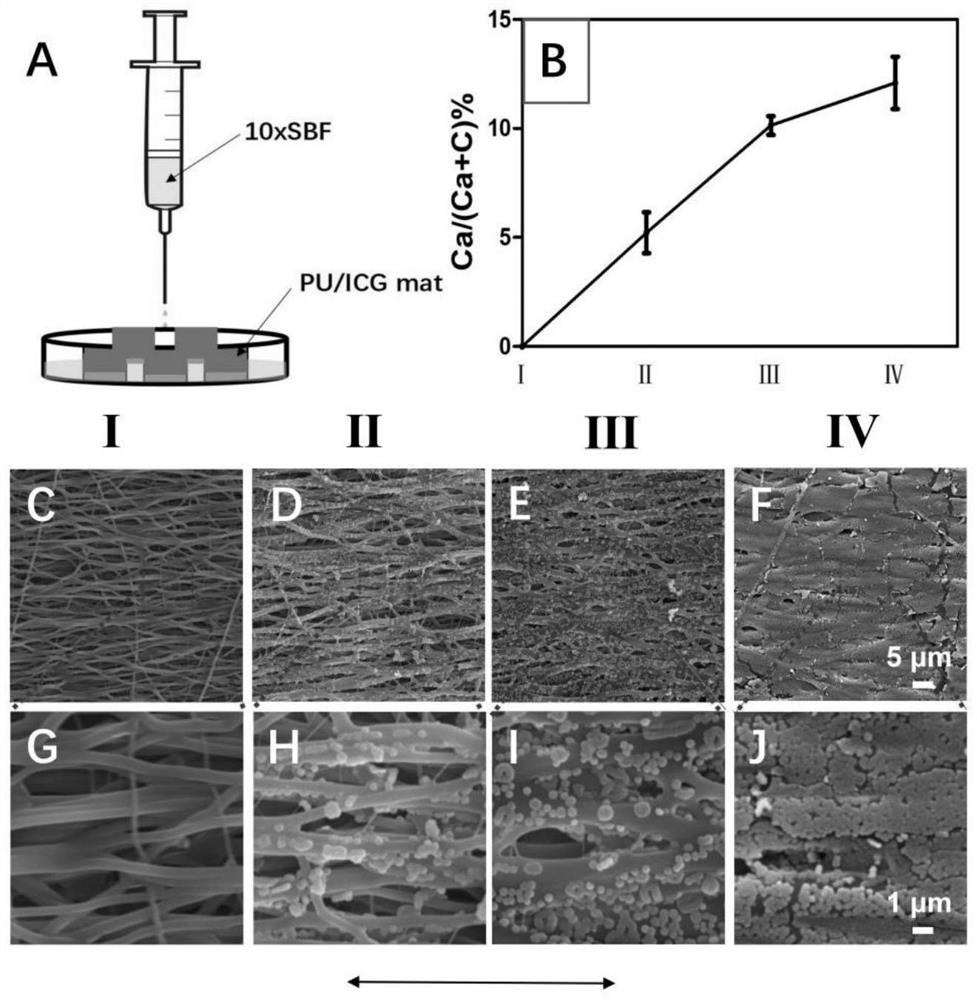
[0033] This embodiment relates to a preparation method of a bionic natural tendon-bone gradient interface patch material, wherein the medical fiber material is polyurethane, and the near-infrared dye is indocyanine green; the specific steps include:
[0034] (1) Accurately weigh 0.8g of polyurethane (PU) and 0.008g of indocyanine green (ICG) with an electronic balance, dissolve them in 10mL of hexafluoroisopropanol, stir overnight with a magnetic stirrer, and stir evenly to obtain PU / ICG Spinning liquid: adopt the electrospinning equipment of prior art to prepare PU / ICG uniaxially oriented nanofiber film, spinning parameter is: the flow rate of spinning liquid is respectively 0.013mL / min, and the distance between nozzle and cylinder is 15cm, The nozzle voltage is 18KV, the drum is grounded, the drum speed is 2500rpm, and the PU / ICG uniaxially oriented nanofiber film is obtained after 5 hours;
[0035] (2) Cut the PU / ICG uniaxially oriented nanofiber membrane obtained in step (...
Embodiment 2
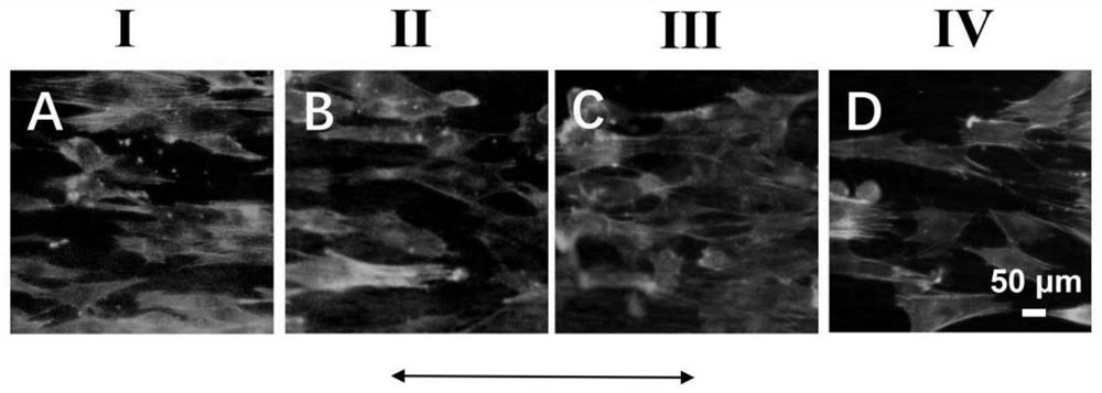
[0043] This example is an experiment on the effect of the bionic natural tendon-bone gradient interface patch material prepared in Example 1 on cell morphology. Tendon stem cells were harvested at 1×10 4 The concentration of each / well was inoculated in a 24-well plate containing the patch material prepared in Example 1. After cultivating for 3 days, F-actin / DAPI staining was used to observe the cell morphology. The specific method is:
[0044] 1. The complete culture medium of tendon stem cells (volume percentage composition: 89% low-sugar DMEM + 10% fetal bovine serum + 1% penicillin / streptomycin) was cultivated for 3 days, the old culture medium was removed, and then phosphate-buffered saline (PBS) wash 3 times;
[0045] 2. Fix the tendon stem cells with 1 mL / well of 3% glutaraldehyde aqueous solution at room temperature for 10 min, remove the glutaraldehyde, and then wash 3 times with PBS;
[0046] 3. Permeabilize with 0.1% Triton X-100 (polyethylene glycol octylphenyl et...
Embodiment 3
[0053] This embodiment relates to the application experiment of the patch material of the bionic natural tendon-bone gradient interface prepared in Example 1, specifically:
[0054] First, construct the injury model, inject pentobarbital sodium (1.5%, 2mL / Kg) into the ear veins of 12 New Zealand white rabbits for general anesthesia, then fix the rabbits sideways on the operating table, and routinely pre-operatively prepare the skin and disinfect , spread a sterile drape, and infiltrate the shoulder with lidocaine for local anesthesia; make a percutaneous incision about 3 cm in length, and separate the subcutaneous fascia layer by layer. After exposing the humeral head, find the subscapularis tendon; between the tendon and bone At the connected position, the subscapularis tendon was cut 50% of the thickness; then the surface stump of the humeral head was cleaned; and then randomly divided into two groups, namely the experimental group and the control group, the experimental grou...
PUM
 Abstract
Description
Claims
Application Information
 - R&D
- Intellectual Property
- Life Sciences
- Materials
- Tech Scout
- Unparalleled Data Quality
- Higher Quality Content
- 60% Fewer Hallucinations
Browse by: Latest US Patents, China's latest patents, Technical Efficacy Thesaurus, Application Domain, Technology Topic, Popular Technical Reports.
© 2025 PatSnap. All rights reserved.Legal|Privacy policy|Modern Slavery Act Transparency Statement|Sitemap|About US| Contact US: help@patsnap.com


