Fluorescent probe for use in detection of brain tumor

A fluorescent probe and brain tumor technology, applied in the field of fluorescent probes, can solve the problems of inability to carry out fluorescent labeling, duration, difficulty in re-administration, and difficulty in improving brain nerve function preservation while taking into account the resection rate, and achieves excellent practicability. , Improve the removal rate, the effect of simple detection steps

Pending Publication Date: 2021-12-24
THE UNIV OF TOKYO
View PDF7 Cites 0 Cited by
  • Summary
  • Abstract
  • Description
  • Claims
  • Application Information

AI Technical Summary

Problems solved by technology

However, the method using 5-ALA has the problem of sensitivity and specificity, the necessity of oral administration before surgery, the duration of metabolism, and the difficulty of re-administration. In addition, in the case of low-risk tumors, there is a problem that fluorescence cannot be performed. Problems of labeling etc. (for example, Non-Patent Documents 1 and 2)
Moreover, traditional tumor visualization methods are often difficult to balance the improvement of resection rate and the preservation of brain nerve function in a trade-off relationship

Method used


Image

  • Fluorescent probe for use in detection of brain tumor
  • Fluorescent probe for use in detection of brain tumor
  • Fluorescent probe for use in detection of brain tumor

Examples


Embodiment 1

[0094] 1. Synthesis of fluorescent probes

[0095]Hydroxymethylrhodamine green (HRMG) with various dipeptide sites was synthesized according to the reaction scheme described in Example 1 of International Publication WO2016 / 006678. Compounds with different dipeptide sites were obtained by making various changes to the Fmoc-amino acid used in the synthesis of compound A7 in Example 1.

[0096] In the synthesized compound, R in the above general formula (I) 1 , R 2 , R 3 , R 4 , R 5 , R 6 , R 7 , R 8 and R 9 Both are hydrogen atoms, X=O, Y=methylene, and P1 and P2 are the following combinations.

[0097] Table 1

[0098] compound P1 P2 YK190 Arginine residues proline YK213 Histidine Glycine YK19 Tyrosine Glycine YK281 methionine alum Acetylleucine

Embodiment 2

[0100] 2. Screening by fluorescent probes

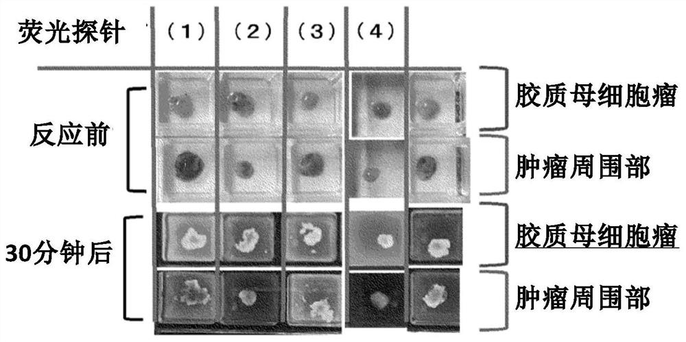
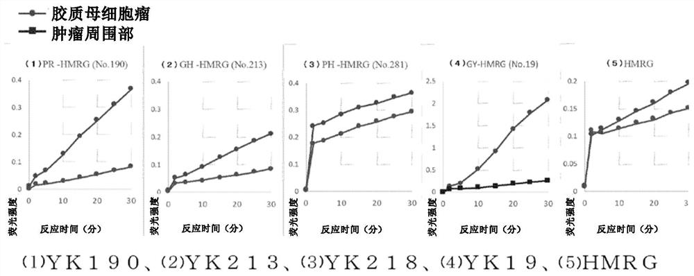
[0101] The fluorescent probe compound group containing YK190, YK213, YK19 and YK281 synthesized in Example 1 was added to the fresh samples of the tumor and the surrounding part of the tumor, and the fluorescence intensity was compared. The sample used was glioblastoma. As a comparative example, hydroxymethylrhodamine green (HRMG) without a dipeptide site was used.

[0102] Dissolve 0.5 μL DDMSO solution (10 mM) of various fluorescent probes in 100 μL of RPMI1640 (phenol red-free) (the final concentration of the probe is 50 μM), drop 200 μL to each sample, and use Maestro InVivo Imaging The System Ex measures fluorescence intensity over time.

[0103] Excitation wavelength: 465nm

[0104] ·Emission wavelength: 515nm

[0105] Such as figure 1 and figure 2 As shown in the results, it was found that (1) YK190 and (2) YK213 were tumor-specific fluorescently labeled compared with the peripheral tissue, while (3) YK218 was not much ...

Embodiment 3

[0108] 3. Protease Identification

[0109] Next, proteases associated with the fluorescent response obtained in Example 2 were identified.

[0110] Cathepsin D, cytosolic non-specific dipeptidase, calpain 1, cytosolic aminopeptidase were identified using DEG (Diced electrophoresis gel) assay and LC MS / MS by peptide mass fingerprinting for protease analysis as candidate enzymes.

[0111] For these candidate enzymes, as a result of measuring the changes in fluorescence intensity using inhibitors (SNJ-1945 and Amastatin), it was found that the fluorescence of calpain 1 and cathepsin D was inhibited by SNJ-1945.

[0112] For these calpain 1 and cathepsin D, when measuring the change in fluorescence intensity due to the reaction with the fluorescent probe YK190, as shown in 4, a significant increase in fluorescence intensity was observed, indicating that the fluorescence of glioblastoma in Example 2 The response is caused by the reaction of these enzymes with fluorescent probes. ...


PUM


Abstract

[Problem] To provide a novel fluorescent probe which can be used in a spray mode, has excellent specificity to sensitivity, also has immediacy, and enables the detection of brain tumor. [Solution] A fluorescent probe for use in the detection of brain tumor, which comprises a compound represented by formula (I) or a salt thereof: (in the formula, P1 represents an arginine residue, a histidine residue or a tyrosine residue, and P2 represents a proline residue or a glycine residue, wherein P1 is liked to an adjacent N atom to form an amide bond, and P2 is linked to P1 to form an amide bond; R1 represents 1 to 4 same or different substituents independently selected from the group consisting of a hydrogen atom and an alkyl group, a carboxyl group, an ester group, an alkoxy group, an amide group and an azide group each of which may be substituted; R2, R3, R4, R5, R6 and R7 independently represent a hydrogen atom, a hydroxyl group, an alkyl group which may be substituted, or a halogen atom; R8 and R9 independently represent a hydrogen atom or an alkyl group; X represents O, Si(Ra)(Rb), Ge(Ra)(Rb), Sn(Ra)(Rb), C(Ra)(Rb) or P(=O)(Ra); Ra and Rb independently represent a hydrogen atom, an alkyl group or an aryl group; and Y represents a C1-C3 alkylene group).

Description

technical field [0001] The present invention relates to fluorescent probes for the detection of brain tumors. In more detail, it relates to a fluorescent probe capable of specifically detecting and labeling brain tumors by coating or spraying a tissue sample, and a detection method using the fluorescent probe. Background technique [0002] Gliomas (gliomas) account for about 30% of primary brain tumors, and the median survival of the most malignant glioblastomas is about 1.5 years. Even low-grade gliomas are rarely completely cured, and most deteriorate within a few years and die within 5-10 years. In glioma surgery, although total resection is a favorable prognostic factor, extensive resection of the surrounding brain cannot be performed due to functional limitations, and visualization of the tumor has been attempted to achieve safer and maximized resection. [0003] To date, 5-aminolevulinic acid (5-ALA) is the only insurance coverage for use as a fluorescent reagent for...

Claims


Application Information

Patent Timeline
Patent Type & Authority Applications(China)
IPC IPC(8): C07K5/06C07K5/062C07K5/078G01N21/64G01N21/78C12Q1/37C09B11/28G01N33/48
CPCG01N2333/96466G01N2333/96472C09B11/24C07K5/06026C07K5/06165C12Q1/37A61K49/0041G01N2800/7028A61K49/0056A61K49/006C07K5/06G01N33/533G01N33/57484
Inventor 北川阳介田中将太齐藤延人栗木优五神谷真子浦野泰照
Owner THE UNIV OF TOKYO
Why Eureka
Try Eureka