Stimuli-responsive hydrogel microdomes integrated with genetically engineered proteins for high-throughput screening of pharmaceuticals

a technology of genetic engineered proteins and hydrogels, applied in the field of stimuli-responsive hydrogels integrated, can solve the problems of ineffective window control of drug release, limited number of biomaterials and limited number of biomaterials that are suitable for biomedical applications. , to achieve the effect of excellent selectivity for ligands

Inactive Publication Date: 2005-09-22
UNIV OF KENTUCKY RES FOUND
View PDF18 Cites 23 Cited by
  • Summary
  • Abstract
  • Description
  • Claims
  • Application Information

AI Technical Summary

Benefits of technology

[0014] The stimuli-responsive hydrogels of the present invention have an integrated biological recognition element that provides excellent selectivity for ligands of interest. The stimuli-sensitive hydrogels reproducibly swell upon addition of a target molecule, which controls the release of a biomolecule. The amount of swelling of these hydrogels was attributed to release of non-covalent bonds within the polymer network.

Problems solved by technology

Currently, there are a limited number of biomaterials that are suitable for biomedical applications.
Traditional drug release tablets cannot control the amount of drug released efficiently.
The drug concentration may extend beyond the therapeutic window risking ineffectiveness or toxicity.
A major limitation in the usefulness of controlled release systems is that conventional implantable devices release drugs at a predetermined rate(s).
However, in certain instances, it is required that drugs are administered either at a life-threatening moment or repeatedly at a certain critical time of day.
Currently, laborious combinatorial techniques are used to screen analytes.
These techniques are taxing on both time and resources.
The conventional hydrogel synthesis does not permit exact control of three-dimensional structure of the hydrogel microdome, making it difficult to predict hydrogel performance and hindering the potential for biomedical applications.

Method used


Image

  • Stimuli-responsive hydrogel microdomes integrated with genetically engineered proteins for high-throughput screening of pharmaceuticals
  • Stimuli-responsive hydrogel microdomes integrated with genetically engineered proteins for high-throughput screening of pharmaceuticals
  • Stimuli-responsive hydrogel microdomes integrated with genetically engineered proteins for high-throughput screening of pharmaceuticals

Examples


example 1

Preparation of CaM Protein

[0119] The CaM with a unique cysteine at the C-terminus is prepared as follows. The gene for CaM was obtained from the plasmid pVUC-1 using PCR. The CaM gene was modified by site-directed mutagenesis and digested with NdeI and SapI. The digested DNA fragment was ligated into the vector pTWIN-1 to yield the pSD1009 vector. DNA sequencing was performed at the Macromolecular Center (University of Kentucky). Bacteria (E. coli, strain ER 2566) were transformed with the pSD1009 vector. Native CaM with a unique cysteine at the C-terminus was expressed using E. coli and purified by affinity chromatography with chitin beads (New England BioLabs, Beverly, Mass.). Tris-(2-cyanoethyl) phosphine was stirred with the purified CaM before reaction with the BMPS-allylamine to ensure a free sulfhydro group on cysteine.

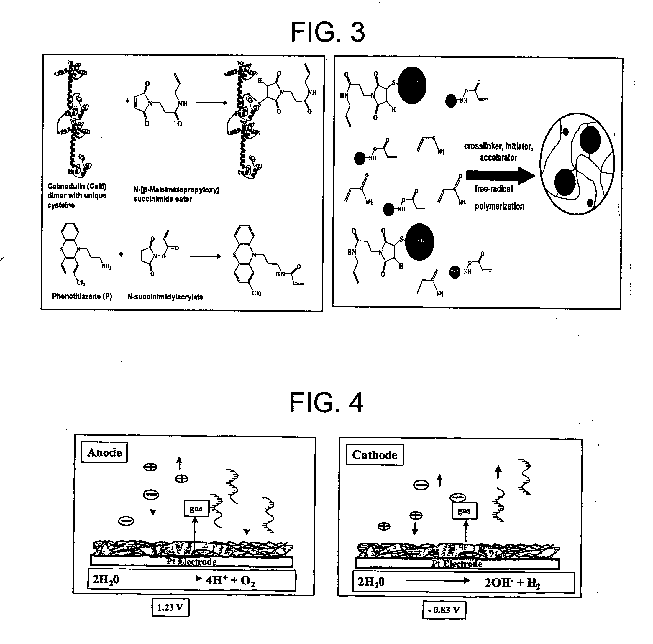
[0120] Tris-(2-cyanoethyl) phosphine was stirred with the purified CaM before a linker containing a vinyl group was attached to the free sulfhydryl residue o...

example 2

Synthesis of CaM-Phenothiazine Hydrogel and Hydrogel Arrays

[0121] Protein-integrated hydrogels were synthesized by free-radical polymerization of the modified CaM, vinyl-containing 3-(trifluorometyl-phenothiazin-10-yl) propyamine (TAPP), acrylamide (AAm), and N,N′-methylenebis(acrylamide) (MBAA). TAPP and CaM were preincubated before polymerization, and as a result, immobilized TAPP is non-covalently bound to CaM creating additional crosslinking within the hydrogel network. Modified CaM (3 nmol), vinyl-containing TAPP (3 nmol), acrylamide (AAM) (0.014 mol), N,N′-methylenebis-(acrylamide) (MBAA) (6.5 μmol, 1% w / w AAm), 20 μL potassium persulfate (KPS) (0.1 M), and 40 μL N,N,N′,N′-tertramethylethylenediamine (TEMED) (0.4 M) were mixed in 1 mL 10 mM HEPES buffer containing 10 mM CaCl2.

[0122] The stimuli-responsive hydrogel microdomes can be reproducibly synthesized using a “microspotting” technique. This technique comprises pipetting 1 μL one or more droplets of a mixture of CaM, TAP...

example 3

Making of a Calmodulin Dimer Protein

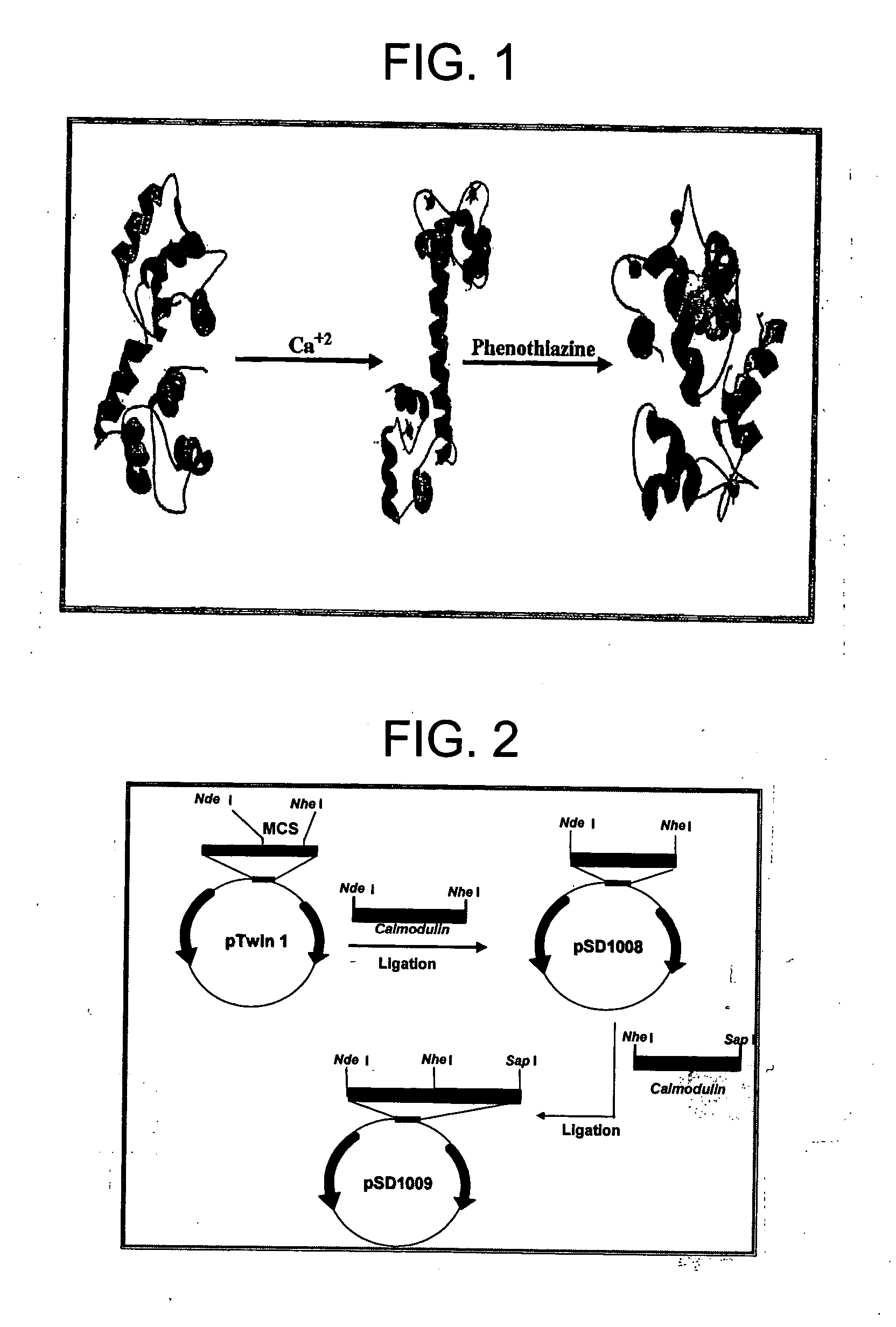
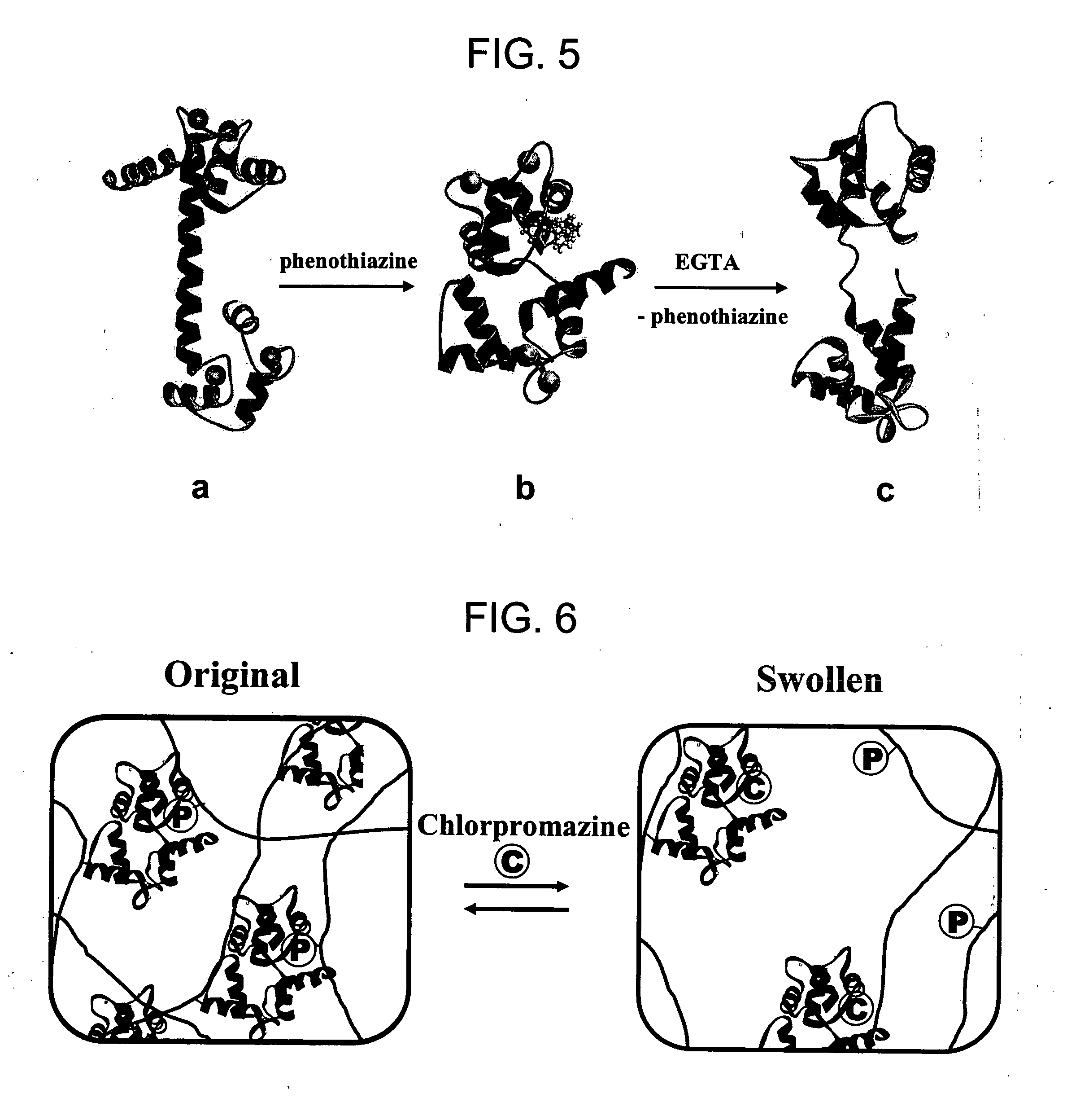
[0124] CaM is a calcium-binding protein that also binds phenothiazines. When CaM binds calcium, it undergoes a conformational change. This conformational change allows CaM to interact with CaM binding proteins, peptides, and drugs such as trifluoropiperazine and phenothiazine. Such a conformational change will be larger when single CaM molecules are linked or fused together to yield a polymeric CaM molecule comprised of at least two single CaM molecules. This example describes preparation of a CaM dimer, a single molecule comprised of two single CaM molecules.

[0125] A CaM dimer protein is made by fusing two CaM-encoding genes together, end-to-end. Such gene fusion techniques are well known to those experienced in the art. The CaM dimer fusion gene is then cloned into a plasmid that will allow expression of the gene in bacteria. The plasmid is used to transform E. coli and bacterial colonies that contain the plasmid are selected. The transformed ...


PUM

PropertyMeasurementUnit
temperaturesaaaaaaaaaa
volumeaaaaaaaaaa
thickaaaaaaaaaa

Abstract

A hydrogel microdome that can swell in response to a stimuli or target molecule is formed by polymerizing a mixture comprising a monomer capable of forming a hydrogel with a biopolymer. An array of hydrogel microdomes can be formed on a substrate by microspotting the mixture and polymerizing. The array can be used for high-throughput screening of analytes as well as for use as an actuator and biosensor using the swelling property of the hydrogel.

Description

CROSS-REFERENCE TO RELATED APPLICATIONS [0001] This application is a continuation-in-part of application Ser. No. 09 / 905,041 filed Jul. 13, 2001, published Jun. 6, 2002 as Publication No. US 2002 / 0068295, which claims priority to U.S. Provisional Application Ser. No. 60 / 218,036, filed Jul. 13, 2000, which is incorporated herein by reference.GOVERNMENT INTEREST IN THE INVENTION [0002] This invention was made with Government support under Grant No. EB003072 awarded by the National Institutes of Health. The Government may have certain rights in this invention.FIELD OF THE INVENTION [0003] This invention is directed to a stimuli-responsive hydrogels integrated with genetically engineered proteins for use for high-throughput screening of analytes as well as for use as an actuator and biosensor. BACKGROUND OF THE INVENTION [0004] Proteins and DNA are information rich molecules with structural and electrical properties which make their incorporation into the human manufacturing arsenal an ...

Claims


Application Information

Patent Timeline
Patent Type & Authority Applications(United States)
IPC IPC(8): G01N33/543G01N33/84
CPCG01N33/84G01N33/54373
Inventor DAUNERT, SYLVIADEO, SAPNA KAMLAKAREHRICK, JASON DOUGLASBROWNING, TYLER WILLIAMBACHAS, LEONIDAS G.
Owner UNIV OF KENTUCKY RES FOUND
Why Eureka
Try Eureka