

Methods and devices for repair or replacement of heart valves or adjacent tissue without the need for full cardiopulmonary support
a heart valve and adjacent tissue technology, applied in the direction of prosthesis, catheters, blood vessels, etc., can solve the problems of reduced mortality rate, stiff valves and less ability to fully open, and limited range of procedures that have been developed for an endovascular approach to date, and achieve the goal of repairing the coronary arteries.
- Summary
- Abstract
- Description
- Claims
- Application Information
AI Technical Summary
Benefits of technology
Problems solved by technology
Method used
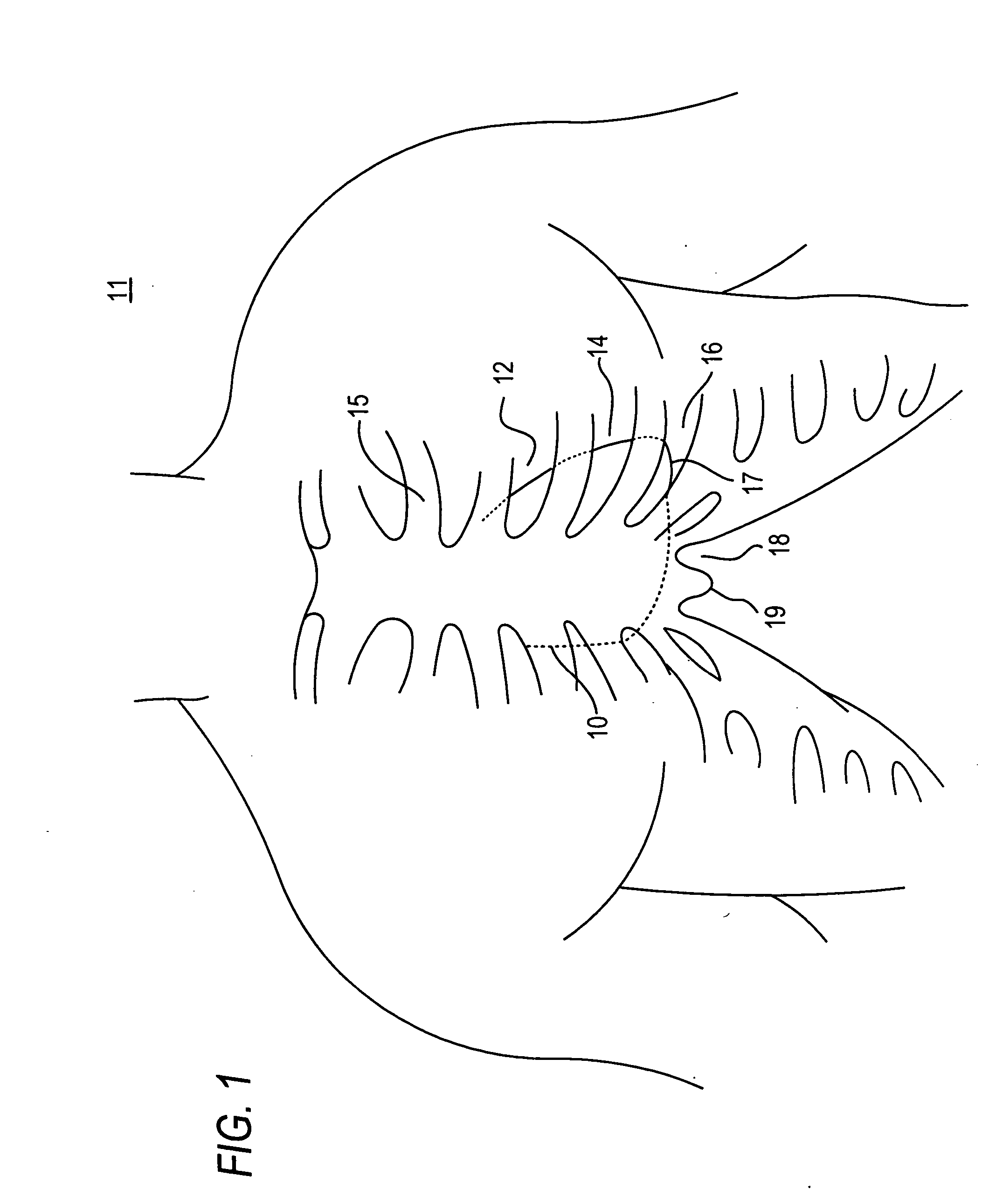
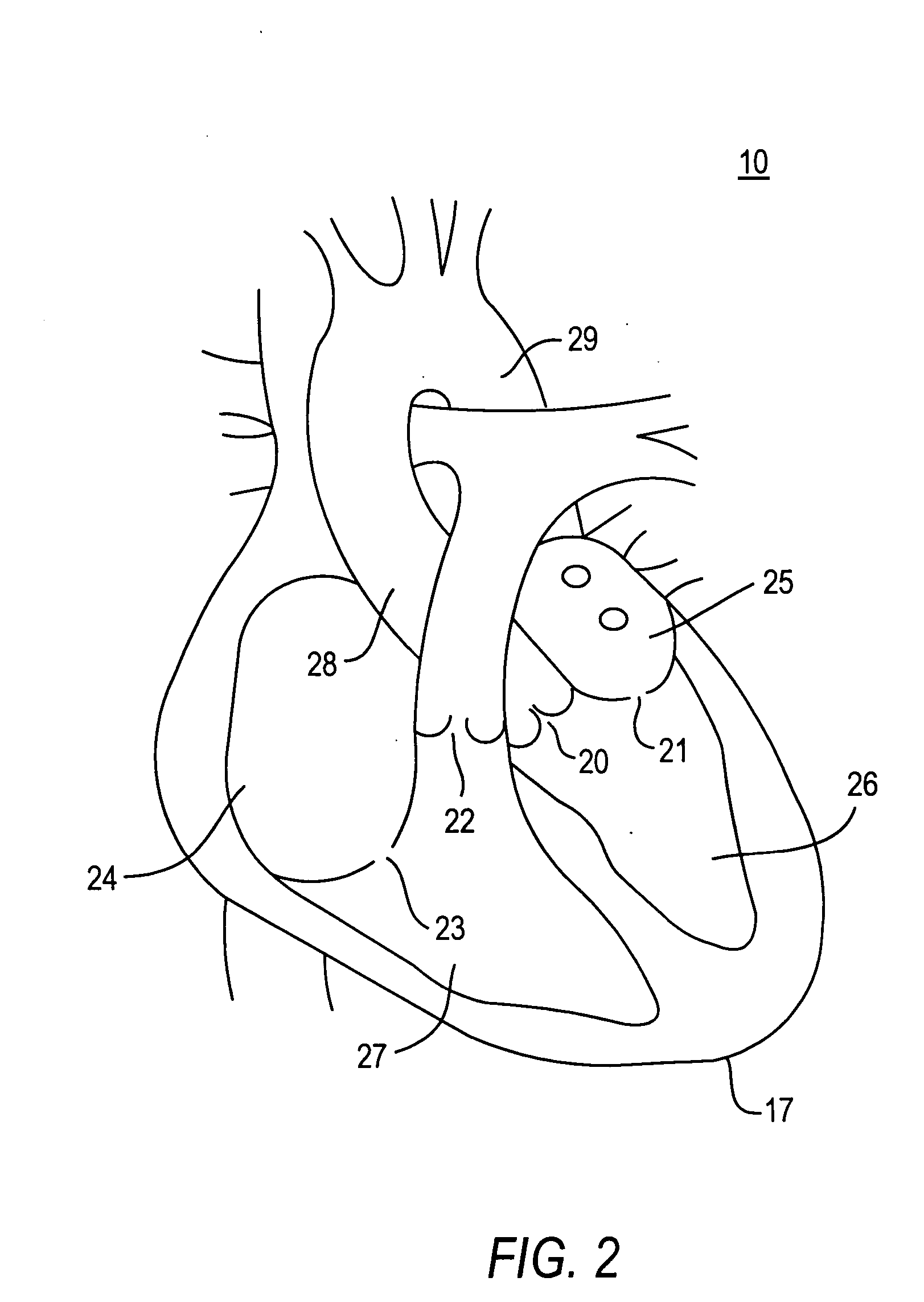
Image

Examples
Embodiment Construction
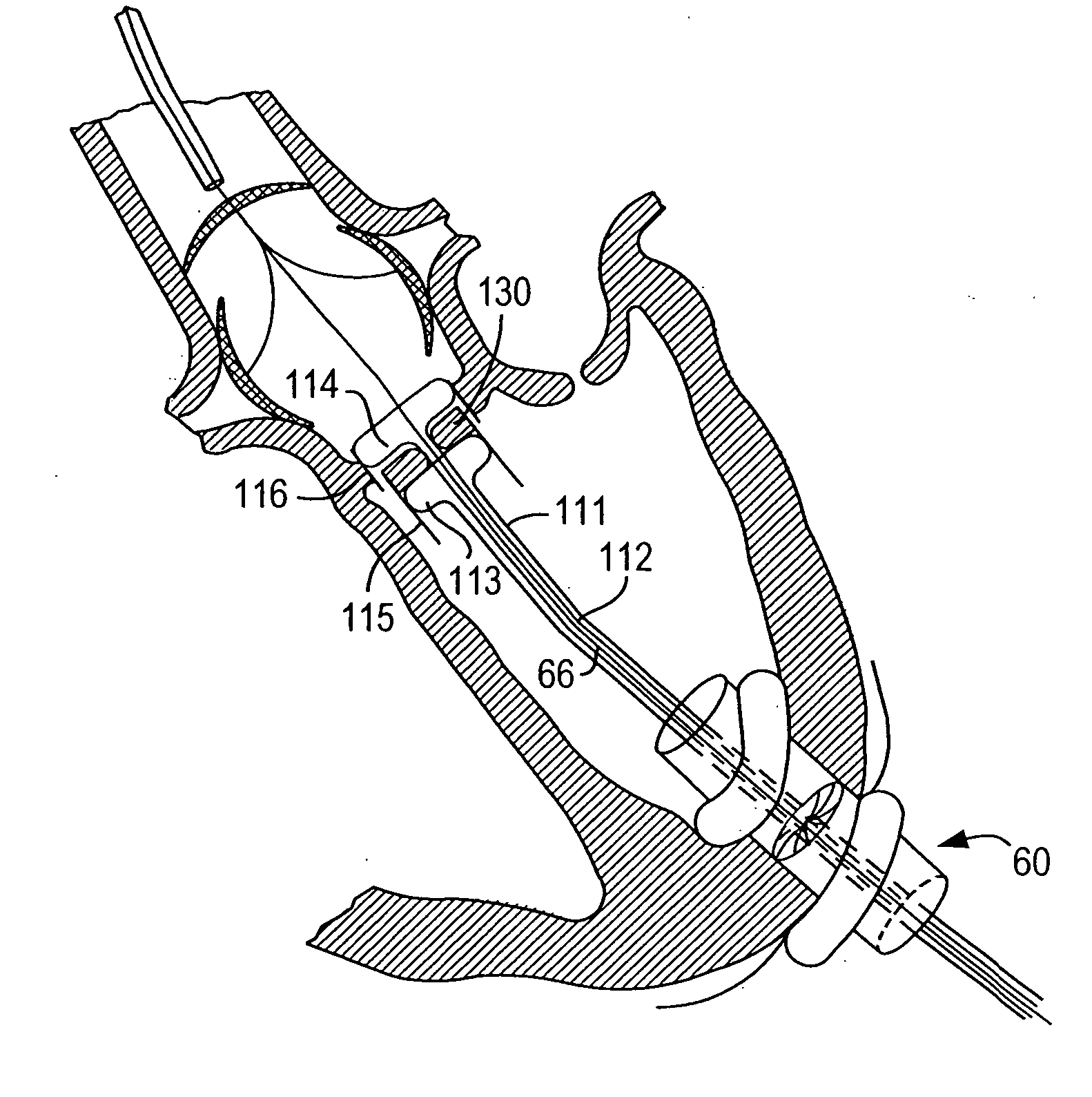
[0070] Because the present invention has a number of different applications, each of which may warrant some modifications of such parameters as instrument size and shape, it is believed best to describe certain aspects of the invention with reference to relatively generic schematic drawings. To keep the discussion from becoming too abstract, however, and as an aid to better comprehension and appreciation of the invention, references will frequently be made to specific uses of the invention. Most often these references will be to use of the invention to resect and replace or implant an aortic valve with an antegrade surgical approach. It is emphasized again, however, that this is only one of many possible applications of the invention.
[0071] Assuming that the invention is to be used to resect and replace or implant an aortic valve, the procedure may begin by setting up fluoroscopy equipment to enable the surgeon to set and use various reference points during the procedure. The surge...
PUM
 Abstract
Description
Claims
Application Information