Primarily cultured adipocytes for gene therapy

Active Publication Date: 2006-06-08
EISIA R&D MANAGEMENT CO LTD
10 Cites 4 Cited by

AI-Extracted Technical Summary

Problems solved by technology

2001 Jan, 115(1):73-82), removing the biological barrier of the skin in the process of isolating the primary culture is problematic considering infection risk.
Patient pain during extirpation and implantation is predicted to be severe, and re-e...

Method used

[0122]FIG. 7 shows (pro)insulin production by plasmid transfection into primary cultured adipocytes. Each of pcDNA3.1Myc-His vectors individually incorporating the native human insulin gene (native) and the site1/site2/B10-modified form (s1s2B10), or an empty vector (mock), was transfected into adipocytes derived from the adipose tissue surrounding the C57BL/6 mice epididymis. Human (pro) insulin was detected in the culture supernatant of resistant cells obtained by G418 selection. This showed that stable gene transfer to primary cultured adipocytes is also possible using a plasmid vector.
[0144]FIG. 11 shows the change in plasma AP activity in mice implanted with AP-expressing primary cultured adipocytes. Individuals that received an implant of cells subjected to differentiation-inducing stimulation for three days before implantation (“with differentiation induction”) showed less fluctuation in the continued expression than individuals that received an ...

Benefits of technology

[0011] (7) adipocytes are widely recognized as superfluous and obs...

Abstract

The present invention relates to primary cultured adipocytes for gene therapy, where the adipocytes stably maintain a foreign gene encoding a protein that is secreted outside of cells. This invention provides cells suitable for gene therapy, which can replace bone marrow cells and liver cells used for conventional ex vivo gene therapy. The present invention established methods for transferring foreign genes into primary cultured adipocytes, which are suitable for ex vivo gene therapy; can be easily collected and implanted; and can be removed after implantation. Specifically, the present invention established these methods that use retroviral vectors. The present invention also established primary cultured adipocytes for gene therapy, where the adipocytes stably maintain a foreign gene encoding a protein that is secreted outside of cells.

Application Domain

Technology Topic

HepatocyteMyelocyte +6

Image

  • Primarily cultured adipocytes for gene therapy
  • Primarily cultured adipocytes for gene therapy
  • Primarily cultured adipocytes for gene therapy

Examples

  • Experimental program(11)

Example

EXAMPLE 1
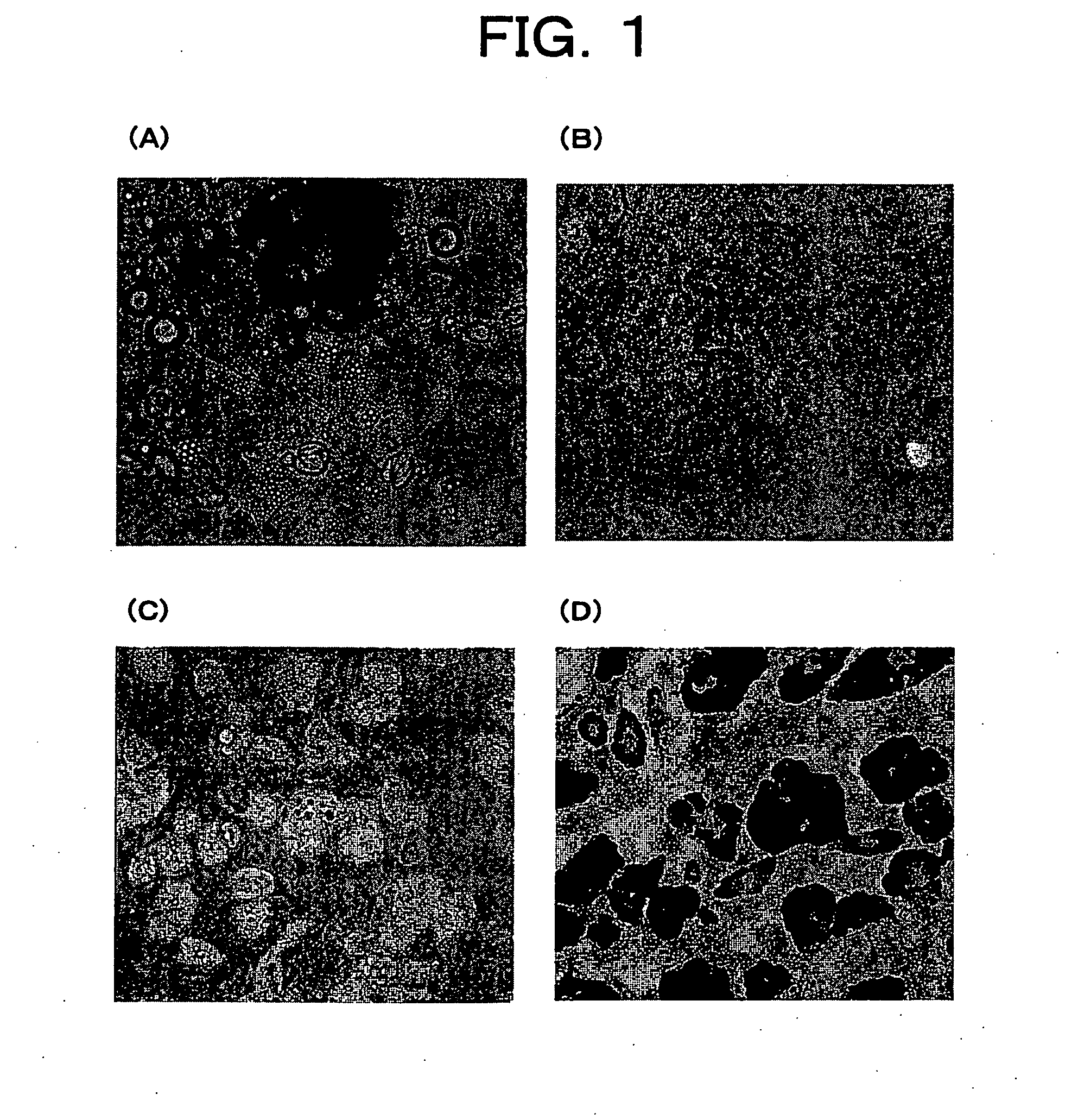
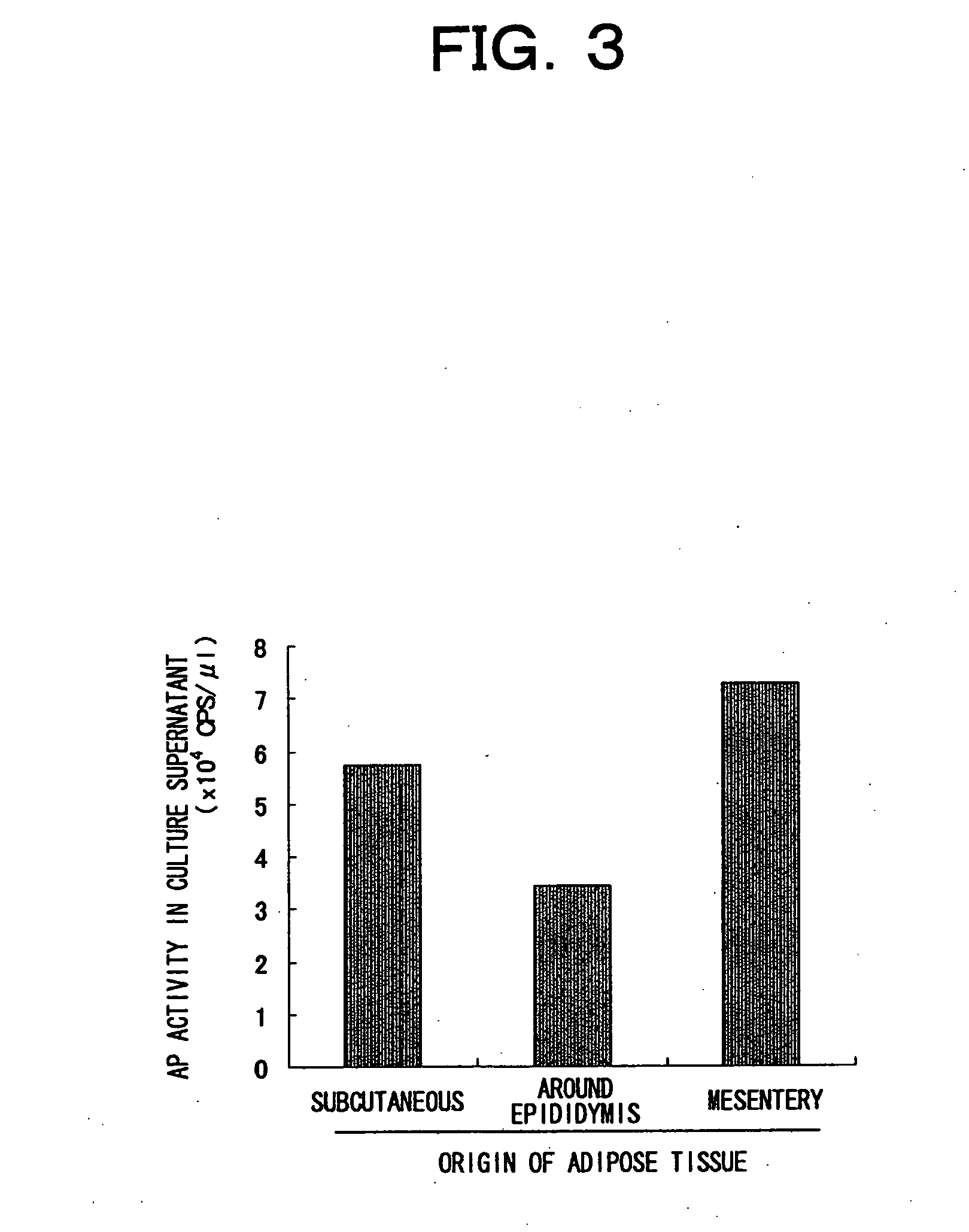
Primary Culture of Murine Adipocytes
[0086] [Methods]
[0087] Three-week old male ICR mice or four- to five-week old male C57BL/6 mice (both from Charles River) were anesthetized with diethyl ether, and sacrificed by collection of whole blood from the heart. Next, inguinal subcutaneous fat, or fat surrounding the epididymis, and mesenteric adipose tissue were individually extirpated under sterile conditions. The extirpated tissues were washed with PBS, and then morcellated using a pair of scissors or a surgical knife. This morcellated tissue was digested with shaking at 37° C. for 20to 60 minutes in normal medium (DMEM-high glucose/SIGMA, 10% FCS) comprising 1 mg/mL of collagenase (S1 fraction/Nitta gelatin), and then separated into precipitate and suspended layer by centrifugation (300 g, five minutes).
[0088] The floating layer was further centrifuged once or twice to remove the collagenase by dilution, and then added to a T-25 flask (IWAKI) filled with medium. Bubbles were removed, and this was cultured under a 5% CO2 atmosphere in a CO2 incubator at 37° C. so the conventional culture surface was upside (ceiling culture). Ten to 14 days after culturing, the cells adhering to the ceiling surface were collected by trypsin treatment and transferred to a normal culturing system. Subculturing was then performed at a ratio of 1:3 to 1:10.
[0089] To induce differentiation, the medium of cells cultured to confluency in a 6-well plate was transferred to an induction medium (normal medium supplemented with 0.5 mM IBMX, 0.25 μM dexamethasone, and 10 μg/mL insulin). This stimulation was continued for 48 hours. Next, the cells were differentiated in a maturation medium (normal medium supplemented with 10 μg/mL insulin). The maturation medium was exchanged every three days.
[0090] Oil red O staining solution was prepared by mixing a stock solution, prepared by mixing 0.3 g of oil red O in 100 mL isopropanol (99%), with distilled water in a 3:2 ratio at the time of use. The cells were washed with PBS and then fixed with 10% neutral formalin solution (WAKO). After washing again with PBS, the cells were stained with oil red O staining solution at room temperature for ten minutes. The cells were washed with PBS again, and then examined by microscope. [0091] [Results]
[0092]FIG. 1 is a set of microphotographs of primary cultured adipocytes isolated from the subcutaneous fat of three-week old ICR mice. After 14 days of ceiling culture, adhesion of adipocytes carrying lipid droplets was observed on the ceiling-side culture surface (A). When these cells were transferred to a normal culturing system, they showed fibroblast-like growth, as shown in (B). However, when differentiation was induced by IBMX, dexamethasone, and insulin, the cells again differentiated into mature adipocytes that carry lipid droplets (C). Stored fat was stained red with oil red O staining (D). Cells isolated by this method were shown to be primary cultured adipocytes comprising the ability to differentiate.

Example

EXAMPLE 2
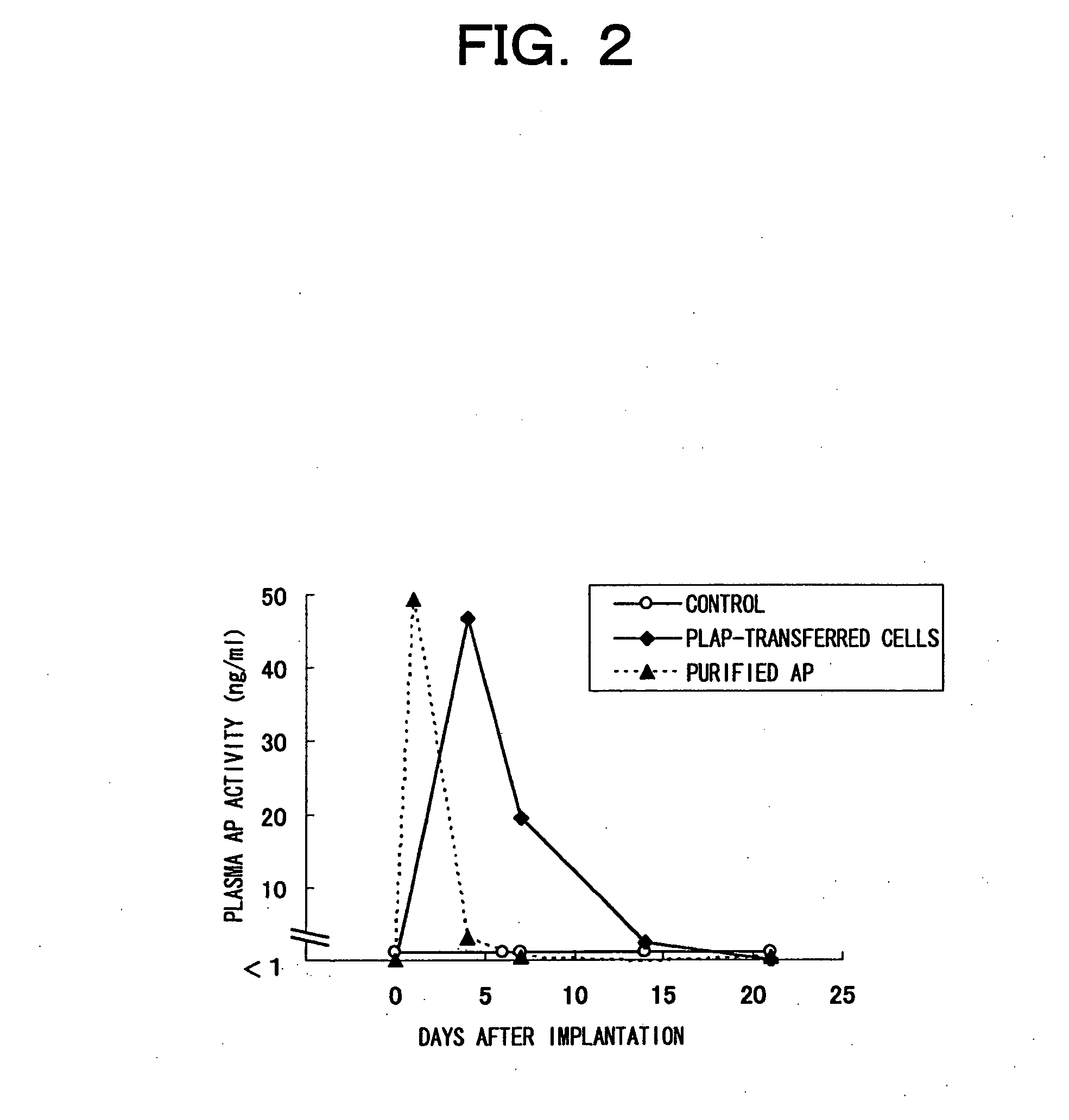
[0093] Transient transfer of thermostable secretory alkaline phosphatase (AP) gene into primary cultured adipocytes, and implantation of transfected adipocytes into mice
[0094] As a model system for gene expression, AP gene, more specifically, SEAP gene (Clontech) or PLAP gene (Goto, M. et al. Mol. Pharmacol. vol. 49 860-873 (1996)) was transferred to primary cultured adipocytes, and changes in AP activity were examined. (Both AP gene products are thermostable and can be easily distinguished from endogenous alkaline phosphatases by thermal treatment.) [0095] [Methods]
(1) Production of Primary Cultured Adipocytes Transiently Transfected with the SEAP Gene
[0096] AP-expressing plasmid (pcDNA3.1-SEAPmh) was constructed by inserting the SEAP sequence, obtained by double digestion of pSEAP2-basic vector (Clontech) with restriction enzymes HindIII-XbaI, into the HindIII-XbaI site of pcDNA3.1Myc-HisA (Invitrogen), which is a vector for expression in mammalian cells.
[0097] For every gene transfer to a 10-cm dish, 500 μL of FCS-free DMEM medium and 15 μL of Fugene 6 reagent (Roche) were mixed, then 5 μg of pcDNA3.1-SEAPmh was added. This mixture was left to stand at room temperature for 15 minutes. This mixture was added to primary cultured cells (derived from ICR subcutaneous fat) cultured to 70 to 80% confluency in a 10-cm dish. This was then cultured for 24 hours in a CO2 incubator.
(2) Implanting Mice with Alkaline Phosphatase Gene-Transferred Primary Cultured Adipocytes
[0098] Gene-transferred cells were collected by trypsin treatment, and washed twice with PBS by centrifugation. The cells were then suspended in PBS at 1×10⁷ cells/mL. The animals (ICR nude mice, five-weeks old at the time of operation) were anesthetized by intraperitoneal administration of 50 mg/kg of sodium pentobarbital (Nembutal; Dainippon Pharmaceutical). After disinfecting the area to be operated with dilute Hibitane solution (Sumitomo Pharmaceuticals), a 3 mm to 5 mm or so incision was made to the skin near the base of the right hind leg, and the inguinal subcutaneous fat was exposed. 0.55 mL of the prepared cell suspension solution (5.5×10⁶ cells/head) was loaded into a 1-mL syringe, and this was injected into the subcutaneous fat using a 22 G injection needle. As a control, PBS was injected to the same site. To compare this to the protein supplementation method, 1 μg of purified AP (Roche) was dissolved in PBS under sterile conditions, and this was injected in a similar manner. The incised skin was sutured and the operated site was disinfected with surgical Isodine (Meiji Seika).
[0099] Blood was collected using a heparin-coated capillary (Dramond) from the postorbital venous plexus before implantation (day 0) and after implantation over time. Plasma was obtained from the whole blood by centrifugation at 2000 g for 15 minutes. AP activity in this plasma was measured using an assay kit (SEAP reporter gene assay kit, Roche) by following the attached instructions. [0100] [Results]
[0101]FIG. 2 shows the plasma AP activity achieved by implanting mice with primary cultured cells, which have been transiently transfected with alkaline phosphatase (AP) -expressing plasmid pcDNA3.1-SEAPmh. For purposes of comparison, mice were administered with 1 μg of purified AP protein (Roche) by injection. Seven days after administration the blood AP activity in these mice decreased to the level of the control. On the other hand, blood AP activity in mice that received an implant of cells holding transiently transferred genes was confirmed to peak on the fourth day after implantation, and the duration of expression was 14 days. The duration of in vivo expression by implanting cells carrying transiently transferred gene was short, and the concentration in the blood was found to vary greatly, although it was maintained longer than by injecting protein.

Example

EXAMPLE 3
Production, by Using a Viral Vector, of Adipocytes that Stably Express AP
[0102] [Methods]
(1) Construction of AP- and Control GFP-Expression Vectors
[0103] The PLAP gene was excised from pTK-PLAP using HindIII and BglII, as described in the literature (Goto, M. et al. Mol. Pharmacol. vol.49, 860-873 (1996)). The SEAP gene was obtained by double digestion of pcDNA 3.1-SEAPmh with HindIII/PmeI. The GFP gene was excised from pEGFP-N2 using NotI-NcoI.
[0104] The plasmid, pBabeCLXI2G, used for viral vector production, was produced based on pBabePuro (Morgenstern, J. P. et al. Nucleic Acids Res. vol.18, 3587-3596 (1990)), by excising its SV40 promoter and neomycin resistance genes using SalI-ClaI, and blunting those ends with Klenow fragments, then replacing these with the internal ribosome re-entry site (IRES) of encephalomyocarditis virus (EMCV), which was excised from pIRES2-EGFP by HincII-HincII, and the green fluorescent protein (GFP); then replacing the portion from the long terminal repeat (LTR) to the foreign gene insertion site (multicloning site) (SspI-BamHI) with a sequence corresponding (SspI-BamHI) of pCLXSN (IMGENEX). Furthermore, pBabeCLXIP, in which the IRES-GFP portion of pBabeCLXI2G had been replaced with IRES-puromycin resistance gene, was also used.
[0105] Each of the DNA fragments of the above-mentioned PLAP, SEAP, and GFP were blunted with Klenow fragments, then inserted into pBabeCLXIP or pBabeCLXI2G vector cleaved with Hpa I, yielding pBabeCL(PLAP)IP, pBabeCL(SEAPmh)I2G, and pBabeCL(GFP)IP, respectively.
(2) Production of Viral Vectors
[0106] Each gene transfer to a 10-cm dish was performed as follows: 30 μL of plasmid transfection reagent TransIT (MIRUS) was mixed into 500 μL of FCS-free DMEM medium, and left to stand at room temperature for five minutes (mixed DMEM/TransIT solution). In a separate tube, 3.3 μg of a vector encoding VSV-G (pCALG, modified according to Arai, T. et al., J. Virol., 1998, 72, pp 1115-21), 3.3 μg of a vector encoding Gag-Pol (pCLAmpho/RetroMax system (IMGENEX)), and 3.3 μg of a vector comprising a packaging signal and the transferred gene (pBabeCL(PLAP)IP, pBabeCL(SEAPmh)I2G, or pBabeCL(GFP)IP), were mixed, totaling 9.9 μg (plasmid solution). The plasmid solution was added to the mixed DMEM/TransIT solution, thoroughly mixed, and then left to stand at room temperature for 15 minutes. This was then added to 293-EBNA cells (Invitrogen), cultured overnight from 2×10⁶ cells/10-cm dish on the previous day.
[0107] Medium was exchanged eight hours after addition, and the culture supernatant was collected after culturing for another two days. The collected culture supernatant was centrifuged (300 g, five minutes) or filtered through a 0.45 μm syringe filter (Millipore) to remove contaminants, and this supernatant was used as the virus solution (MLV(VSV)/pBabeCL(PLAP)IP, MLV(VSV)/pBabeCL(SEAPmh)I2G, and MLV(VSV)/pBabeCL(GFP) IP, respectively). Some of the virus solution was concentrated by ultracentrifugation (19,500 rpm, 100 minutes) and then used.
(3) Gene Transfer to and Culturing of Primary Cultured Adipocytes
[0108] Adipocytes to be used for gene transfer (derived from subcutaneous fat, fat surrounding the epididymis, and mesenteric fat of ICR mice, and the subcutaneous fat of C57BL/6 mice) were prepared in 6-well or 96-well plates so that they were 50 to 80% confluent by the day before transfection. The medium was discarded, and equal amounts of 4 μg/mL Polybrene (SIGMA) solution and virus solution were added to the cells to transduct the viral vector. Eight hours after transduction, the medium was changed to a normal medium, and further culturing and subculturing were performed. The AP activity of a portion of the cells was measured by collecting the 24-hour culture supernatant on day four after transfection (FIG. 3).
[0109] Subculturing was performed according to the method of Example 1 on a 10-cm-dish scale. Cells were cultured for four to seven days, and medium was exchanged on reaching confluence. AP activity was measured in the culture supernatant 17 hours later. These cells were continuously subcultured and by appropriately performing similar manipulations, maintenance of expression was examined (FIGS. 5 and 6). AP activity was not measured every time subculturing was performed.
[0110] Differentiation was induced in 6-well plates according to the method of Example 1. However, treatment was performed for three days with induction medium, which was replaced with maturation medium every three days thereafter. The AP activity of the culture supernatant was measured using the culture supernatant obtained every three days, and the x-axes in the figures show the day on which the supernatant was collected. For the GFP-transfected cells, microphotographs were taken under appropriate GFP light (FIGS. 4 and 6). Non-differentiation-inducing conditions refer to conditions in which culturing is continued in a normal medium instead of an induction medium or mature medium. [0111] [Results]
[0112]FIG. 3 is a comparison of the gene transfer efficiency for each kind of tissue-derived cell when using retroviral vectors. AP activity was confirmed in the culture supernatant of all cells when gene transfer was performed on the primary cultured adipocytes isolated from each of the adipose tissues existing in the inguinal subcutaneous tissue, area around the epididymis, and mesentery of ICR mice. This showed that retroviral vectors can transfer genes regardless of the site of cell origin.
[0113]FIG. 4 is a set of microphotographs showing images of the differentiation induction of cells transducted with a GFP-expressing retroviral vector. Differentiation induction was initiated 13 days after gene transfer, and the photographs were taken three weeks later. GFP fluorescence was observed in cells containing lipid droplets, which showed that the viral vector can transfer genes into preadipocytes that possess the ability to differentiate, and that gene transfer by the vector does not affect their ability to differentiate.
[0114]FIG. 5 shows the continuity of expression in the subcultures of primary cultured adipocytes transfected with an AP-expressing viral vector. AP activity was measured in culture supernatant taken 17 hours after cells reached confluency in a 10-cm dish. Continuous AP production was confirmed over the 87 days for which primary cultured adipocytes derived from C57BL/6 mice subcutaneous fat were examined (A), and over the 63 days for which primary cultured adipocytes derived from ICR mice subcutaneous fat were examined (B). These results showed that transduction of the viral vector to primary cultured adipocytes can produce stable expression cells that maintain the foreign genes in the daughter cells produced after division.
[0115]FIG. 6 is a set of photographs and a graph showing changes of expression in differentiation-induced gene-transferred adipocytes. GFP-expressing adipocytes derived from ICR subcutaneous fat showed strong GFP expression under both normal culture conditions (A), and differentiation-inducing conditions (B). Furthermore, AP-expressing adipocytes derived from ICR subcutaneous fat showed continuous expression of AP under both non-differentiation-inducing conditions (non-differentiation) and differentiation-inducing conditions (differentiation) (C). The primary cultured adipocytes that were gene transferred by the viral vectors were found to stably express genes at any phase, not only under the proliferation conditions described in FIG. 5, but also under non-differentiation-inducing conditions, or more specifically under non-proliferative conditions or mature conditions.

PUM

PropertyMeasurementUnit
Time1728000.0s
Flow rate
Therapeutic
tensileMPa
Particle sizePa
strength10

Description & Claims & Application Information

We can also present the details of the Description, Claims and Application information to help users get a comprehensive understanding of the technical details of the patent, such as background art, summary of invention, brief description of drawings, description of embodiments, and other original content. On the other hand, users can also determine the specific scope of protection of the technology through the list of claims; as well as understand the changes in the life cycle of the technology with the presentation of the patent timeline. Login to view more.

Similar technology patents

Nitride semiconductor laser chip

ActiveUS20100316082A1Easily obtainSufficient reliabilityOptical wave guidanceLaser detailsResonatorLaser
Owner:SHARP FUKUYAMA LASER CO LTD

Plant information display apparatus and method

InactiveUS20090228839A1Good operabilityEasily obtainProgramme controlTotal factory controlData selectionSoftware selection
Owner:YOKOGAWA ELECTRIC CORP

Classification and recommendation of technical efficacy words

Why Eureka
Try Eureka