Porous biodegradable polymeric materials for cell transplantation

a biodegradable polymer and cell technology, applied in the field of polymeric materials, can solve the problems of loss of organ function, injury or disease, congenital defects, etc., and achieve the effect of enhancing cell attachmen

Inactive Publication Date: 2006-06-29
CHILDRENS MEDICAL CENT CORP +1
View PDF22 Cites 14 Cited by
  • Summary
  • Abstract
  • Description
  • Claims
  • Application Information

AI Technical Summary

Benefits of technology

[0011] Polymeric materials are used to make a pliable, non-toxic, implantable porous template for vascular ingrowth and into which cells can be injected. The pore size, usually between approximately 100 and 300 microns, allows vascular and connective tissue ingrowth throughout approximately 10 to 90% of the matrix following implantation, and the injection of cells uniformly throughout the implanted matrix without damage to the cells or patient. The introduced cells attach to the connective tissue within the matrix and are fed by the blood vessels. The preferred material for forming the matrix ...

Problems solved by technology

Loss of organ function can result from congenital defects, injury or disease.
Many times treatment with drugs or surgery is not in itself sufficient and the patient dies or is severely disabled.
However, there is a tremendous shortage of donor organs, most of which must come from a recently deceased individual.
One of the problems with implanting dissociated cells into the body is that they do not form three dimensional structures and the cells are lost by phagocytosis and attrition.
While this method can sometimes maintain viable functioning cells, the cells do not form organs or structures and rarely result in long ter...

Method used


Image

  • Porous biodegradable polymeric materials for cell transplantation
  • Porous biodegradable polymeric materials for cell transplantation
  • Porous biodegradable polymeric materials for cell transplantation

Examples


example 1

Preparation of Devices

Materials and Methods

Materials

[0036] The homopolymer, poly(L-lactic acid) (PLLA), and the copolymers, poly(DL-lactic-co-glyColic) (PLGA) (85:15) and PLGA (50:50), were supplied by Medisorb (Cincinnati, Ohio). (The ratios 85:15 and 50:50 stand for the copolymer ratio of lactic acid to glycolic acid). The polymer molecular weights were measured by gel permeation chromatography as Mn=104,800 (Mw / Mn=11.13) for PLLA, as Mn=121,100 (Mw / Mn=1.16) for PLGA (85:15), and as Mn=82,800 (Mw / Mn=1.14) for PLGA (50:50) (where Mn is the number average molecular weight and Mw is the weight average molecular weight). Granular sodium chloride (Mallinckrodt, Paris, Ky.) was ground with an analytical mill (model A-10 Tekmar, Cincinnati, Ohio). The ground particles were sieved with ASTM sieves placed on a sieve shaker (model 18480, CSC Scientific, Fairfax, Va.). Chloroform was furnished by Mallinckrodt.

[0037] Seven groups of transplantation devices were prepared by a two-step pr...

example 2

Implantation and Harvest of Devices

Implantation into Rats

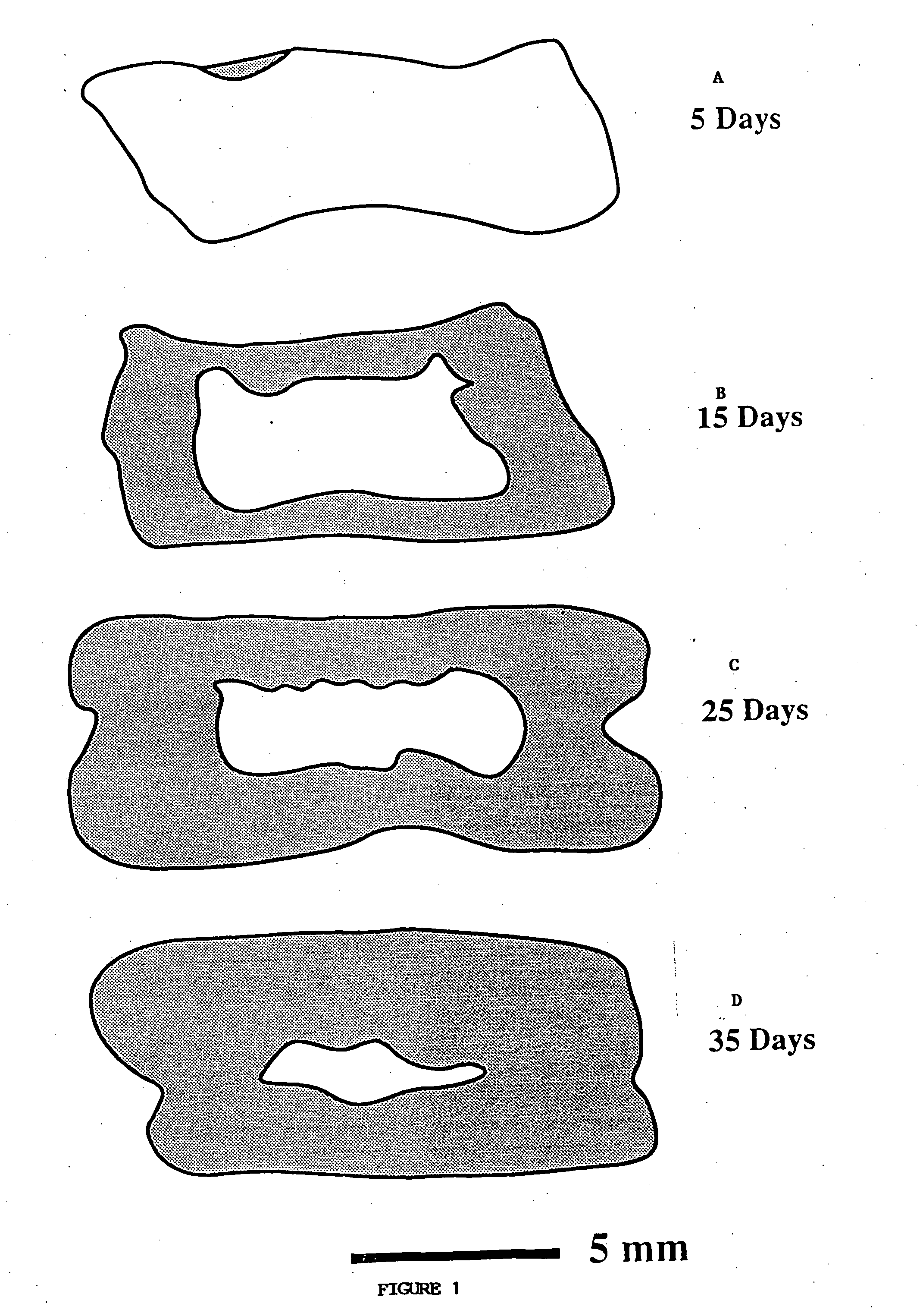
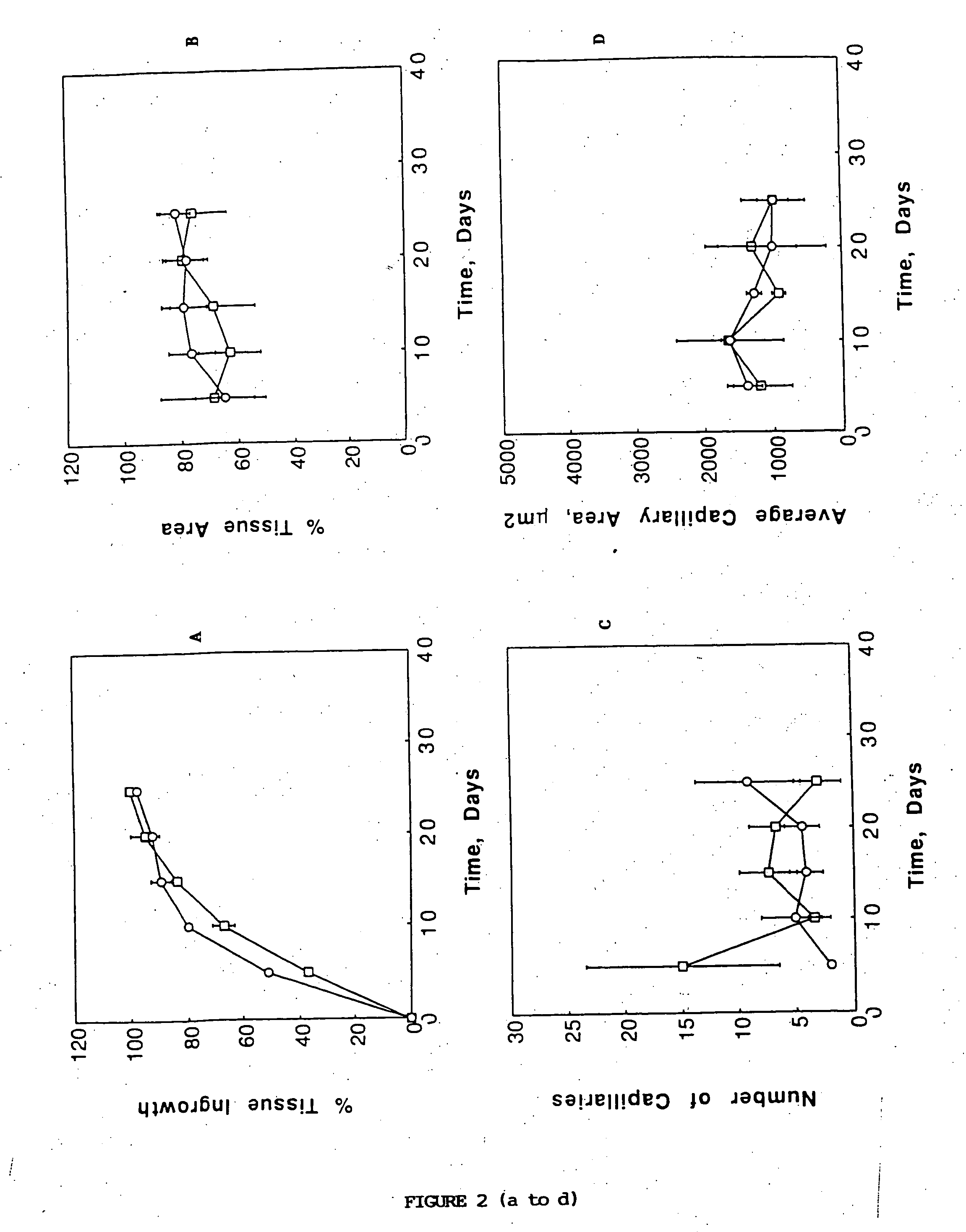
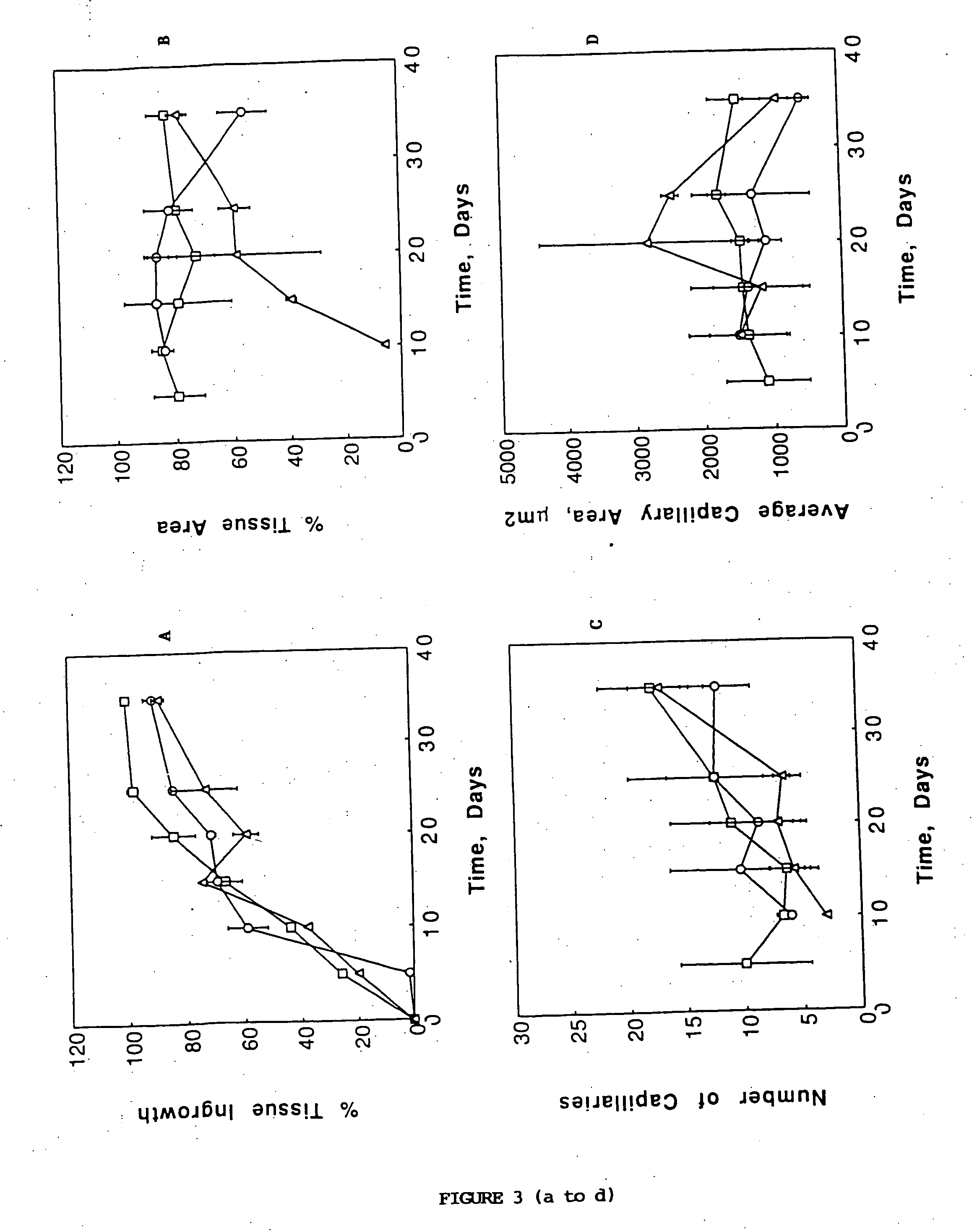
[0041] Devices were implanted in the mesentery of male 15 syngeneic Fischer 344 rats (Charles River, Wilmington, Mass.). In a typical experiment, the rat was anesthetized using methoxyflurane (Pitman Moore, Mundelein, Ill.) and its abdominal wall was incised. The mesentery, which is a thin fatty layer of tissue supplying blood to the small bowel, was displayed on sterile gauze carefully to avoid traumatizing the blood vessels or the tissue. The sterile device was placed onto the unfolded mesentery. Then, the mesentery was folded back over the device so as to envelope it, and put back into the rat. The device was sutured on the mesentery using non-absorbable surgical suture (Prolene, Ethicon, Sommerville, N.J.). The laparectomy was closed separately for the muscle layer and the skin with a synthetic absorbable suture (Polyglactin 910, Ethicon). Two devices were implanted in each 200 g rat, one proximally and one distally. Fo...


PUM

PropertyMeasurementUnit
Pore sizeaaaaaaaaaa
Pore sizeaaaaaaaaaa
Pore sizeaaaaaaaaaa

Abstract

Polymeric materials are used to make a pliable, non-toxic, injectable porous template for vascular ingrowth. The pore size, usually between approximately 100 and 300 microns, allows vascular and connective tissue ingrowth throughout approximately 10 to 90% of the matrix following implantation, and the injection of cells uniformly throughout the implanted matrix without damage to the cells or patient. The introduced cells attach to the connective tissue within the matrix and are fed by the blood vessels. The preferred material for forming the matrix or support structure is a biocompatible synthetic polymer which degrades in a controlled manner by hydrolysis into harmless metabolites, for example, polyglycolic acid, polylactic acid, polyorthoester, polyanhydride, or copolymers thereof. The rate of tissue ingrowth increases as the porosity and/or the pore size of the implanted devices increases. The time required for the tissue to fill the device depends on the polymer crystallinity and is less for amorphous polymers versus semicrystalline polymers. The vascularity of the advancing tissue is consistent with time and independent of the biomaterial composition and morphology.

Description

BACKGROUND OF THE INVENTION [0001] This is a continuation of continuation of U.S. Ser. No. 08 / 669,760, filed Sep. 26, 2000, which is a continuation of U.S. Ser. No. 08 / 052,387, filed Apr. 23, 1993, now abandoned, which is a continuation-in-part of U.S. Ser. No. 08 / 012,270, filed Feb. 1, 1993, now issued as U.S. Pat. No. 5,514,378.[0002] This invention is generally in the field of polymeric materials, and in particular in the area of biocompatible artificial matrices for implantation of cells. [0003] Loss of organ function can result from congenital defects, injury or disease. Many times treatment with drugs or surgery is not in itself sufficient and the patient dies or is severely disabled. One approach for treatment has been to transplant donor organs or tissue into the patient. Drugs such as cyclosporin can be used to prevent tissue rejection. However, there is a tremendous shortage of donor organs, most of which must come from a recently deceased individual. There have been a num...

Claims


Application Information

Patent Timeline
IPC IPC(8): A61K35/12G02B27/00G03F7/40H01L21/027H01L21/3213
CPCA61L27/18A61L27/56A61L2400/06G02B27/0031G03F7/40H01L21/0273H01L21/32139C08L67/04
Inventor MIKOS, ANTONIOS G.INGBER, DONALD E.VACANTI, JOSEPH P.LANGER, ROBERT S.
Owner CHILDRENS MEDICAL CENT CORP
Why Eureka
Try Eureka