Photochemical therapy to affect mechanical and/or chemical properties of body tissue

a photochemical therapy and chemical technology, applied in the field of ocular medicine, can solve the problems of increased astigmatism, myopia, and higher-order aberrations that become difficult to correct by spectacles or contact lenses, and achieve the effect of reducing the risk of post-operative corneal deformation and treating or reducing the risk of ocular deformation

Active Publication Date: 2008-05-15
CALIFORNIA INST OF TECH +1
View PDF4 Cites 64 Cited by
  • Summary
  • Abstract
  • Description
  • Claims
  • Application Information

AI Technical Summary

Benefits of technology

[0047]whereby the photoinitiator compound directly treats or reduces the risk of the ocular deformation condition.
0048] wherein the ocular tissue includes at least a portion of the cornea, the method is performed substantially concurrently with a corneal operation and the photoinitiator is provided in a therapeutically effective amount which directly reduces the risk of a post operative corneal deformation condition.

Problems solved by technology

The progressive distortion of corneal shape usually becomes noticeable in early adulthood, causing increasingly severe astigmatism, myopia, and higher-order aberrations that become difficult to correct by spectacles or contact lenses.
Glyceraldehyde has been used as a corneal crosslinking agent, but it has significant toxicity.
Riboflavin / UV requires painful removal of the epithelium with risk of long-term ulceration; it uses UV light, with inherent risk of damage to ocular tissues; it requires 30 minutes of irradiation which is clinically undesirable and increases the risk of corneal infection.
Because this stretching and thinning occurs preferentially in the posterior pole and involves the macula, eyes with degenerative myopia are subject to visual loss.
As the mechanical properties of the sclera are altered in myopia, the eye is prone to stretching due to the load effect of intraocular pressure.
As the macular tissues stretch, retinal cells atrophy, causing irreversible visual loss.
Although a large population is affected by this disease worldwide, there is currently no effective method to arrest progression and reduce the rate of visual loss.
Refractive errors induced by progressive myopia are readily corrected by spectacles, contact lenses, corneal refractive surgery, or intraocular lenses, these modalities do not prevent visual loss induced by stretching of chorioretinal tissues.
Furthermore, current means to treat choroidal neovascularization in degenerative myopia, such as photodynamic therapy, are minimally effective (Blinder, K. J., Blumenkranz, M. S., Bressler, N. M., Bressler, S. B., Donati, G., Lewis, H., Lim, J. I., Menchini, U., Miller, J. W., Mones, J. M., Potter, M. J., Pournaras, C., Reaves, A., Rosenfeld, P., Schachat, A. P., Schmidt-Erfurth, U., Sickenberg, M., Singerman, L. J., Slakter, J., Strong, H. A., Virgili, G.
Largely because these modalities remain unproven in well controlled clinical trials, none has been widely adopted to manage patients with degenerative myopia.
As the mechanical properties of the sclera are altered in myopia, the eye is prone to stretching due to the load effect of intraocular pressure.
Currently, there are no proven means to prevent the excessive ocular enlargement that occurs in degenerative myopia.
Efforts have been made to support the macular region with an external donor scleral or synthetic polymer band placed around the eye, but this has not been proven to be effective.
Since they are not light-activated, the authors report that it might be difficult to spatially control the cross linking effects of both glutaraldehyde and glyceraldehyde.
This approach utilizes chemical crosslinkers, some with undesirable toxicity profiles, by caging the chemicals in photo-labile structures which are disrupted to release the crosslinker chemicals in a controlled manner, thereby limiting the area of exposure and in some instances toxicity effects.

Method used


Image

  • Photochemical therapy to affect mechanical and/or chemical properties of body tissue
  • Photochemical therapy to affect mechanical and/or chemical properties of body tissue
  • Photochemical therapy to affect mechanical and/or chemical properties of body tissue

Examples


example 1

Poly(ethylene-glycol)dimethacrylate (PEGDM) Polymerization Using Eosin Y / TEOA Photoinitiation

[0172]Following the teachings of U.S. Published Application No. 20050271590, the Inventors Eosin Y / TEOA as an alternative photoinitiator of PEGDM crosslinking for reinforcement of scleral tissues. It was surprisingly discovered that Eosin Y / TEOA was comparably effective in the absence of a target crosslinking chemical (e.g. PEGDM) for enhancing the strength of scleral tissues. It was particularly unexpected that a Type II photoinitiator was capable of strengthening the sclera to a therapeutically useful degree with illumination periods of approximately 30 minutes or less. Without intending to be bound by the theory, it is believed that Eosin Y may sufficiently promote the direct crosslinking of collagen and other scleral components to enhance the strength of scleral tissue directly. While this dose not preclude the co-use of crosslinking chemicals such as PEGDM, the results with Eosin Y sugg...

example 2

Dose Response and Mechanical Properties of Treated Tissue

[0173]A number of different techniques were used to initially evaluate scleral tissue strengthening including tensile tests, oscillatory shear measurements and the eye expansion assay discussed more fully below.

[0174]The ability of initiator with or without crosslinking compounds to strengthen the sclera was initially evaluated using rheometric analysis of tissue sections. Sclera sections (8 mm diameter) were cut from porcine eyes and tested on a TA Instruments AR1000 rheometer (using a cleated parallel plate geometry) to quantify changes in modulus pre- and post-treatment. Two example photoinitiators were selected for initial evaluation. IRGACURE® 2959 (2-hydroxy-1-[4-(2-hydroxyethoxy)phenyl]-2-methyl-1-propanone) and Eosin Y with triethanolamine (Eosin Y / TEOA). IRGACURE® 2959 (12959) is a UV light activated photoinitiator that has shown low toxicity in cell encapsulation studies. Eosin Y / TEOA was selected as an example photo...

example 3

Ocular Shape in Inflation Model of Myopia and Keratoconous

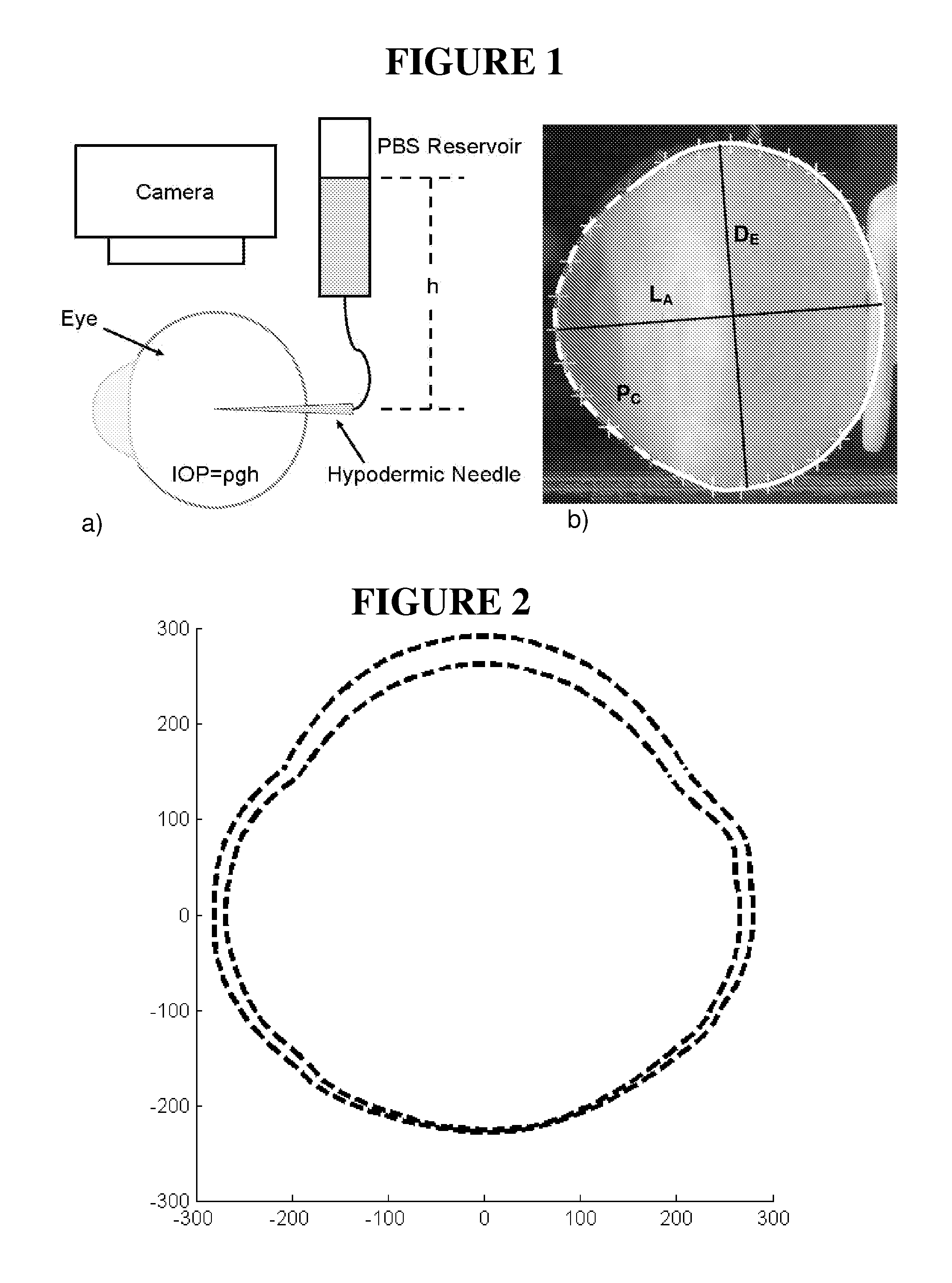
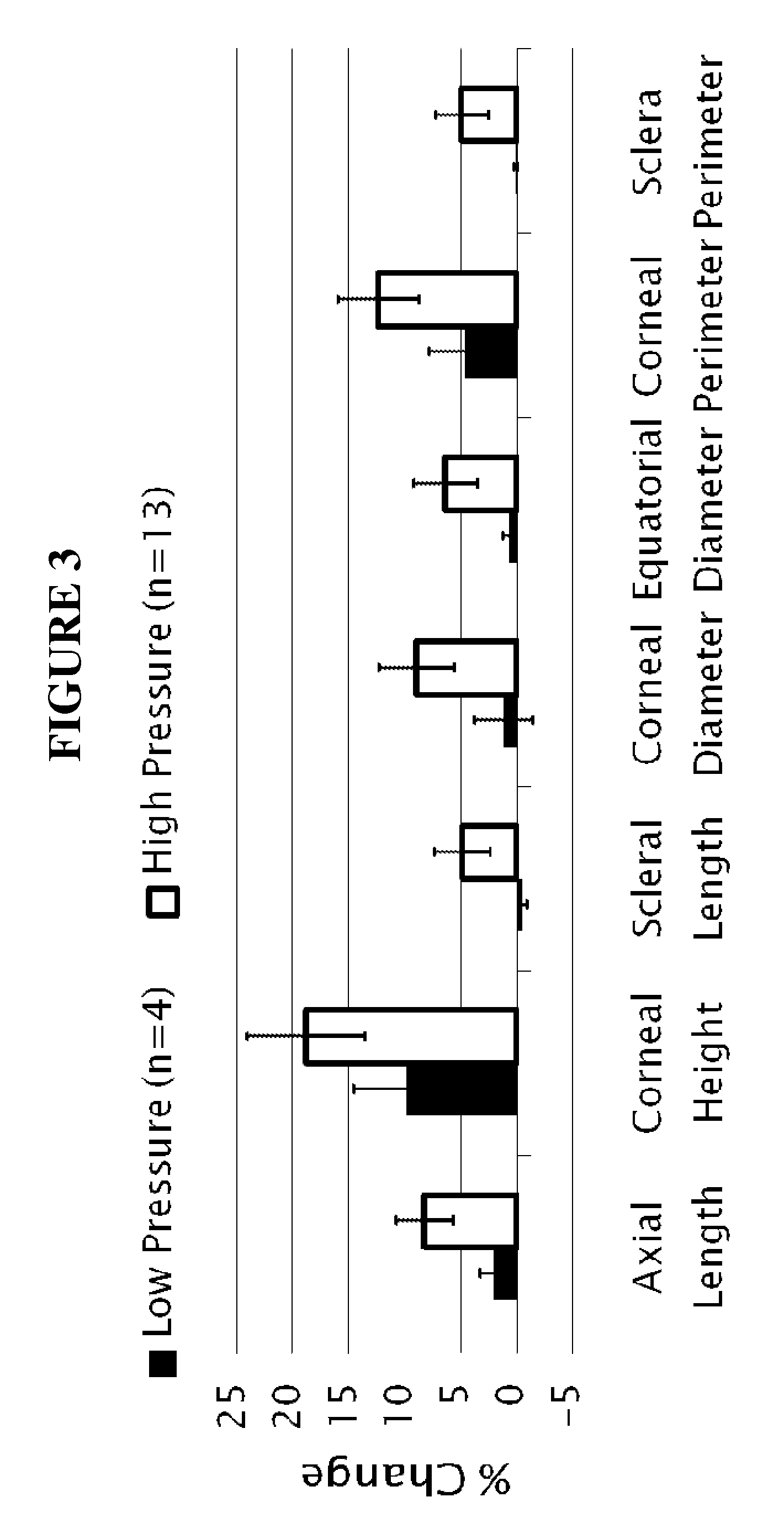
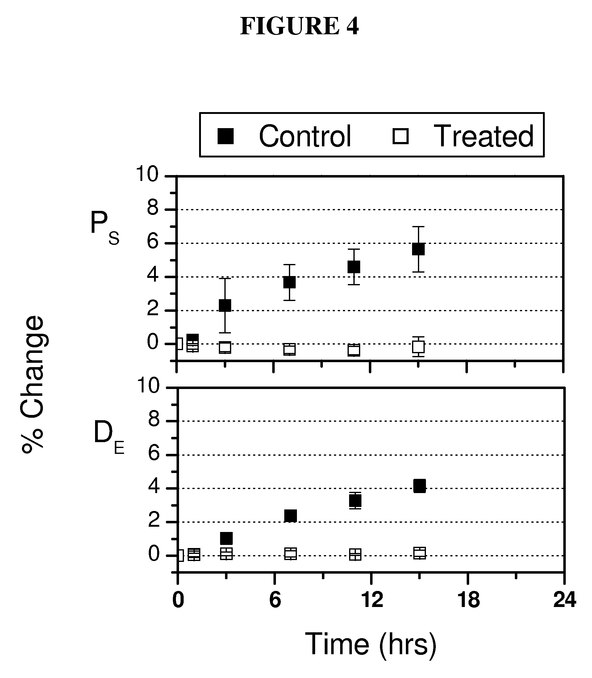
[0175]Stabilization of ocular shape is evaluated in vitro using pairs of enucleated eyes from young (˜1-2 weeks) New Zealand White Rabbits, one eye treated and the fellow eye as control. To mimic and accelerate aspects of the loading geometry present in vivo, the intact globes are mechanically tested by imposing an elevated intraocular pressure: a syringe connects the intraocular fluid to an elevated reservoir of Dulbecco's phosphate buffered saline (DPBS) while the exterior of the eye is immersed in DPBS at ambient pressure (FIG. 1). Enucleated eyes from young rabbits provide a model of distensible corneal and scleral tissue: when subjected to elevated intraocular pressure in vitro, the cornea progressively bulges outward and the scleral wall expands (FIGS. 2 and 3).

[0176]The size and shape of the globes are simultaneously recorded from two orthogonal perspectives over time. To prepare eyes for the tests, the epithelium is o...


PUM


Abstract

The disclosure concerns altering the mechanical and/or chemical property of a body tissue, particularly an ocular tissue. In specific cases, it concerns altering or stabilizing the shape of the cornea, such as in a subject suffering or at risk for ectasia or keratoconus. In other specific cases, it concerns strengthening the sclera in a subject suffering or at risk for myopia. The invention employs light irradiation of a photoactivatable compound, such as one that applies crosslinking to the tissue, for example.

Description

RELATED APPLICATIONS[0001]This application claims the benefit under 35 U.S.C. 119(e) of U.S. Provisional Patent Application No. 60 / 853,949 filed 24 Oct. 2006 and the benefit under 35 U.S.C. 119(e) of U.S. Provisional Patent Application No. 60 / 954,541 filed 7 Aug. 2007.STATEMENT REGARDING FEDERALLY SPONSORED RESEARCH OR DEVELOPMENT[0002]The present invention was developed at least in part with funds from National Institutes of Health grant R41 EY017484. The United States Government may have certain rights in the invention.TECHNICAL FIELD[0003]The fields of the invention concern medicine, anatomy, cell biology, or molecular biology, for example. In particular, the field of the invention concerns ocular medicine, including opthalmology.BACKGROUND OF THE INVENTION[0004]Keratoconus[0005]Keratoconus (Rabinowitz, 1998; Krachmer et al., 1994; Bron, 1988) is the most common corneal dystrophy, affecting 1 in 2000 persons. Keratoconus results in corneal thinning and is named for the conical sh...

Claims


Application Information

Patent Timeline
Patent Type & Authority Applications(United States)
IPC IPC(8): A61M5/00
CPCA61K9/0048A61K9/0051A61K9/19A61K31/00A61F2009/00893A61F9/0079A61F2009/00865A61F2009/00872A61F9/008A61P27/02A61P27/06A61P27/10A61K31/352
Inventor MATTSON, MATTHEW S.KORNFIELD, JULIA A.SCHWARTZ, DANIEL M.MALONEY, ROBERT K.
Owner CALIFORNIA INST OF TECH
Why Eureka
Try Eureka