Method of Detection, Separation and Identification for Expressed Trace Protein/Peptide

a technology of expressed protein and trace amount, applied in the field of detection, separation and identification of trace amount of expressed protein and/or peptide, to achieve the effect of easy and simultaneous comparison, easy and accurate quantification, and easy quantification of trace protein

Active Publication Date: 2008-11-13
IMAI KAZUHIRO
View PDF11 Cites 8 Cited by
  • Summary
  • Abstract
  • Description
  • Claims
  • Application Information

AI Technical Summary

Benefits of technology

[0008]An object of the present invention is to provide a method for detecting, separating and identifying the above-mentioned expressed protein and / or peptide present in trace amounts capable of detection separation and identification with high sensitivity using a simple method for measuring trace expressed proteins and / or peptides expressed through a gene.
[0155]As has been described in detail above, the present invention relates to a method and system for detecting, separating and identifying trace expressed protein and / or peptide. According to the present invention, protein and / or peptide expressed through a gene can be detected separated and identified with high sensitivity using a simple method and means. According to the method of the present invention, trace expressed protein and / or peptide unable to be detected with methods of the prior art can be detected, separated and identified with high sensitivity and in a short period of time. In addition, the present invention is also able to provide a system for detecting, separating and identifying trace amounts of trace expressed protein and / or peptide used in the above-mentioned detection, separation and identification method. The present invention is useful for providing proteome platform technology.

Problems solved by technology

However, this method had problems with reproducibility of the method due to the complex procedure Separation and identification methods using multi-dimensional high-performance liquid chromatography (multi-dimensional HPLC), and techniques using ICAT have recently been proposed to overcome this problem (see Non-patent Document 2).

Method used


Image

  • Method of Detection, Separation and Identification for Expressed Trace Protein/Peptide
  • Method of Detection, Separation and Identification for Expressed Trace Protein/Peptide
  • Method of Detection, Separation and Identification for Expressed Trace Protein/Peptide

Examples


example 1

Separation and Identification of Proteins / Peptides Containing Thiol Islets of Langerhans in Rat Pancreatic Tissues

(1) Derivatization of Proteins / Peptides Containing Thiol in Islets of Langerhans

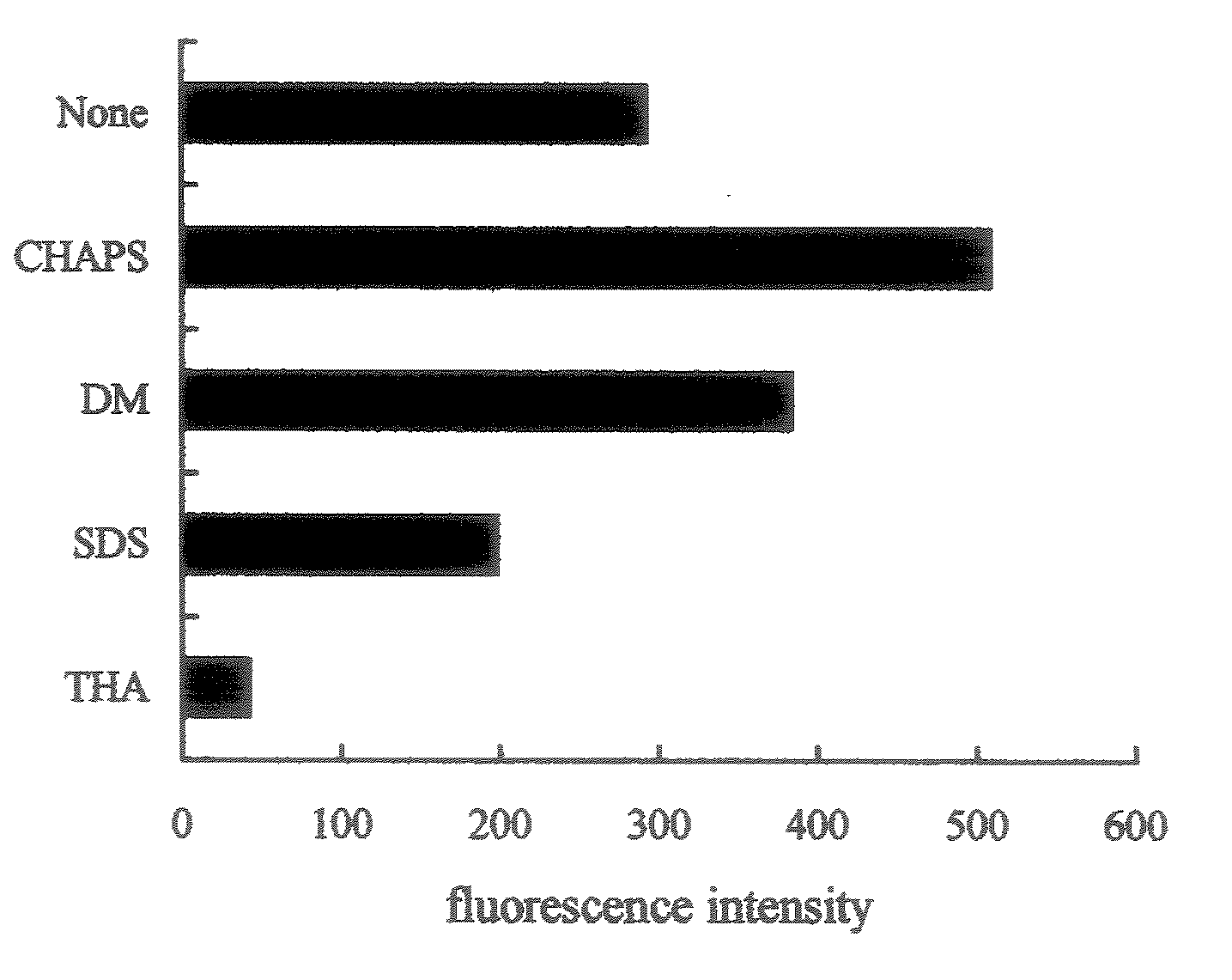
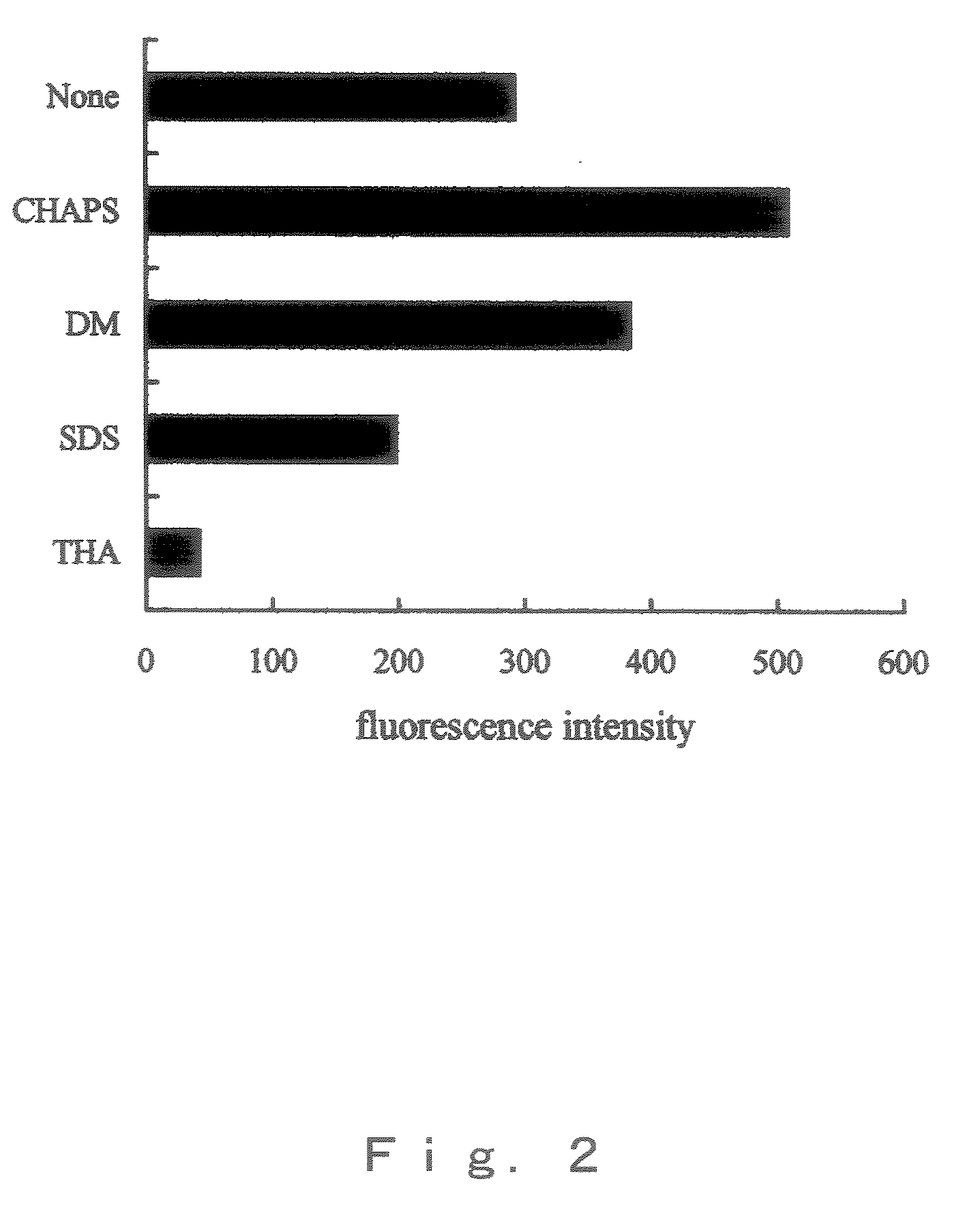
[0041]The islets of Langerhans were solublized by adding 50 μl of a 6 M guanidine chloride dissolved in 0.1 M boric acid buffer (pH9.0), and 50 μl of each 17.5 mM TCEP, 17.5 mM SBD-F, 10 mM EDTA and 50 mM CHAPS dissolved in 6 M guanidine chloride solution were added thereto respectively, and were mixed. The mixture was reacted at 40° C. for 3 hr to derivatize them.

(2) Primary Separation Using Ion Exchange HPLC

[0042]The above reaction solution was subjected to ion exchange column, and fluorescence proteins / peptides were eluted by NaCl gradient (0, 0.04, 0.08, 0.12 and 0.3 M) to separate 5 fractions. The detection of fluorescence proteins / peptides was performed by fluorescence of SBD skeleton. HPLC conditions are shown below.

(HPLC Conditions)

[0043]Column: TSKgel DEAE-5PW 7.5×75 mm (Toso)[0044]G...

example 2

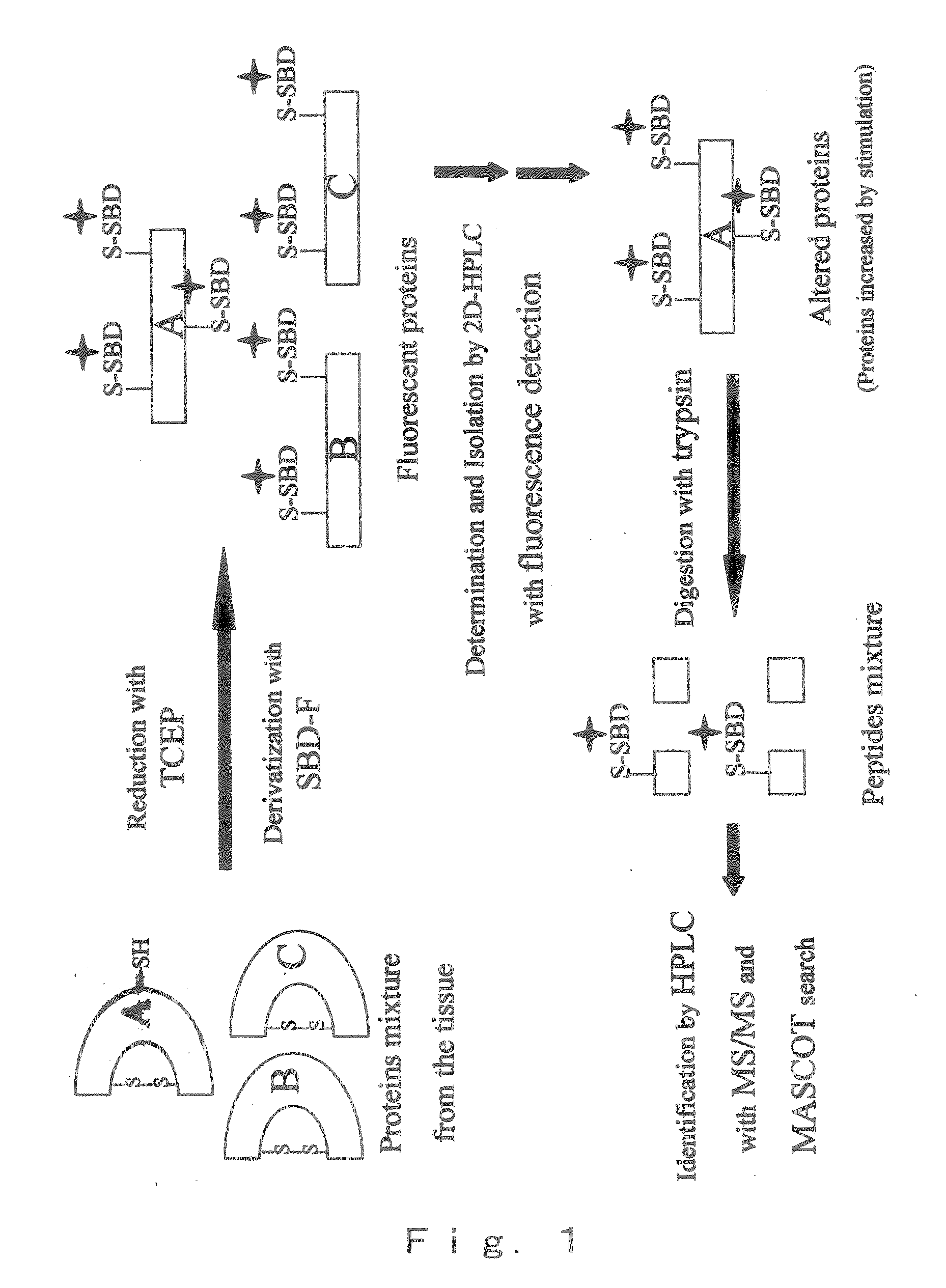
[0075]The derivatized BSA with SBD-F was digested with trypsin by the process as depicted in FIG. 1, the resulting peptides mixture was separated by reversed phase liquid chromatography (RPLC), and detected by fluorescence detector. Then, each peptide was subjected MS / MS analysis by ESI mass spectromometer. Theoretically, by trypsin digestion, BSA should generate 25 Cys-containing peptides and 35 non-Cys-containing peptides of more than 4 amino acid residue. In the present Example, more than 27 fluorescent peptides were detected fluorometically so that the quantitative derivatization has been performed (FIG. 4, A).

[0076]Eleven Cys-containing peptides and 17 non-Cys-containing peptide were detected in the mass chromatogram (FIG. 4, B). FIG. 5 shows the tandem mass spectrum derived by collision-induced dissociation (CID) of the (M+2H)2+ precursor, m / z=873.4 (marked with an arrow in FIG. 4).

[0077]Database-searching with MASCOT, which adopts probability-based protein identification algo...

example 3

[0078]The applicability of the method was tested for rat pancreas with or without dexamethasone (Dex) administration.

[0079]Dex induces type 2 diabetes, a predominant type in human diabetes, through the increase in the hepatic glucose production and induction of insulin resistance. Actually, at 24 h after the Dex treatment, the blood glucose levels reached 209.8 mg / dL, which were significantly above the pretreatment value of 118.3 mg / dL (p<0.05). In the present Example, islet of Langerhans (around 60 islets) were collected from the rat pancreas treated with or without Dex for two days and derivated with SBD-F. An important aspect for the application of the method to biological samples is the isolation of a target protein(s) by HPLC from the protein mixture. In the present Example, the fluorescent proteins were first separated via ion exchange chromatography (IEC) on the basis of varieties of negative changes generated by SBD-F and amino acids moieties.

[0080]IEC was performed with the...


PUM

PropertyMeasurementUnit
excitation wavelengthaaaaaaaaaa

Abstract

A method of detection separation and identification for expressed trace protein / peptide; and a system therefor. There is provided a method of detecting, separating and identifying a minute amount of expressed protein and / or peptide, characterized in that a fluorescent derivative of protein and / or peptide contained in a test subject sample having been labeled with a fluorescence reagent is applied to HPLC; a fluorescent fraction is collected and subjected to enzymatic hydrolysis; mass-spectrometry of the resultant fluorescence-labeled fragments and non-fluorescence-labeled fragments is carried out; and the thus obtained ion molecular weight information on each of the fragments is collated with an available protein and / or peptide fragment database to thereby accomplish a structural analysis. Further, there is provided an identification system therefor.

Description

TECHNICAL FIELD[0001]The present invention relates to a method for detecting separating and identifying trace amounts of expressed protein and / or peptide, and more particularly to a novel method for detecting, separating and identifying trace amounts of protein and / or peptide produced in the body by expression of a gene, which enables trace amounts of protein and / or peptide to be detected and identified easily and with high sensitivity and to an identification system of the same.[0002]The present invention is useful for providing a novel detection, separation and identification technology for proteome technology, which is expected to play an important role in the post-genome area through comprehensive analysis of expressed proteins and / or peptides.BACKGROUND ART[0003]An important objective in the post-genome area is the detect-ion of trace amounts of expressed protein / peptide expressed through genes and the separation and identification thereof. In the past, peptide fingerprinting f...

Claims


Application Information

Patent Timeline
Patent Type & Authority Applications(United States)
IPC IPC(8): C12Q1/37C12Q1/00C12M1/40B01DG01N21/64G01N21/77G01N21/78G01N27/447G01N27/62G01N30/02G01N30/72G01N30/88G01N33/483G01N33/533G01N33/68
CPCC07D285/10C07K1/13C07K1/36G01N21/6428G01N27/447G01N33/6851G01N33/533G01N33/6848G01N2030/027G01N33/582G01N30/7233
Inventor IMAI, KAZUHIRO
Owner IMAI KAZUHIRO
Why Eureka
Try Eureka