Joining and/or Sealing Tissues Through Photo-Activated Cross-Linking of Matrix Proteins

Active Publication Date: 2009-12-10
COOK MEDICAL TECH LLC
View PDF11 Cites 6 Cited by
  • Summary
  • Abstract
  • Description
  • Claims
  • Application Information

AI Technical Summary

Benefits of technology

[0125]Reactions contained 25 μg of bovine fibrinogen (Sigma); 2 mM (Ru(bpy)3]Cl2 (Aldrich) all in 25 μl PBS. (SPS: sodium persulfate; APS: ammonium persulfate).
[0126]Reactions were exposed to 300W incoherent light from Quartz Halogen dichroic source for 1 min (FIG. 5) demonstrating efficacy across a range of concentrations.
[0127]Preliminary testing involved the examination of the sealant properties of FBTS using porcine fibrinogen to repair incisions made in abattoir specimens of various bovine tissues (see FIGS. 6 and 7). These appeared to form rather strong tissue welds, as in some cases the surrounding tissue was ruptured in attempting to disrupt the tissue seal.
[0128]This example demonstrates an in vivo

Problems solved by technology

The preparation and application of fibrin sealants are somewhat complicated.
Nevertheless there remains the difficulty that the seal takes a significant period of time to form and reach strength sufficient to hold when under pressure.
Typically 70% of its ultimate strength is reached in 10 minutes, and full strength in approximately 2 hours, which means that fibrin sealants are unsuitable for many applications (for example, sealing an incision in a blood vessel, since prolonged clamping would be required) and inconvenient in many other applications compared to traditional methods.
They found that a fibrinogen conjugate with chlo

Method used


Image

  • Joining and/or Sealing Tissues Through Photo-Activated Cross-Linking of Matrix Proteins
  • Joining and/or Sealing Tissues Through Photo-Activated Cross-Linking of Matrix Proteins
  • Joining and/or Sealing Tissues Through Photo-Activated Cross-Linking of Matrix Proteins

Examples


example 1

Photochemical Cross-Linking of Bovine Fibrinogen

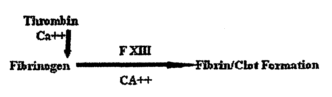
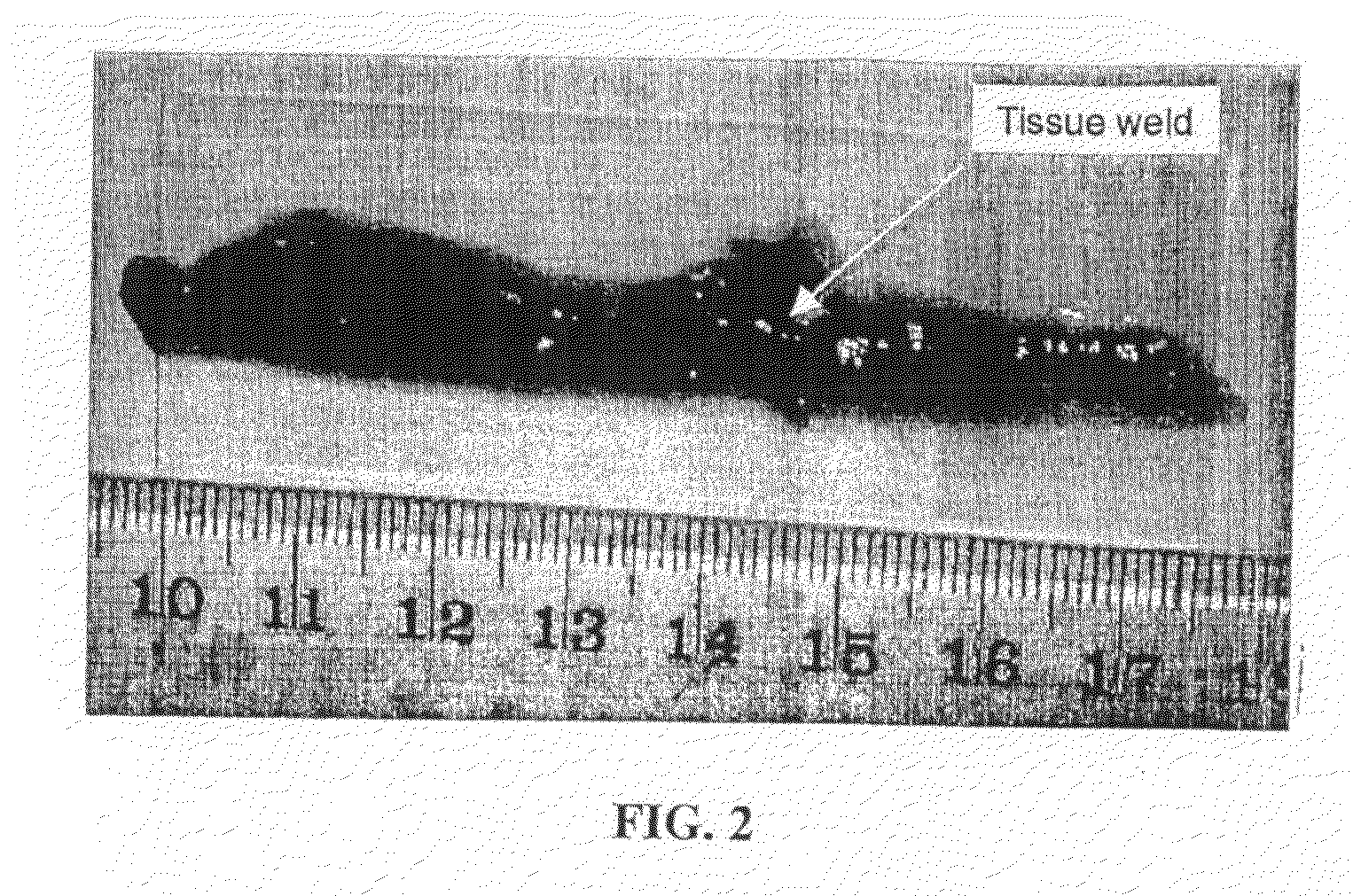
[0099]A photochemical method was used to cross-link the soluble fibrinogen into a solid biomaterial and to effect the covalent cross-linking of the fibrinogen matrix to the proteins contained in the extracellular matrix surrounding the muscle tissue. Two small strips of bovine longissimus dorsi (LD) were dissected and the opposing surfaces coated in the sealant solution (200 mg / ml bovine fibrinogen was dissolved in PBS, with 2 mM (Ru(bpy)3]Cl2, 10 mM ammonium persulfate). Following 10 sec of irradiation, the two pieces of muscle were firmly attached at the position shown in FIG. 2. The light source chosen for the present study was a 600-W tungsten-halide source (2×300-W lamps; GE #38476). The spectral output showed a broad peak from 300 nm-1200 nm. Bovine fibrinogen (Fraction I, Sigma) (200 mg / ml) was dissolved in PBS, with 2 mM [Ru(bpy)3]Cl2, 10 mM ammonium persulfate) and photochemically cross-linked (600 W at 10 cm for 10 s).

example 2

[0100]Degradation of and Tissue Response to Polymerised Fibrinogen Biopolymers in vivo

[0101]The solid fibrinogen biomaterial to be evaluated in the rat subcutaneous implant study is derived from a purified soluble fibrinogen protein which was crosslinked using a photochemical method involving Tris(bipyridyl) Ruthenium (II) chloride (2 mM final concentration) and ammonium persulphate (20 mM final concentration). The light source chosen for these studies was a 600-W tungsten-halide source (2×300-W lamps; GE #38476). The spectral output showed a broad peak from 300 nm-1200 nm. Preliminary in vitro cytotoxicity evaluations on this purified expressed protein biomaterial had been encouraging however further, standard cell culture methods to screen for direct and leachable potential toxic components were required prior to implantation in animals. Therefore the fibrinogen biomaterials were evaluated in rats for tissue responses as well as the extent of degradation of the material over a 36 ...

example 3

Strength of Fibrinogen-Based Tissue Sealant (FBTS).

[0115]The comparative strength of tissues joined using FBTS and tissues joined using fibrin glue was measured using de-fleeced adult sheep skin. Skin was de-haired and the dermal surface scraped clean using a wire brush. The skin was soaked in phosphate-buffered saline (PBS) and “dumbbells” (ca. 10 cm×1 cm) were cut from the re-hydrated skin for tensile testing. An incision was made across the width of a dumbbell shaped strip of skin, and the two halves then “glued” or sealed (at room temperature) following photochemical treatment of fibrinogen in accordance with the present invention. The adhesive properties of FBTS was compared with a commercial fibrin-based tissue sealant (Tisseel™-Baxter), used as instructed by the manufacturer. The glued skin was then mounted in a tensile testing instrument (Instron) and tested for load to break. The units are Newtons (N). The cut dumbbells were either overlapped by 1 cm at the join or were but...


PUM

PropertyMeasurementUnit
Molar densityaaaaaaaaaa

Abstract

A method of joining and/or scaling tissues in a surgical procedure or medical treatment, comprising the steps of: (1) applying a matrix protein, a photoactivatable metal-ligand complex and an electron acceptor to a tissue portion; (2) irradiating said tissue portion to photactivate the photoactivatable metal-ligand complex: thereby initiating a cross-linking reaction of the matrix protein to seal said tissue portion or join said tissue portion to an adjacent tissue portion.

Description

TECHNICAL FIELD [0001]The present invention relates to a method of joining and / or sealing tissues in surgical procedures and medical methods, and compositions for use in said methods.BACKGROUND ART [0002]Tissue adhesives have been suggested as alternatives in surgical procedures to physical means of connecting tissues such as sutures and staples. Tissue adhesives will hold cut or separated areas of tissue together to allow healing and / or serve as a barrier to leakage, depending on the application. The adhesive should break down or be resorbed and it should not hinder the progress of the natural healing process. Ideally, the agent should promote the natural mechanism of wound healing and then degrade.[0003]Tissue adhesives are generally utilized in three categories:[0004]i) Hemostasis (for example, by improving in vivo coagulation systems, tissue adhesion itself has a hemostatic aim and it is related to patient clotting mechanisms)[0005]ii) Tissue sealing: primary aim is to prevent l...

Claims


Application Information

Patent Timeline
IPC IPC(8): A61B17/03A61K38/36A61K33/24A61L15/16A61K9/00A61F13/20
CPCA61L24/10C09J189/00A61L24/106
Inventor BROWNLEE, ALAN GEORGEELVIN, CHRISTOPHER MALCOLM
Owner COOK MEDICAL TECH LLC
Why Eureka
Try Eureka