
Extracellular matrix-binding proteins from staphylococcus aureus

a staphylococcus, extracellular matrix technology, applied in the field of microbiology and molecular biology, can solve the problems of thrombogenic and prone to bacterial colonization, invasive infections, and vascular grafts, and achieve the effect of simple survey method

Inactive Publication Date: 2010-01-14
TEXAS A&M UNIVERSITY
View PDF1 Cites 2 Cited by
  • Summary
  • Abstract
  • Description
  • Claims
  • Application Information

AI Technical Summary

Benefits of technology

The patent describes the discovery and characterization of extracellular matrix-binding proteins from S. aureus, designated ClfB, SdrC, SdrD, and SdrE. These proteins can be used as preventive, inhibitory, or diagnostic tools for S. aureus infections. They can also be used as research tools to identify S. aureus binding sites on the extracellular matrix and to develop antibacterial therapies. The patent also describes the use of these proteins or their consensus or variable motifs as immunogens or antigens to elicit immunity against S. aureus infections. The nucleic acid sequences of these proteins can also be used as probes to identify other S. aureus extracellular matrix-binding proteins or as polynucleotides for gene expression. The immunization with gene rather than its gene product has advantages such as the relative simplicity of the immunogen and the potential for the immunogen to enter the MHC class.

Problems solved by technology

Initial localized infections of wounds or indwelling medical devices can lead to serious invasive infections such as septicemia, osteomyelitis, and endocarditis.
Vascular grafts, intravenous catheters, artificial heart valves, and cardiac assist devices are thrombogenic and prone to bacterial colonization.

Method used


Image

  • Extracellular matrix-binding proteins from staphylococcus aureus
  • Extracellular matrix-binding proteins from staphylococcus aureus
  • Extracellular matrix-binding proteins from staphylococcus aureus

Examples


example 1

Gene Cloning, Sequencing and Expression

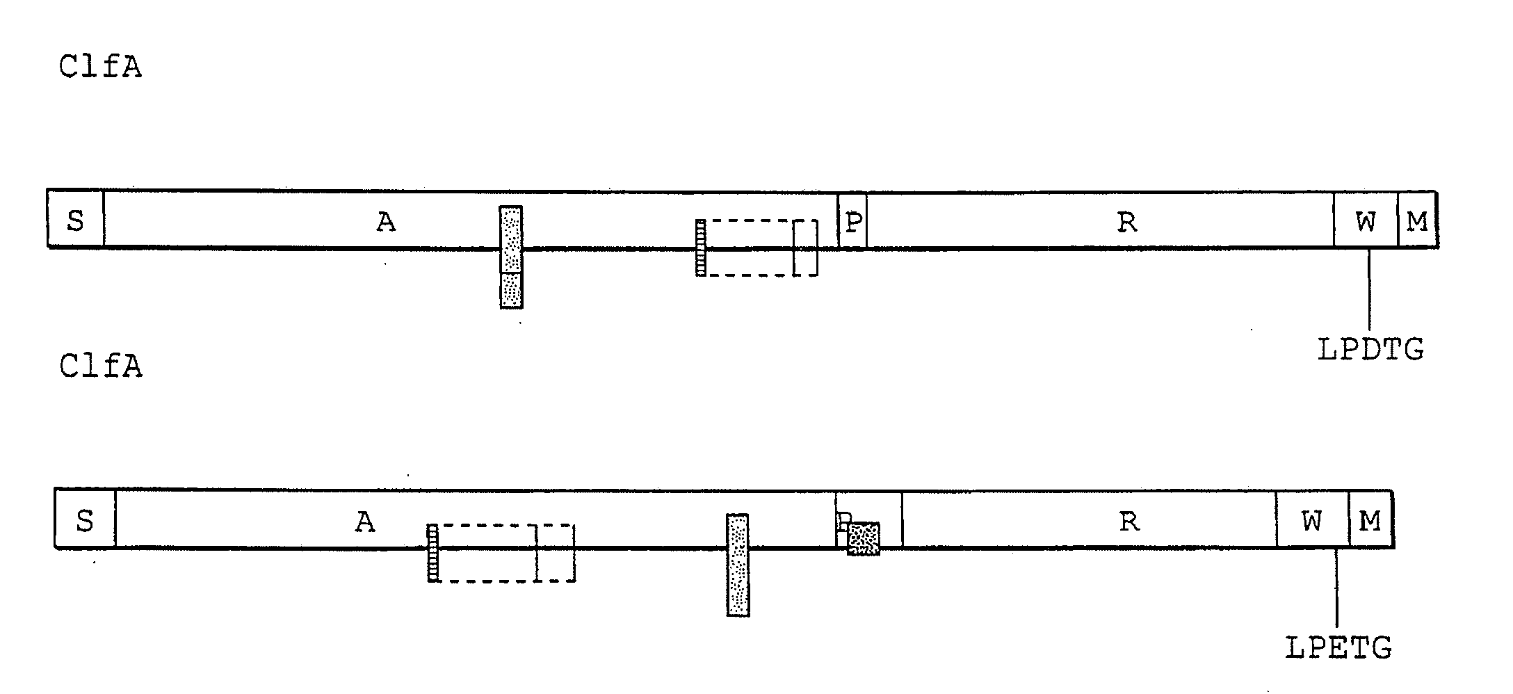
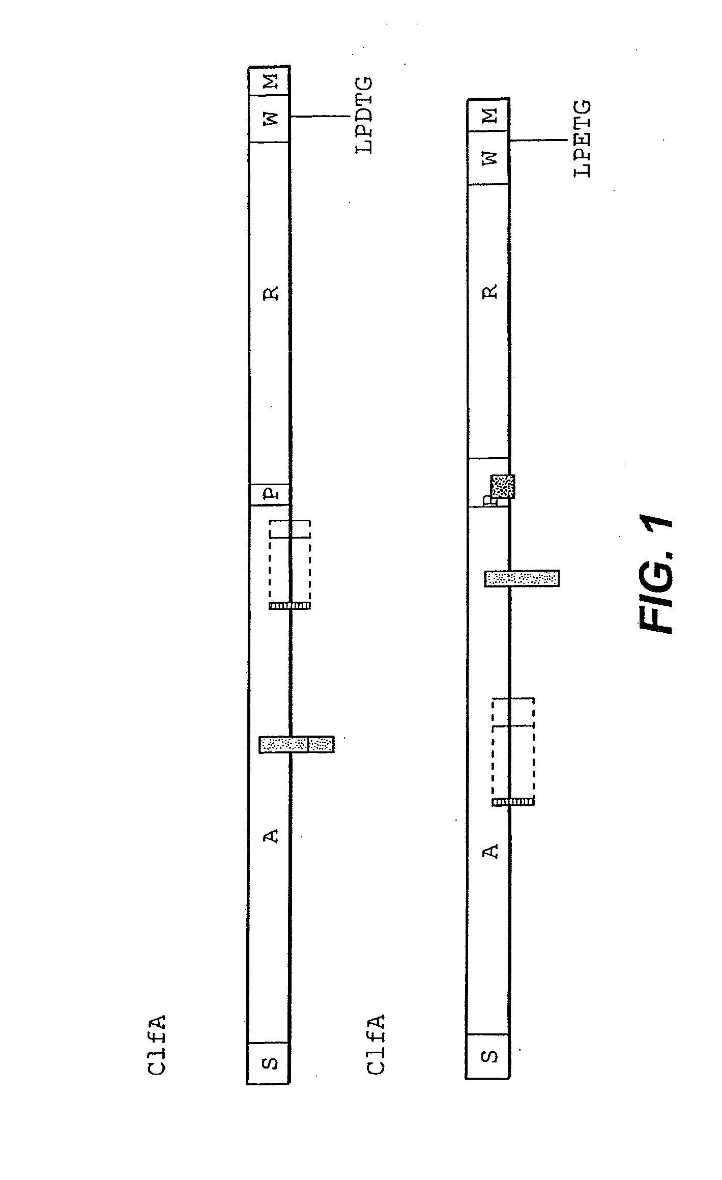
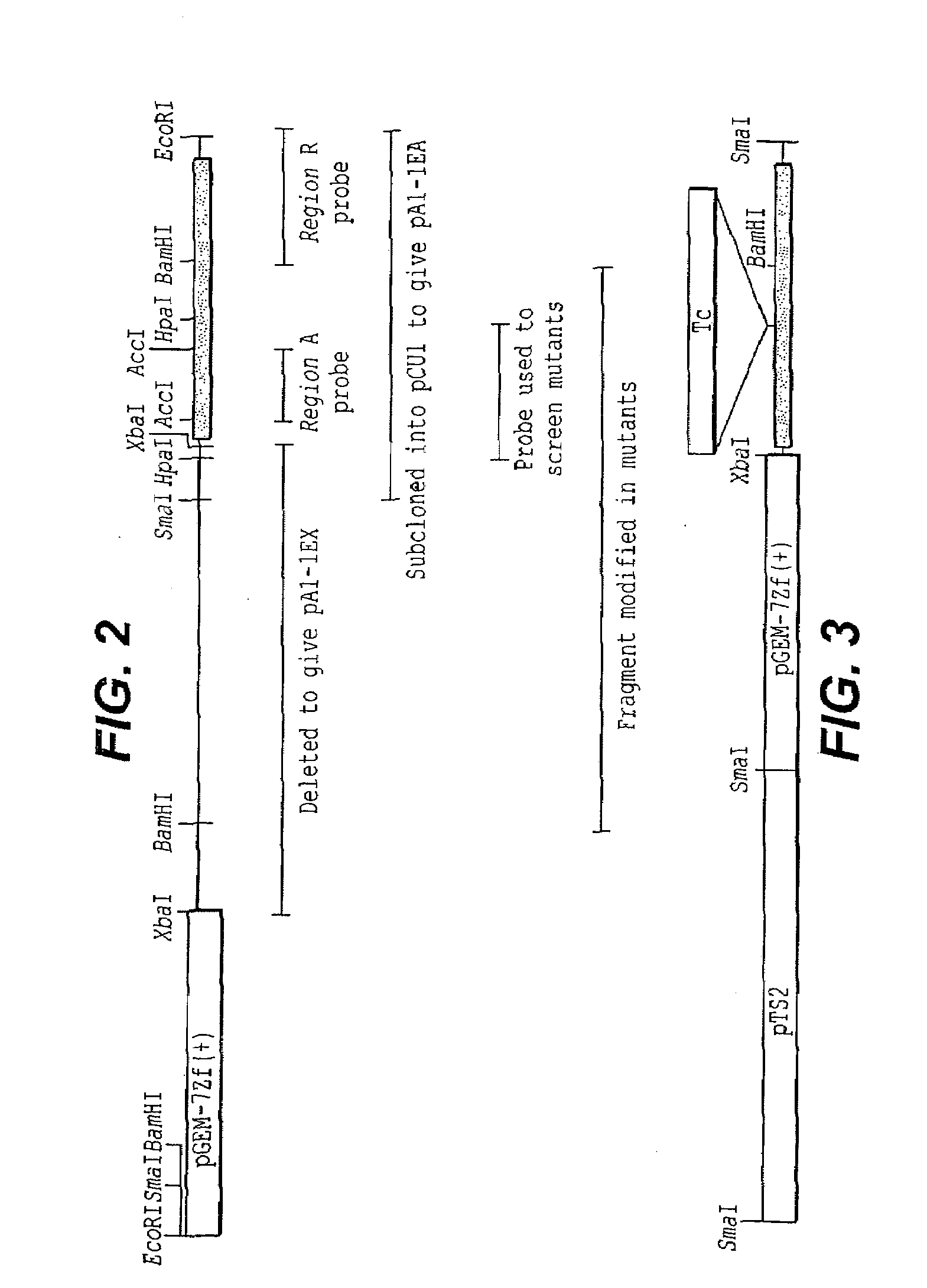
[0140]A fibrinogen-binding protein gene, designated clfB, was isolated, cloned and sequenced as follows:

[0141]Bacterial Strains and Growth Conditions

[0142]The E. coli and S. aureus strains used for the cloning and sequencing of clfB are listed in Table 1, below. Escherichia coli was routinely grown on L-broth or agar. S. aureus was routinely grown on trypticase soy broth (Oxoid) or agar. The following antibiotics were incorporated into media where appropriate: ampicillin (Ap), 100 μg / ml; tetracycline (Tc), 2 μg / ml; chloramphenicol (Cm), 5 μg / ml; erythromycin (Em) 10 μg / ml.

BacterialRelevant properties / StrainGenotypeUse in present studySource / referenceC600F−, lacY1, leuB6, supE44,Propagation of lambdaAppleyard, Geneticsthi-1, thr-1, tonA21recombinants39: 440-452 (1954)DH5αF−, ø80dlacZM15, deoR,Recombination deficient, hostHanahan et al, J. Mol.endA1, gyrA96, hsdR17,strain for plasmids and for DNABiol. 166: 557-580(rk−, mk+), (lacZYA-argF)sequenci...

example 2

Production of Anti-ClfB Serum

[0176]Antibodies to recombinant region A were raised in two young New Zealand white rabbits (2 kg) showing no prior reaction with E. coli or S. aureus antigens in Western blots. Injections, given subcutaneously, contained 25 μg of the antigen, diluted to 500 ml in phosphate buffered saline (PBS) emulsified with an equal volume of adjuvant. The initial injection contained Freund's complete adjuvant; the two to three subsequent injections, given at two-week intervals, contained Freund's incomplete adjuvant. When the response to the recombinant protein was judged adequate, the rabbits were bled, serum recovered, and total IgG purified by affinity chromatography on protein A sepharose (Sigma Chemical Co., St. Louis, Mo.).

[0177]SDS-PAGE and Western Blotting

[0178]Samples were analyzed by SDS-PAGE in 10 or 12% acrylamide gels. Isolated proteins and E. coli cell lysates were prepared for electrophoresis by boiling for five minutes in denaturation buffer. For S. ...

example 3

Immunoassay for ClfB Using Biotinylated Recombinant ClfB Region A

[0182]The DNA encoding region A of clfB (encoding residues S45 to N542) was amplified from genomic DNA of S. aureus Newman using the following primers:

Forward:(SEQ ID NO: 17)5′ CGAAAGCTTGTCAGAACAATCGAACGATACAACG 3′Reverse:(SEQ ID NO: 16)5′ CGAGGATCCATTTACTGCTGAATCACC 3′

[0183]Cleavage sites for HindIII and BamHI (underlined) were appended to the 5′ ends of the respective primers to facilitate cloning of the product into the His-tag expression vector pV4. Cloning employed E. coli JM101 as a host strain. The recombinant region A was purified by nickel affinity chromatography.

[0184]Enzyme Linked Immunosorbent Assay (ELISA)

[0185]Immulon 1™ plates (Dynatech™, Dynal, Inc., Great Neck, N.Y.) were coated overnight with 100 μl of 10 μg / ml human fibrinogen (Chromogenix). They were then blocked with 200 μl of 5 mg / ml bovine serum albumin (BSA) for one hour. The plates were then incubated for three hours with 100 μl biotinylated Cl...


PUM

PropertyMeasurementUnit
apparent molecular weightaaaaaaaaaa
molecular weightaaaaaaaaaa

Abstract

An isolated extracellular matrix-binding protein, designated as SdrC and its corresponding amino acid and nucleic acid sequences and motifs are described. The proteins, peptides, fragments thereof or antigenic portions thereof are useful for the prevention, inhibition, treatment and diagnosis of S. aureus infection and as scientific research tools. Further, antibodies or antibody fragments to the proteins, peptides, fragments thereof or antigenic portions thereof are also useful for the prevention, inhibition, treatment and diagnosis of S. aureus infection. In particular, the proteins or antibodies thereof may be administered to wounds or used to coat biomaterials to act as blocking agents to prevent or inhibit the binding of S. aureus to wounds or biomaterials.

Description

CROSS REFERENCE TO RELATED APPLICATIONS[0001]The present application is a divisional application of U.S. application Ser. No. 10 / 744,616, filed Dec. 24, 2003, which is a divisional application of U.S. application Ser. No. 09 / 200,650, filed Nov. 25, 1998, now U.S. Pat. No. 6,680,195, and claims the benefit of U.S. Provisional Application Nos. 60 / 066,815, filed Nov. 25, 1997 and Ser. No. 60 / 098,427, filed Aug. 31, 1998, all of said applications incorporated herein by reference.[0002]The U.S. Government has rights in this invention arising out of National Institutes of Health grant number A120624.FIELD OF THE INVENTION[0003]The present invention is in the fields of microbiology and molecular biology. The invention includes the isolation and use of extracellular matrix-binding proteins and genes that express the proteins from Staphylococcus aureus to inhibit, prevent and diagnose S. aureus infection.BACKGROUND OF THE INVENTION[0004]In hospitalized patients Staphylococcus aureus is a maj...

Claims


Application Information

Patent Timeline
Patent Type & Authority Applications(United States)
IPC IPC(8): A61K39/085C07K14/00A61P31/12G01N33/569A61K38/00A61K39/00C07K14/31
CPCA61K38/00A61K39/00G01N2469/00G01N33/56938C07K14/31Y10S530/825Y10S530/81Y10S435/975A61P31/12
Inventor PATTI, JOSEPH M.FOSTER, TIMOTHY J.JOSEFFSON, ELIZABETEIDHIN, DEIDRE NIHOOK, MAGNUS A. O.PERKINS, SAMUEL E.
Owner TEXAS A&M UNIVERSITY
Learn More