[0011]The illustrated embodiment of the present invention is a simple-to-operate device which uses a spatially shaped elongated laser focused spot for micro-dissection of living / nonliving microscopic objects with high
throughput. In the past, either a scanning stage or
scanning mirror was used to scan a region in a single
cell or group of cells for microprocessing and / or
microdissection. Besides being expensive, the prior art approach requires complex control of the
scanning beam via computer. Most importantly, the above mentioned techniques require large
processing time for groups of cells. This reduces the
throughput of the laser
microbeam system. Further, this may lead to the requirement of immobilization (by optically
trapping or otherwise) of the motile object(s) for precise micro-dissection.
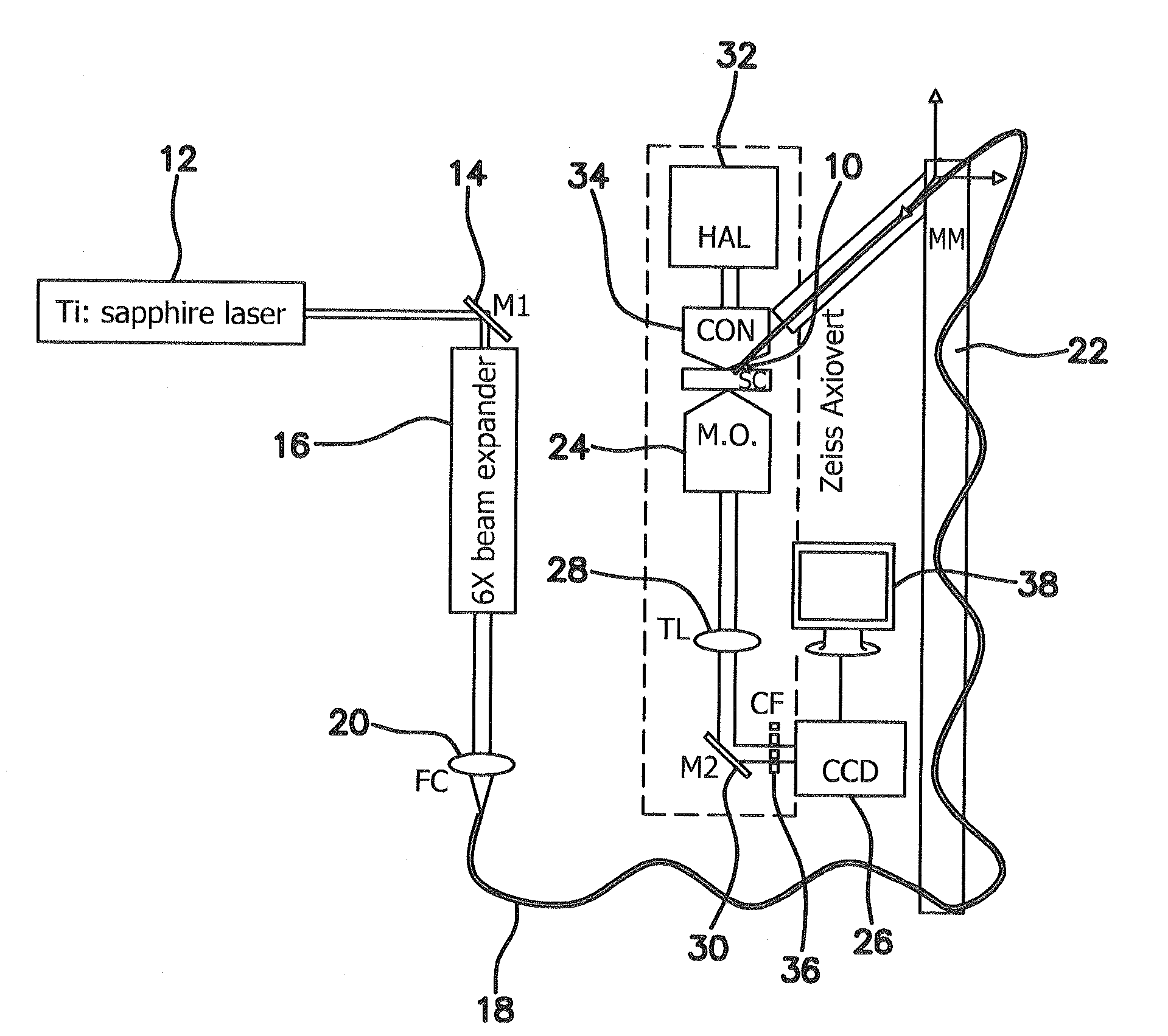
[0012]The illustrated embodiment of the invention is a device and method that uses a spatially shaped laser beam (generated simply by
insertion of a
cylindrical lens) for manipulation, micro-dissection, alteration /
ablation, and excitation of living or nonliving microscopic objects with high throughput. To achieve the spatial shaped laser beam, the N2 pulsed UV-laser beam was focused to a linear profile by use of a
cylindrical lens. For optical
trapping of the objects in suspension, a CW NIR laser beam was also aligned with the
UV laser scissors beam by use of a dichroic mirror. This elongated UV (and or NIR) laser beam was relayed to the back aperture of a high NA Zeiss
microscope objective via the epi-
fluorescence port. The
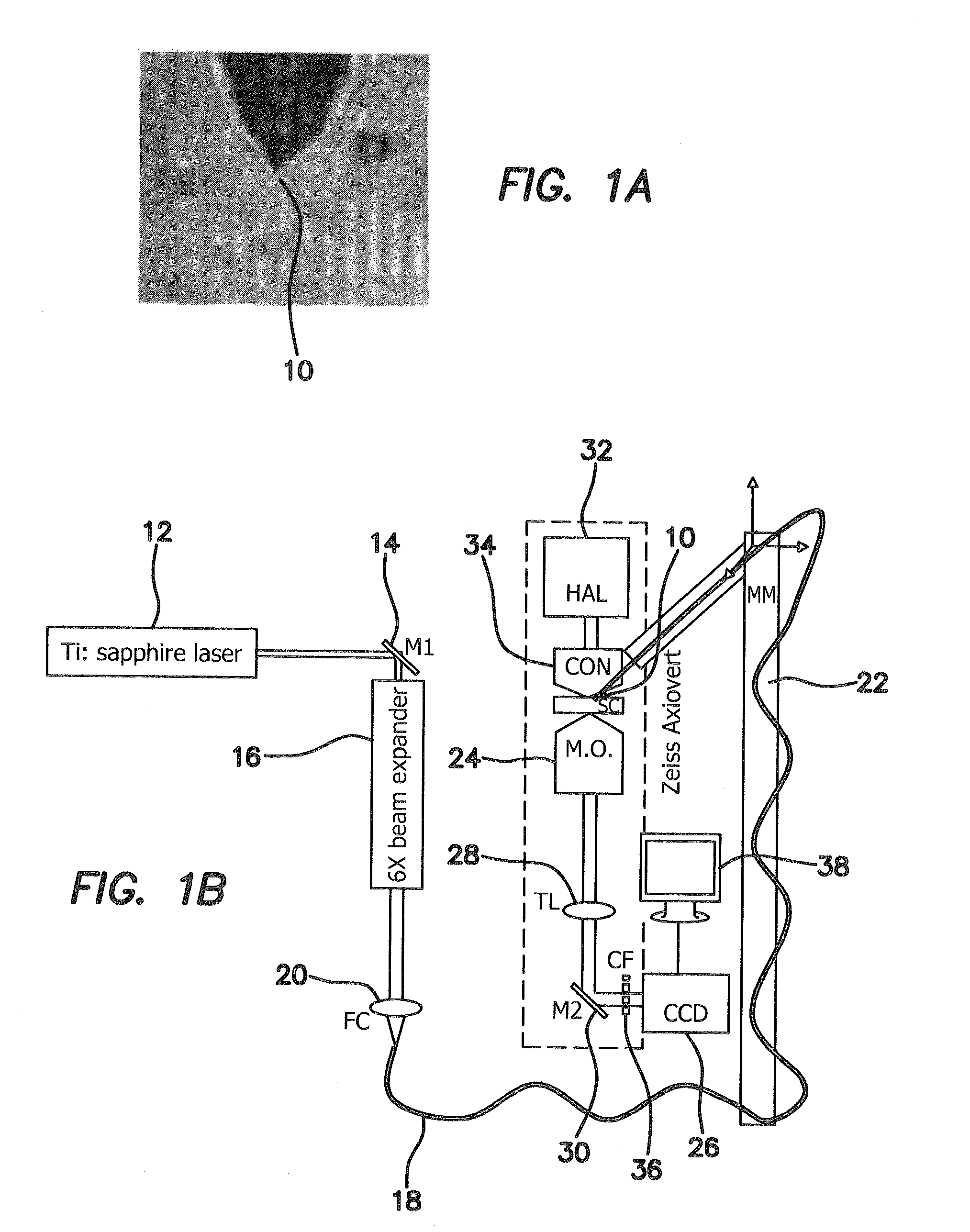
cylindrical lens was mounted on a motorized rotational stage so as to enable controlled orientation of the direction of elongation in the x-y plane. A variable aperture (VAP) was placed at the focal plane of the cylindrical lens in order to control the length of the focused spot. For
fluorescence imaging, light from a mercury lamp was collimated and coupled to the
microscope by use of a dichroic mirror that reflects UV (and or NIR), but transmits the visible region of the mercury spectrum. The
excitation filter was placed in a
filter wheel and another dichroic mirror was used to reflect the
fluorescence excitation light, the 337 nm laser scissors beam and optionally the laser
tweezers beam. This dichroic mirror transmitted the emitted fluorescence as well as the transmitted
halogen light from the sample in the
sample chamber. The emission filter also blocked the
UV laser scissors and NIR laser
tweezers beam. The dimensions of the generated elliptical focused spot determine the length over which the object(s) can be dissected and / or trapped. Use of a 50 mm
focal length cylindrical lens in our setup led to a focal spot length of ˜40 / μm at the object plane. The intensity pattern of the elliptically focused
UV laser at the focal plane of the objective was monitored by fluorescence excitation of a suitable dye on a coverslip. Control of energy / pulse was achieved by orientation of a
polarizer in the beam path. Predetermined numbers of pulses could be delivered through external triggering of the laser by use of National Instrument's
data acquisition and control board. While the dimension of the line scissors could be easily varied by controlling the size of the aperture (VAP), control on orientation of the cuts required rotation of the cylindrical lens manually or by use of a rotatable
stepper motor.
[0013]One of the purposes of the illustrated embodiment of the invention is to develop a device for fast patterned linear
ablation of microscopic objects which is simple to operate and provides high throughput in a uniform manner.
[0015]The fundamental principle exploited is that by shaping the
pulsed laser beam into a linear profile via a cylindrical lens, line scissors can be achieved that provides simple and fast line patterned ablation as compared to scanning the beam with motorized stage or
scanning mirror. While the dimension of the line scissors can be easily varied by controlling the size of a circular aperture in the beam path, control of orientation of the line cuts requires rotation of the cylindrical lens achieved by use of a rotating
mount.
[0016]The advantages of the present invention include (1) fast patterned linear ablation of microscopic objects, (2) simple operation, (3) high throughput in a uniform manner, and (4) less-expense. Uses of the invention include, but are not limited to: (i) induction of
DNA damage, (ii) dissection of neuronal axons, (iii) micro-dissection of organelles such as microtubules, (iv)
thinning of
zona pellucida for use in
reproductive medicine, (v) optoporation of exogenous material into cells.

