
System and method for monitoring cardiomyocyte beating, viability and morphology and for screening for pharmacological agents which may induce cardiotoxicity or modulate cardiomyocyte function

a cardiomyocyte and monitoring system technology, applied in the field of cell-based assays, can solve the problems of affecting drug discovery and development projects, increasing their cost, and lack of reliable monitoring monitoring methods, so as to improve the efficiency of potential throughput, improve cell characterization, and improve the effect of characterization

Active Publication Date: 2011-02-17
AGILENT TECH INC
View PDF0 Cites 33 Cited by
  • Summary
  • Abstract
  • Description
  • Claims
  • Application Information

AI Technical Summary

Benefits of technology

[0020]The present invention discloses devices, systems and methods for performing extracellular recording of excitable cells, such as cardiomyocytes, in vitro. Specifically, the devices and methods provide improved characterization of excitable cells in response to compound administration to assess cardiotoxic effects, while increasing efficiency of throughput for potential drug candidates. The invention further provides improved characterization of cells during differentiation processes related to development of cardiomyocytes.
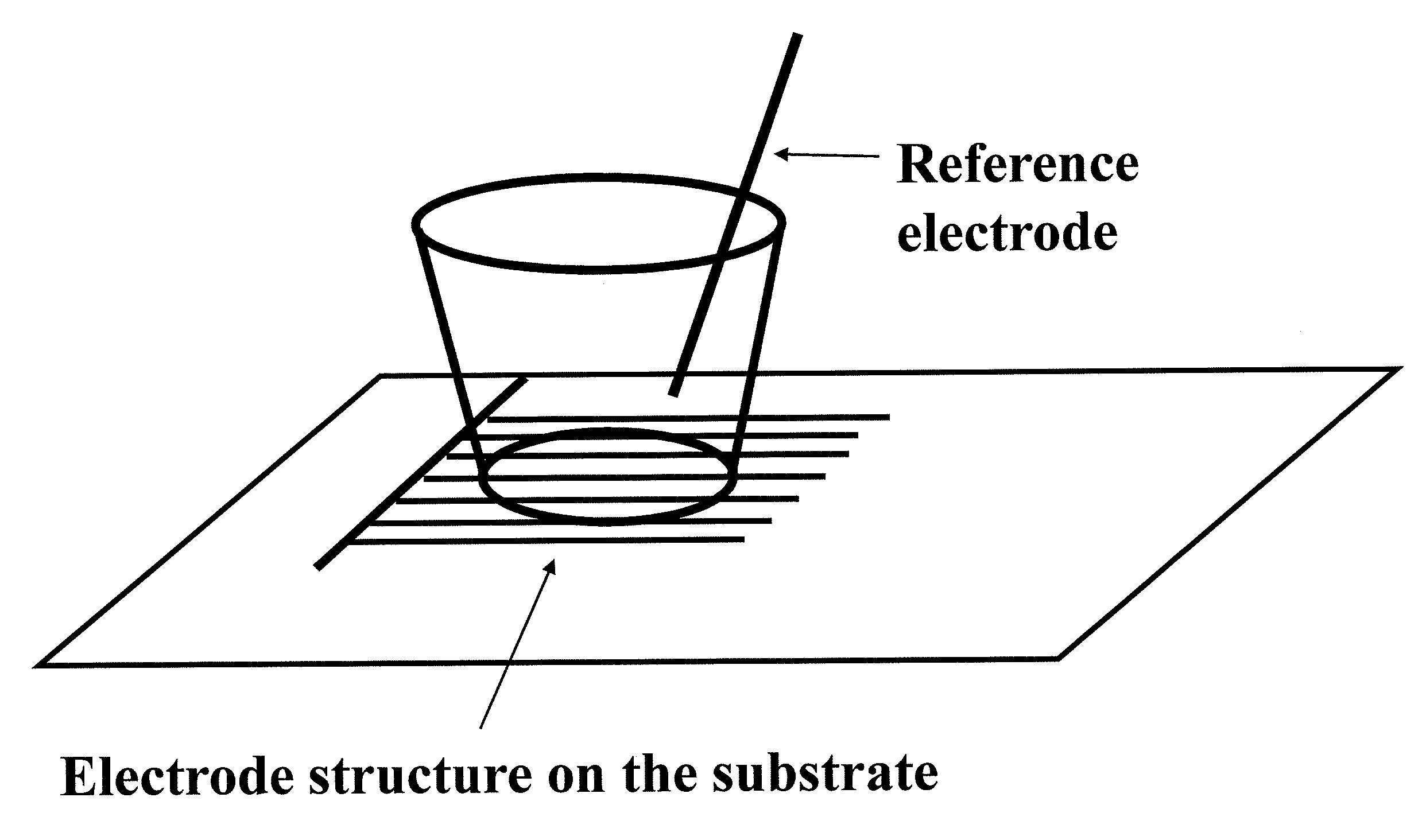
[0024]In some embodiments the extracellular recording electrode is a unitary or unbranched electrode and may be of a simple geometry such as a circle, a square and the like. In other embodiments, the extracellular recording electrode has a branched configuration, which may permit extracellular recording over a larger cell population. Increasing the measured cell population for extracellular recording may increase measurement accuracy or reproducibility of the recorded signals. In one exemplary embodiment, a device for performing extracellular recordings of excitable cells in vitro includes a) a nonconductive substrate; b) one or more wells on the substrate; c) one or more electrode structures fabricated on the substrate, each electrode structure associated with one of the one or more wells; and d) one or more reference electrodes external to the substrate, each of which can be inserted into one of the one or more wells, wherein for each of the one or wells, the corresponding electrode structures and reference electrodes form an electrode pair. In further embodiments the substrate has a surface suitable for attachment of excitable cells, wherein the attachment of excitable cells on the substrate can result in a detectable extracellular recording potential between each electrode pair. In a further embodiment, which is also a preferred embodiment, each electrode structure comprises multiple electrode elements. In another preferred embodiment, each electrode structure comprises multiple electrode elements, forming half of an interdigitated electrode array. In another preferred embodiment, each electrode structure comprises multiple electrode elements which have different electrode element shapes, including rectangular shape, or sinusoidal shape, or circle-on-line shape.
[0031]In present invention, we describe label-free methods for monitoring cardiomyocytes in vitro. In one aspect of the present invention, extracellular-recording measurement, or parallel impedance and extracellular recording measurement of cardiomyocytes on microelectrodes is used to non-invasively monitor cardiac cell viability in vitro in long term experiments. This method will allow the continuous monitoring of cardiomyocyte viability overtime and can monitor the interaction of compounds which ultimately result in promoting loss of cardiomyocyte viability.
[0037]The system of the present invention having the capability of parallel impedance-based monitoring as well as extra-cellular recording based monitoring of cardiomyocytes fills a major technological gap in monitoring cardiomyocytes in vitro. At present to our knowledge there are only a few technologies that can monitor cardiomyocyte population function in vitro, especially with regards to cardiotoxicity and are limited in their throughput. In addition to functional monitoring of cardiomyocyte beating the current invention offers several other major benefits which are worth discussing. Among these include the impedance system described here can monitor cardiomyocytes for short durations, milliseconds to minutes and long durations, several hours to days. Therefore, both short term and long term effects of drugs not only on cardiomyocyte beating, but viability and changes in morphology and adhesion can also be assessed. This feature is especially important because certain compounds such as β-2 adrenergic receptor agonists, well known and characterized modulators of heart function in vivo and in vitro, can induce long term hypertrophic responses in cardiomyocytes, which is associated with elongated morphology of the cells. In addition, the systems of the present invention can also record cardiomyocyte action potentials or field potentials, allowing detailed electrophysiological analysis and assessment of cardiomyocytes for relatively short duration at a time.

Problems solved by technology

Cardiotoxicity has been cited as the reason for 30 percent of all failed drug compounds during development and is a major cause of compound attrition.
The late detection of cardiotoxic side effects caused by pharmacological compounds can impede drug discovery and development projects, and consequently increase their cost.
The core of the current issue in pharmacological safety assessment and drug development is the lack of a reliable screening methodology capable of monitoring potential drug-mediated cardiotoxicity and distinguishing between different modes of cardiotoxicity.
This approach is extremely labor-intensive, time consuming and costly and at the same time not very amenable to the high throughput demands of pharmaceutical industry.
Use of this method in high throughput requires automation of patch clamping in array format, which even though is available in last several years but is not yet widespread.
The other issue with this approach is that cardiac toxicity may occur by other mechanisms which can easily be missed by this type of targeted approach.
High throughput techniques for short term functional characterization of ion channels and other targets in cardiomyocytes has been rather challenging and limited.
The systems such as automatic patch clamp instrumentation that are available can monitor a single cardiomyocyte at a time and with very limited throughput.
Accordingly, while there exists different approaches to assess cardiotoxicity of various compounds, each suffers from one or more drawbacks.

Method used


Image

  • System and method for monitoring cardiomyocyte beating, viability and morphology and for screening for pharmacological agents which may induce cardiotoxicity or modulate cardiomyocyte function
  • System and method for monitoring cardiomyocyte beating, viability and morphology and for screening for pharmacological agents which may induce cardiotoxicity or modulate cardiomyocyte function
  • System and method for monitoring cardiomyocyte beating, viability and morphology and for screening for pharmacological agents which may induce cardiotoxicity or modulate cardiomyocyte function

Examples


example 1

Extra-Cellular Recording on a Disc-Shaped Microelectrode Array

Medium and Reagents

[0239]The standard cell culture medium was Cor.At® Culture Medium (AXIOGENESIS AG, Cologne, Germany) supplemented with 5% of fetal bovine serum (Hyclone, Logan, USA), 100 μg / ml of puromycin. Fibronectin from bovine plasma (1 mg / mL solution, Sigma, St. Louis, USA) was used as coating (4 hrs or overnight) material before plating the mouse embryonic stem cell. E4031, a class III antiarrhythmic drug which is a specific antagonist for hERG and EAG like channels was obtained from Sigma (St. Louis, USA).

Cardiomyocyte Preparation

[0240]Cardiomyocytes (Cor.At®, AXIOGENESIS AG, Cologne, Germany) are derived from transgenic mouse embryonic stem cells. Each vial contains 1 million viable Cor.At® cardiomyocytes (>99.9% pure). These highly purified cardiomyocyte maintain the phenotype of adult mouse cardiomyocyte and express cardiac-specific connexin-43, which is an indication of the ability for excitation-contraction...

example 2

Extra-Cellular Recording Using One Electrode Structure on the Substrate and One External Reference Electrode

[0246]FIG. 22 shows an extra-cellular field potential recording of mouse stem-cell derived cardiomyocytes obtained using a device of the present invention. The circle-on-line electrode array (with circle diameter 90 micron, line width 30 micron, the electrode gap distance 20 micron, see FIG. 13F for representation of circle-on-line electrodes) consisting of two electrode structures are fabricated on a glass substrate. During extra-cellular field potential recording, one circle-on-line electrode is used as a recording electrode and an external wire electrode is inserted into the well serving as a reference electrode. Cell preparation, reagents, cell culture and extracellular recording setup are the same as those described in to example 1.

[0247]FIG. 22A shows a field potential (after an amplification of 10,000) before treatment for a 3-day cardiomyocyte culture in a well contain...

example 3

Parallel Cell-Impedance Measurement and Extra-Cellular Recording for Cardiomyocytes Prior to and after Qunindine Treatment

[0249]FIG. 23A through 23E show another example of field potential change for cardiomyocytes at different time points before and after the treatment with 3 uM Quinidine, as recorded from a 3-day cardiomyocyte culture in a well containing circle-on-electrodes. Similar to the configuration for data in FIG. 22, an external wire electrode was inserted to the well serving as a reference electrode. Before treatment, cardiomyocytes were beating at a rate of ˜72 beats per minute. As shown in FIG. 23 B through D, corresponding to the field potential recorded at 10 seconds, 50 seconds, 3 minutes and 9 minutes after treatment with 3 uM Quindine, Quindine has a dramatic effect on the field potential of the cardiomyocytes.

[0250]In parallel, impedance of the cardiomyocytes in such a well is monitored. FIG. 24A shows the impedance spikes as monitored on the circle-on-line elect...


PUM

PropertyMeasurementUnit
diameteraaaaaaaaaa
diameteraaaaaaaaaa
timeaaaaaaaaaa

Abstract

Devices and methods for performing extracellular recording of cells, such as excitable cells, cardiomyocytes, and cardiomyocyte precursor cells is provided. An exemplary device includes a nonconductive substrate forming or provided as a base of one or more wells; a recording electrode positioned on the substrate within the well, wherein the recording electrode is accessible to cells when a cell sample is added to the device; and a reference electrode positioned within the well in a cell-free zone, the cell-free zone characterized as free from contact with cells when the cell sample is added to the device, thereby preventing contact between cells and the reference electrode.

Description

CROSS REFERENCE TO RELATED APPLICATIONS[0001]This application is a continuation in part of U.S. patent application Ser. No. 12 / 435,569, filed May 5, 2009, entitled, “Label free monitoring of excitation-contraction coupling and excitable cells using impedance based systems with millisecond time resolution,” which claims priority to U.S. provisional patent application Ser. No. 61 / 191,684, filed Sep. 11, 2008, and U.S. provisional patent application Ser. No. 61 / 126,533, filed May 5, 2008; the contents of each are herein incorporated by reference in their entirety.[0002]This application also claims priority to U.S. patent application Ser. No. 61 / 323,782, filed on Apr. 13, 2010, entitled, “Impedance based monitoring of cardiomyocytes”; U.S. patent application Ser. No. 61 / 310,557, filed Mar. 15, 2010, entitled, “Impedance based monitoring of cardiomyocytes”; and U.S. patent application Ser. No. 61 / 175,566, filed May 5, 2009, entitled, “System and method for monitoring cardiomyocyte beatin...

Claims


Application Information

Patent Timeline
Patent Type & Authority Applications(United States)
IPC IPC(8): C12Q1/02C12M1/34
CPCG01N33/48735
Inventor WANG, XIAOBOOUYANG, WEILI, NANXI, BIAOABASSI, YAMA A.XU, XIAO
Owner AGILENT TECH INC
Learn More