Probe reagent for measuring oxidative stress

a technology of oxidative stress and probe reagents, which is applied in the direction of oxidoreductases, in-vivo radioactive preparations, depsipeptides, etc., to achieve the effect of efficient measurement of ros, prolonged integration time, and poor permeability across cell membranes

Inactive Publication Date: 2011-08-25
JAPAN SCI & TECH CORP +1
View PDF7 Cites 9 Cited by
  • Summary
  • Abstract
  • Description
  • Claims
  • Application Information

AI Technical Summary

Benefits of technology

[0009]An Example of a method of measuring ROS include a method using a chemiluminescent reagent such as isoluminol. This method is to measure a luminescent signal that is accompanied with an oxidation reaction of the reagent with the ROS. This reaction is widely used as a method of evaluating an immunological activity of leukocytes in order to measure ROS released during the attack against a foreign substance that enters a living body (Dahlgren, C. and Karlsson, A., J. Immunol. Meth., 232, 3-14, 1999). Unfortunately, isoluminol has a poor permeability across cell membranes. So, the measurement of ROS released outside cells can be performed, but the method is unsuitable for the measurement of ROS generated inside the cells. In addition, the intensity of this luminescence is weak. Since the method requires a large number of cells for this measurement and a prolonged integration time even for a high-sensitive detector, it is difficult to efficiently measure the ROS.
[0010]A measurement using an organic fluorescent dye has been also carried out. The dye, 2′,7′-dichlorodihydrofluorescein, dihydroethidium, and the like are widely used (Munzel, T., et al., Arterioscler. Thromb. Vasc. Biol., 22, 1761-1768, 2002). Although these are nonfluorescent compounds under non-oxidative condition, these compounds become fluorescent by changing their structures due to an oxidation reaction with ROS. This fluorescent intensity indicates to an amount of the ROS in situ. These reagents can be introduced in cells by passing through cell membranes. However, since these reagents cannot be selectively localized in a particular organelle inside the cells, it is difficult to measure the ROS at a particularly localized area. Moreover, the fluorescence emission of these dyes is irreversible, and these cannot repeat the measurement because once the oxidized dye lose the reactivity with the ROS any more.
[0011]Recently, a fluorescent protein has been isolated from Aequorea victoria. One improved in its feature is widely used as a probe reagent for measuring a physiological activity in cells. A probe reagent that is composed of proteins can be used as a form of the gene. By being incorporated into cells as a plasmid, the expressed probe reagent can be used for the measurement. This method also allows only a specific cell in a living body or a specific organelle in cells to limitedly express the probe reagent, and makes it possible to measure the physiological activity at a more limited region.
[0012]There have been developed oxidative stress probe reagents using fluorescent proteins. Such reagents have a structure in which the fluorescent protein is fused to a marker protein that detects ROS. A protein that changes its structure due to the reaction with the ROS is used as the marker protein. The change in the structure is designed to change the intensity and wavelength of the fluorescence emitted by the fluorescent protein. For example, a probe reagent, what is called “Hyper”, uses a protein reacting specifically with hydrogen peroxide as a sensor protein, and the protein has a structure attached to a fluorescent protein, YFP. The reaction of hydrogen peroxide with the sensor protein results in change in fluorescent intensity of YFP (Belousov, V. V., et al., Nature Methods, 3, 281-286, 2006 Rev., 82, 47-95, 2002). Another example includes an application using fluorescence resonance energy transfer. This probe reagent has a structure in which a sensor protein is sandwiched between two types of fluorescent proteins having different fluorescent wavelength. The change in the structure resulting from the reaction of the sensor protein with the ROS alters the distance between the fluorescent proteins at each end. This change in distance gives a change in the efficiency of the energy transfer. This reagent can monitor ROS by measuring a change in fluorescence spectra (JP Patent Publication (Kokai) No. 2005-95171A). However, the fluorescent change that is based on the structural change of the sensor protein of the reagent is small, and the detection becomes difficult when ROS is slightly produced in particular.
[0013]There is a protein that is expressed for a protective response against oxidative stress when cells are exposed to the stress. The oxidative stress of the cells can be detected by measuring an increase in expression of such a protein. In an example that has measured oxidative stress in muscle, proteins such as TNFα and pIκB have been measured as indicators of the oxidative stress (JP Patent Publication (Kokai) No. 2006-162346A). In this method, the intended proteins are measured using a biochemical technique such as Western blotting. Specifically, proteins in cells are extracted by disrupting the cells and then the proteins are subjected to separation. Thus, because the troublesome procedures are required in the prior art, it is impossible to measure the ROS quickly and continuously in living cells
[0014]As described in Background Art, under the situation in which various disadvantageous effects by ROS are discussed, precisely measuring a change in distribution and amount of ROS generated in living bodies is expected to give an important insight in respect to a mechanism of disease and ageing in which the ROS is involved, and to further contribute largely to the development of the effective prophylaxis and treatment. However, the above-mentioned problems have been pointed out in currently used or proposed methods of measuring ROS that evoke oxidative stress. Hence, there are demands for techniques that can efficiently measure behavior of ROS in vivo to overcome these problems.

Problems solved by technology

However, the above-mentioned problems have been pointed out in currently used or proposed methods of measuring ROS that evoke oxidative stress.

Method used


Image

  • Probe reagent for measuring oxidative stress
  • Probe reagent for measuring oxidative stress
  • Probe reagent for measuring oxidative stress

Examples


example 1

Design of a Probe Reagent

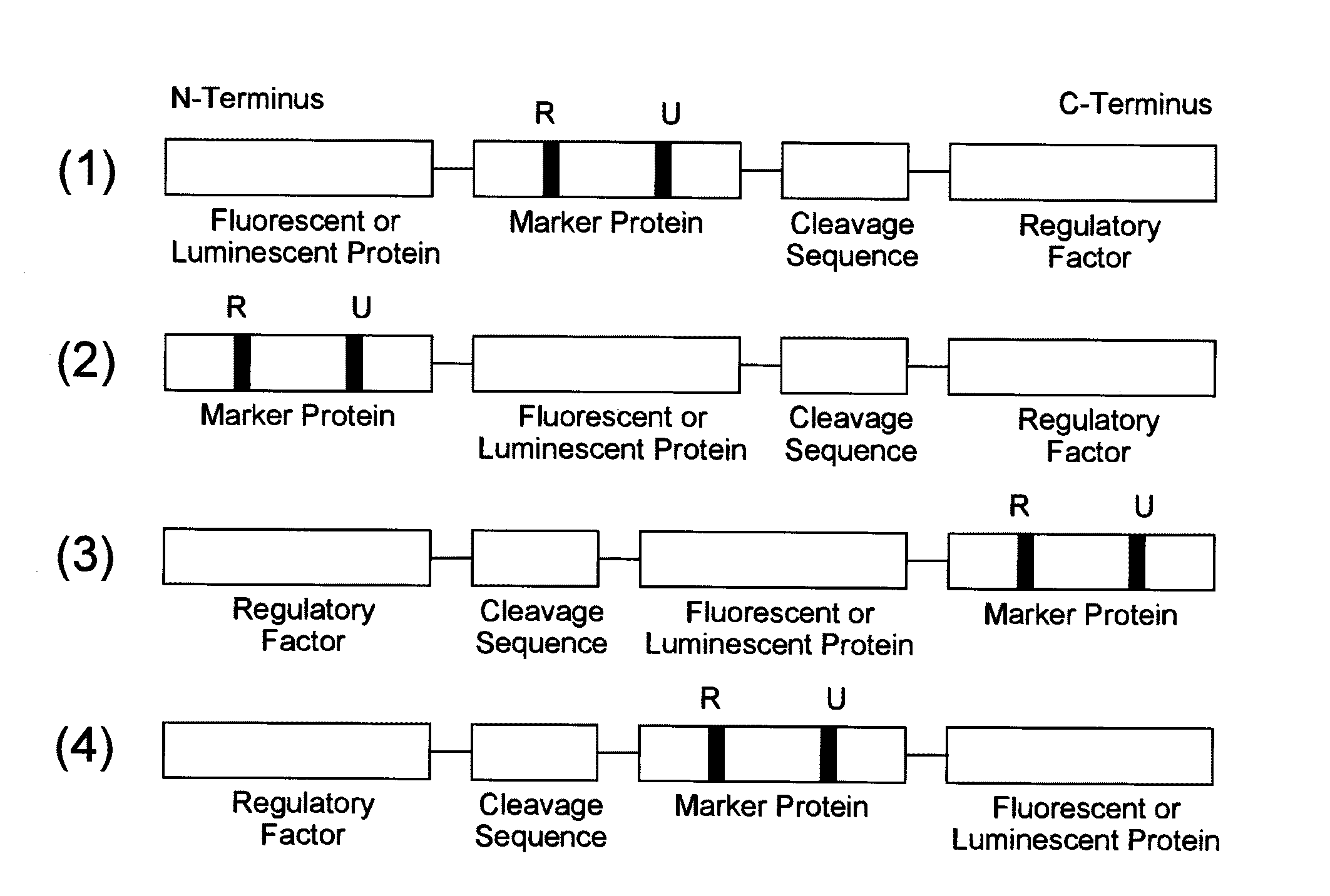
[0149]As an example of a probe reagent having the feature described above, a probe reagent for measuring oxidative stress was prepared using mKO2 as a fluorescent protein, Nrf2 as a marker protein, and Keap1 as a regulatory factor. In this reagent, as oxidative stress increased, the degradation of the probe reagent reduced, thereby increasing the fluorescent intensity.

[0150]First, intrinsic kinetics of Nrf2 and Keap1 in a cell is explained. Nrf2 responds to oxidative stress in a cell, and functions as a transcription factor that induces expression of antioxidative proteins responsible for the defensive functions against this oxidative stress. Under the condition without oxidative stress in a cell, Nrf2 is present in the cytoplasm in the form of binding to Keap1. At this time, Keap1 further binds to a ubiquitin ligase complex, thereby promoting ubiquitination of Nrf2. Since ubiquitinated Nrf2 is degraded by a proteasome that is a protease complex, the increas...

example 2

[0152]Preparation of cDNA

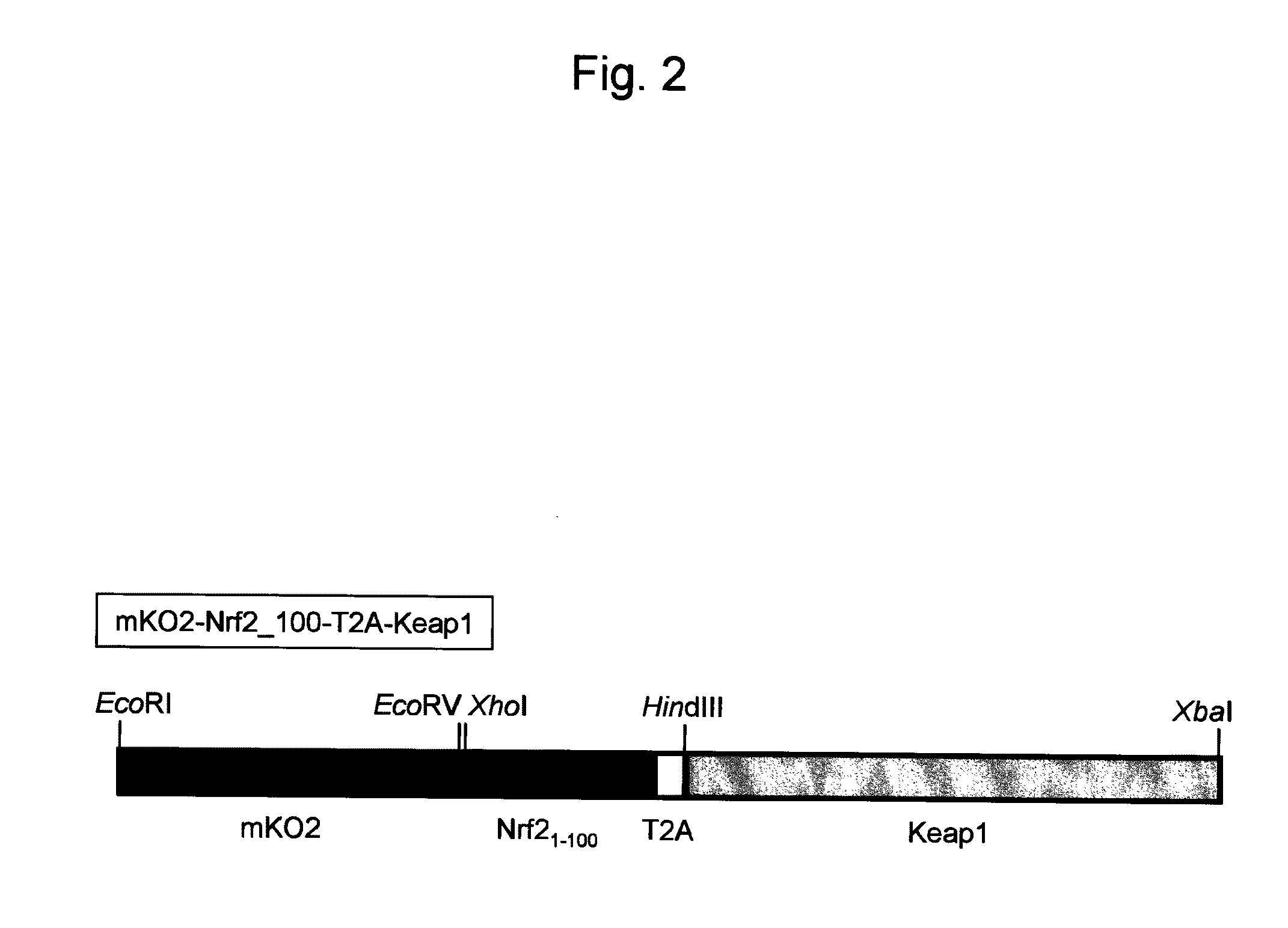
[0153]cDNA encoding a gene of a probe reagent was prepared by the following procedures. This cDNA was inserted into a plasmid for introducing itself into a cell.

[0154]Nrf2 and Keap1 genes were cloned from a human heart cDNA library. Sense primer (SEQ ID NO: 1) and antisense primer (SEQ ID NO: 2), and PrimeSTAR enzyme (manufactured by TAKARA Bio Co., Ltd.) were used to perform a PCR reaction to amplify a nucleic acid encoding the amino acids 1-100 of Nrf2 by using the Nrf2 gene as a template. The sense primer contained a cleavage site for XhoI. The antisense primer contained a nucleotide sequence of T2A peptide and a cleavage site for HindIII. In addition, sense primer (SEQ ID NO: 3) and antisense primer (SEQ ID NO: 4), and PrimeSTAR enzyme were used to perform a PCR reaction using Keap1 gene as a template. The sense primer contained a cleavage site for HindIII. The antisense primer contained a cleavage site for XbaI.

[0155]Both the ends of the PCR product amp...

example 3

Evaluation of a Probe Reagent by Imaging of a Cell

[0157]The prepared cDNA was transfected into HeLa cells that were cultured on a glass bottom dish by using FuGENE HD (manufactured by Roche Applied Science). The medium was replaced by the fresh medium 12 hours after the transfection. After 24 hours, fluorescence was observed with an incubator microscope (Olympus Corporation, LCV 100). This microscope was equipped with a cooled CCD camera so that fluorescent images of a cell were able to be captured over time. Before the experiment, only fluorescence at the background light level was detected even in a cell that expressed the probe reagent. This demonstrates that almost all of the probe reagent in the cell was degraded under stable conditions without being loaded with oxidative stress.

[0158]In order to verify that the constructed probe reagent responded correctly to oxidative stress as intended, diethylmaleate (DEM), which is an oxidative stress inducer, was added at a concentration ...


PUM

PropertyMeasurementUnit
timeaaaaaaaaaa
emission wavelengthaaaaaaaaaa

Abstract

The present invention relates to fluorescent or luminescent probe reagents for measuring oxidative stress in a cell or an organism. Examples of the probe reagents include: a fluorescent or luminescent protein and a marker protein; a fluorescent or luminescent protein, a marker protein, and a regulatory factor; or a fluorescent or luminescent protein, a marker protein, a cleavage sequence, and a regulatory factor. In the probe reagents, the marker protein makes it possible to detect the oxidative stress caused by reactive oxygen species and comprises a regulatory factor-binding site and a ubiquitin-binding site; and the regulatory factor is a protein making it possible to regulate degradation of the marker protein in response to the reactive oxygen species. The present invention also relates to a method of measuring oxidative stress in a cell or an organism, or a method of screening a substance which suppresses or promotes the oxidative stress in a cell or an organism by using the probe reagent.

Description

TECHNICAL FIELD[0001]The present invention relates to fluorescent or luminescent probe reagents for measuring oxidative stress in a cell or an organism.[0002]The present invention also relates to nucleic acids encoding the probe reagents, and vectors comprising the nucleic acids.[0003]The invention further relates to a method of measuring oxidative stress or a method of screening an oxidative stress-regulating substance in a cell or an organism by using the probe reagent or vector.[0004]The present invention further relates to a kit for measuring oxidative stress or screening an oxidative stress-regulating substance, comprising the probe reagent, nucleic acid, or vector.BACKGROUND ART[0005]Recently, an excessive oxidation reaction, what is called oxidative stress, occurring inside cells has been found to exert harmful effects in living bodies. This oxidation reaction is evoked by a molecular, what is called “reactive oxygen species (ROS)” which has a strong oxidative power generated...

Claims


Application Information

Patent Timeline
Patent Type & Authority Applications(United States)
IPC IPC(8): A61K49/00C12N15/63G01N33/53C12Q1/68C07K19/00C12N9/02C07H21/04C07H21/02
CPCC07K14/00C12Q1/28G01N21/6428G01N21/77G01N33/5008G01N33/5014A61K51/088G01N2500/10A61K51/08A61K49/14A61K49/0056A61K49/0017G01N33/84
Inventor MIYAWAKI, ATSUSHISASAKI, KAZUKI
Owner JAPAN SCI & TECH CORP
Why Eureka
Try Eureka