Equipment for infrared vision of anatomical structures and signal processing methods thereof

a technology of infrared vision and anatomical structures, applied in the field of photonics, image acquisition, image processing, vision enhancement and information extraction, can solve the problems of affecting the surgical field, and reducing the surgical field, so as to improve the surgeon's orientation, facilitate the viewing of the vasculature, and reduce the surgical procedure. the effect of invasiveness

Inactive Publication Date: 2011-12-01
FCRB +2
View PDF4 Cites 190 Cited by
  • Summary
  • Abstract
  • Description
  • Claims
  • Application Information

AI Technical Summary

Benefits of technology

[0018]One aim of the present invention is to solve the technical difficulties that currently exist in the surgery for complications of monochorionic twins pregnancies, in order to locate and identify blood vessels coagulated by the use of a laser source for therapeutic purposes, achieving improved safety and repeatability in such surgical operations.
[0029]The ability to navigate or to view vascular characteristics is greatly improved by means of the present invention since the surgeon, in addition to the standard obtained visualization, has at least the above-referred five options or new ways of visualization.
[0033]A further object of the invention is an equipment wherein, alternatively and with the aim to simplify the multimodal images, the light sources are coupled to the video systems by using different channels of the endoscope. The use of more than one channel allows the definition of different light paths, thus simplifying the employment of optical elements in each channel.
[0044]It is important to highlight that the system has the advantage that it does not need contrast agents to carry out the task of representing the vascular map, being that feature an essential property to perform foetal surgery (avoiding the use of substances potentially dangerous for the fetus when administered in a considerable amount or during a long period of time) and reducing, in general, the invasiveness of the rest of the surgical procedures.
[0045]Additionally, the equipment of the invention includes a device that generates a global map of the patient vascular surgical sites; in particular, in operations of complications in monochorionic twins pregnancies it facilitates viewing the vasculature of the placenta thus achieving a better surgeon's orientation.
[0046]The equipment of the invention is also able to report functional information on the anatomical structures giving an enhanced view with rich and relevant data of the field that is being imaged with not only spatial or temporal dependent information but also with information on the functional performance of the anatomical structure such amount of oxygen level in tissues or vessels and to distinguish between arteries and veins amongst others.

Problems solved by technology

Despite endoscopy is an advanced surgical technique which greatly minimizes surgical procedure risks, some problems still persist, like the risk of accidentally cutting a mayor blood vessel.
Even though the use of minimally invasive endoscopy offers clear advantages to the patient, it imposes certain disadvantages to the physicians, like a constrained vision of the surgical field or a poor contrast and / or definition.
The need to accurately identify blood vessels under such conditions may represent a serious challenge to any surgeon, rendering surgery extremely dependent on the surgeon's experience, resulting in prolonged operations due to bleeding episodes, and occasionally resulting in major hemorrhagic complications.
That limitation to a purely diagnostic use is because the injected dye rapidly dissolves into the bloodstream, and the five minutes that it lasts would not allow using it in therapy or surgery, which require much longer duration.
However, another disadvantage with respect to the present invention is that it requires a contrast agent to enhance the visualization of blood vessels, both superficial (that is apparent to the naked eye) and submucosal (running under the mucosa and therefore normally non visible to simple inspection), whereas the present invention makes use of an algorithm to perform such feature, making it less invasive and hence more appropriate in the surgery field.
In addition to the fact that IRI is not designed as an assistance vision system for surgery, the said technique does not provide other additional features like: image segmentation, image mapping of the surgical field, or functional assessment of blood vessels (by obtaining relevant information such as, for example, the amount of oxygen carried by the blood or the coagulation state of the vessels).
A mayor disadvantage of this system compared with the present invention is that it requires of a contrasting agent or dye to be injected to the blood stream of the patient, consequently rendering this system non-usable in any surgical procedure for the reasons above mentioned, namely the rapid dilution of the contrast agent into the bloodstream with the consequent inefficiency to assist physicians in carrying out therapy or surgery.
A further weakness is that it only focuses on the ability of detecting blood vessels and does not provide specific embedded methods as, for example, image segmenting, image mapping and assessing vessel functionality.
A significant disadvantage of said techniques is their technical complexity, since they necessary comprise a scanning unit and a complex optical assembly.
A further disadvantage, due to the small field of view of such small instrument, is that the physician's angle of vision is substantially restricted, thus limiting considerably the feasibility of such system for surgical applications.
A disadvantage of said apparatus is that it can only obtain information to generate an enhanced image for a narrow portion of the area under study; furthermore it does not compose a substantially enhanced image.
The main disadvantage of said device is that it does not include specific image analysis processing intended for segmenting and displaying the information in conjunction with the visual image; it also lacks image reconstruction functions.
Major drawbacks of said patent in comparison with the present invention include the lack of tracking (defined as a signal processing procedure for tracking and localizing of blood vessels between consecutive scenes from the images generated), lack of image mapping, lack of functional analysis such as the analysis of tissue (blood vessel) oxygenation or the lack of use of more than one infrared spectral band.
Additionally, those other state of the art algorithms and platforms are not intended for the service of physicians with the aim of assisting surgical procedures in real time.
Besides, the methods disclosed in the state of the art do not allow to map large vascular areas of interest, and to obtain functional information about vessels which can assist in decision during an operation.

Method used


Image

  • Equipment for infrared vision of anatomical structures and signal processing methods thereof
  • Equipment for infrared vision of anatomical structures and signal processing methods thereof
  • Equipment for infrared vision of anatomical structures and signal processing methods thereof

Examples


Embodiment Construction

[0055]In view of the aforementioned figures and according to the numbering adopted in them, different embodiments of the invention are described hereunder.

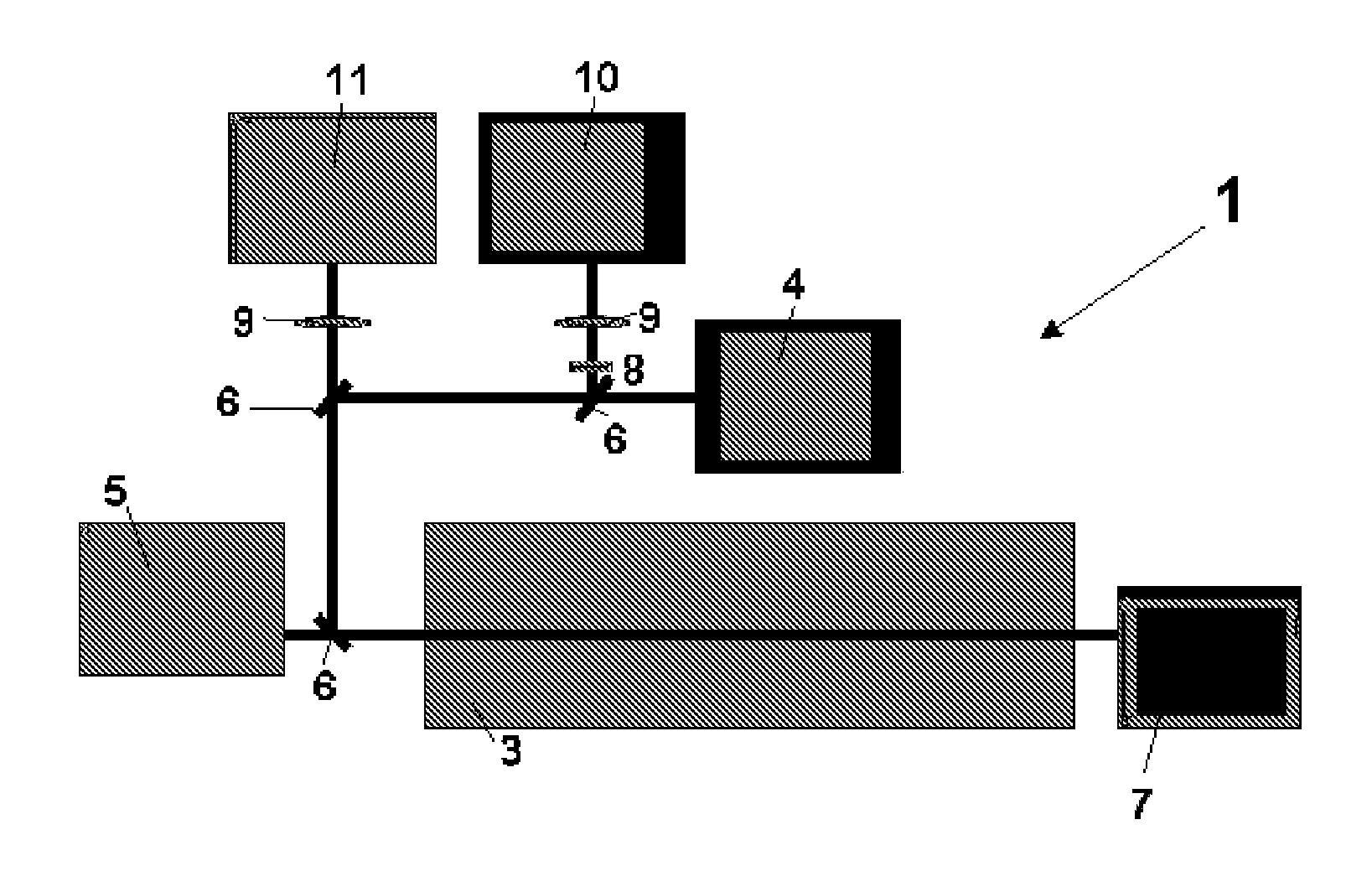
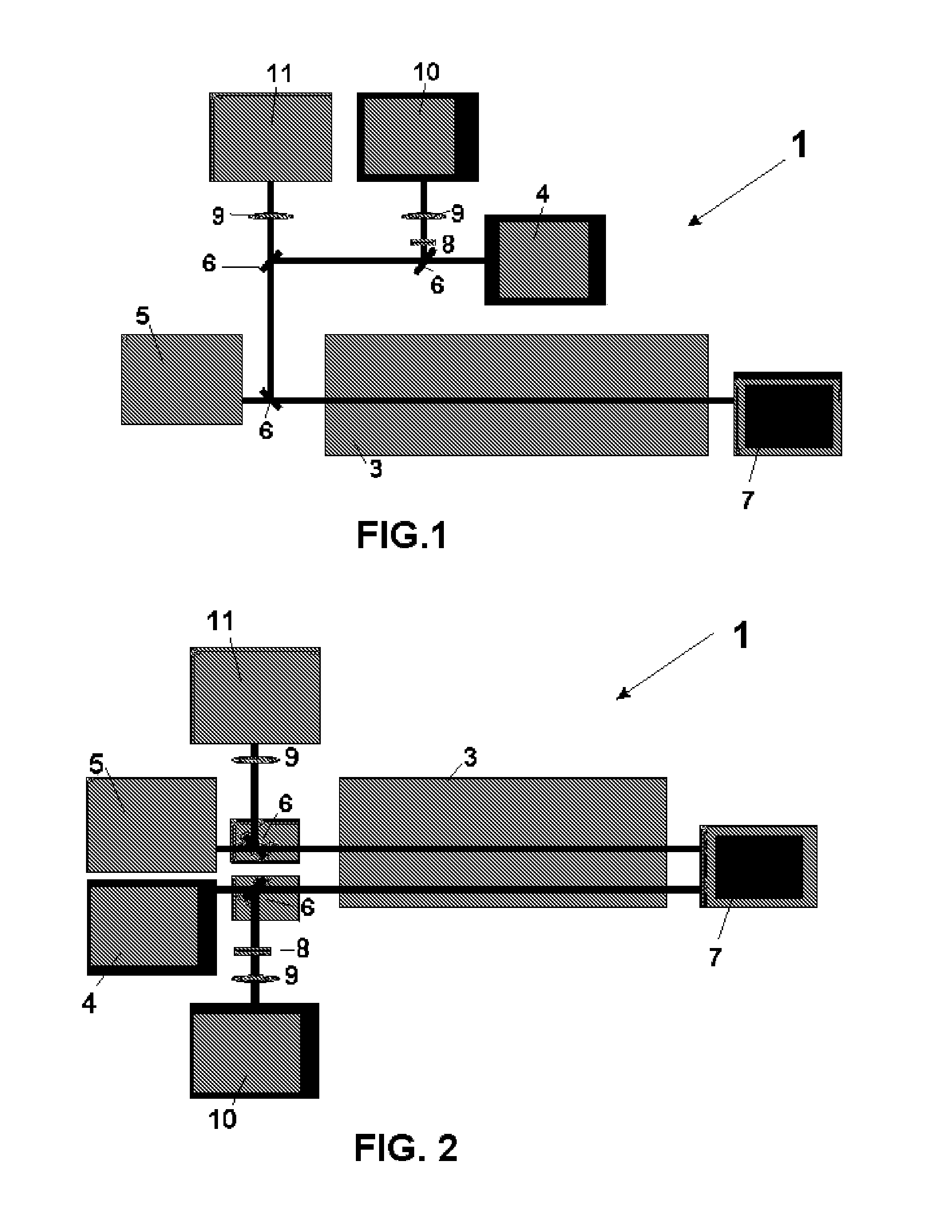
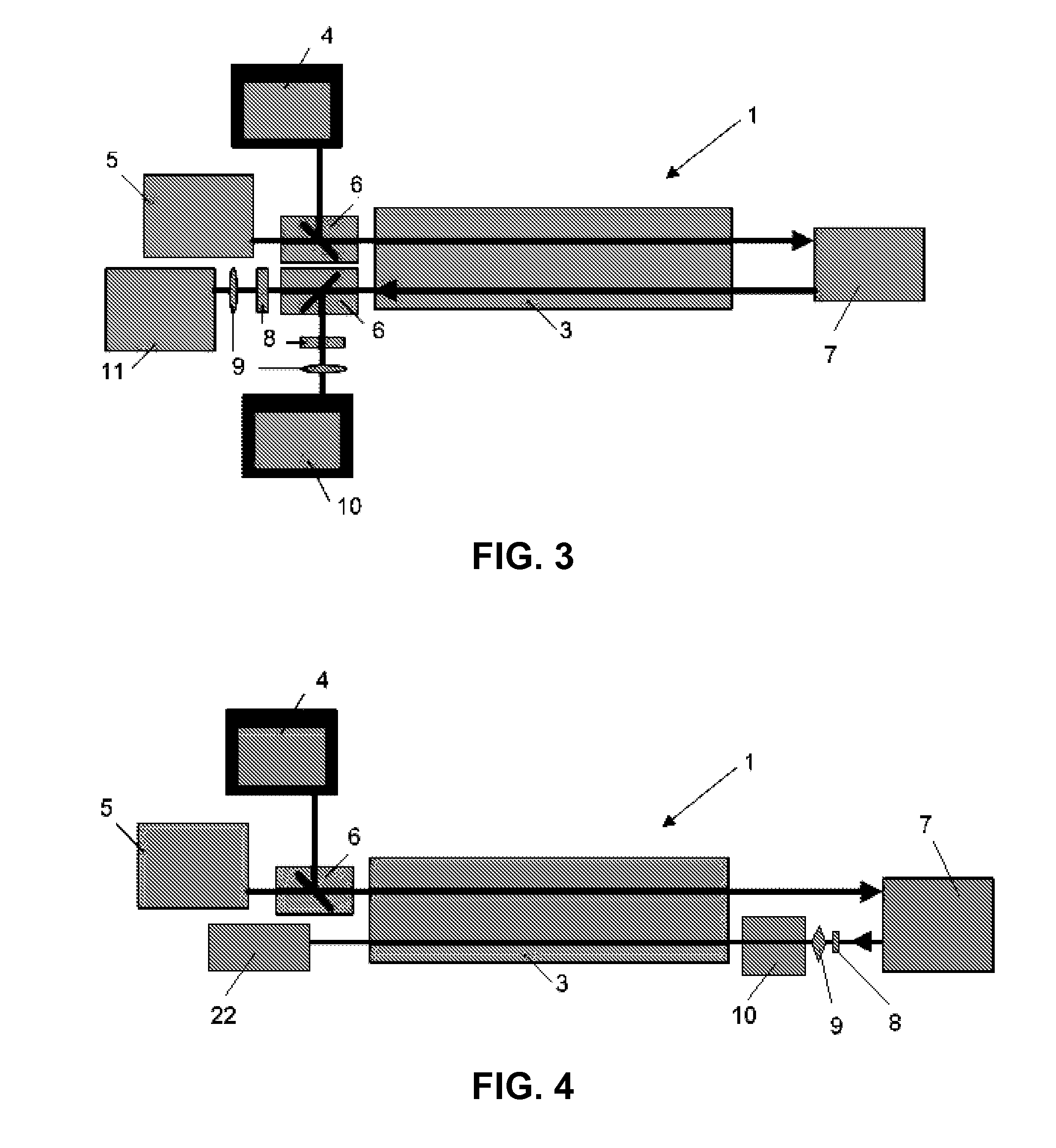
[0056]Thus, as shown in said figures, the equipment comprises a multimodal image acquisition unit (1) and an image processing unit (2).

[0057]The multimodal image acquisition unit (1), whose preferred implementation as shown in FIG. 1 includes an image capturing device, preferably an endoscopic image acquisition device comprising an endoscope, a fetoscope or a laparoscope and additional optical systems, comprising said systems at least one channel from which the video images from the inside of the patient are acquired, and at least one light source to illuminate the observed tissues.

[0058]In a preferred embodiment of the invention, the video channel or channels that are available on the endoscope are coupled to an infrared light source (4) and a white light source (5) or a light source that contain at least three wavelengths within...


PUM


Abstract

Equipment for infrared vision of anatomical structures applicable to assist the physicians in endoscopic, fetoscopic or laparoscopic operations and methods for signal processing to enhance said vision, comprising two units that work together: a multimodal or multispectral imaging unit, constituted by a device comprising an endoscope or fetoscope or laparoscope, and additional optical systems to acquire multimodal images of the interior of the patient's body; and an image processing unit, to which said images are transferred, comprising processing devices with a navigation interface which process said images and display the patient's enhanced anatomical map image and the endoscope location, equipped with hardware and software that apply at least five different vision-enhancing methods, namely normalization, segmentation, tracking, mapping and fusion.

Description

FIELD OF THE INVENTION[0001]The present invention relates to the field of photonics, image acquisition, image processing, vision enhancement and information extraction applied to life sciences, mainly focused on the medical and biomedical fields and specially, but not exclusively, on the fields of endoscopy, fetoscopy and laparoscopy.[0002]More specifically, the invention relates to a medical device or equipment comprising means for acquiring multimodal or multispectral images of a living subject, including illumination means and a digital image processing platform associated to said means, with embedded algorithms to extract and / or enhance specific image information with the objective of assisting physicians in their decisions, for example, when diagnosing, monitoring and / or performing a given therapy or surgical operation.BACKGROUND OF THE INVENTION[0003]In the medical field a number of imaging systems have been devised to display and enhance the visualization of an area under exa...

Claims


Application Information

Patent Timeline
Patent Type & Authority Applications(United States)
IPC IPC(8): A61B1/04
CPCA61B1/00009A61B1/0638A61B1/3132A61B5/0075A61B1/0684A61B5/02007A61B1/00186A61B1/04A61B1/063A61B5/0086A61B1/000094A61B1/044A61B1/046A61B1/07G06T7/00
Inventor GRATACOS SOLSONA, EDUARDAMAT ROLDAN, IVAN
Owner FCRB
Why Eureka
Try Eureka