

Optical mapping of genomic DNA
a technology of genomic dna and optical mapping, applied in the field of polynucleotide mapping with nanometre resolution, can solve the problems of inability to reliably assembly the genome, complicated situation, etc., and achieve the effects of improving the density of targeted (labelled) sites, high degree of confidence in analysis and interpretation of fluorocodes, and improving the precision of determining
- Summary
- Abstract
- Description
- Claims
- Application Information
AI Technical Summary
Benefits of technology
Problems solved by technology
Method used
Image

Examples
example 1
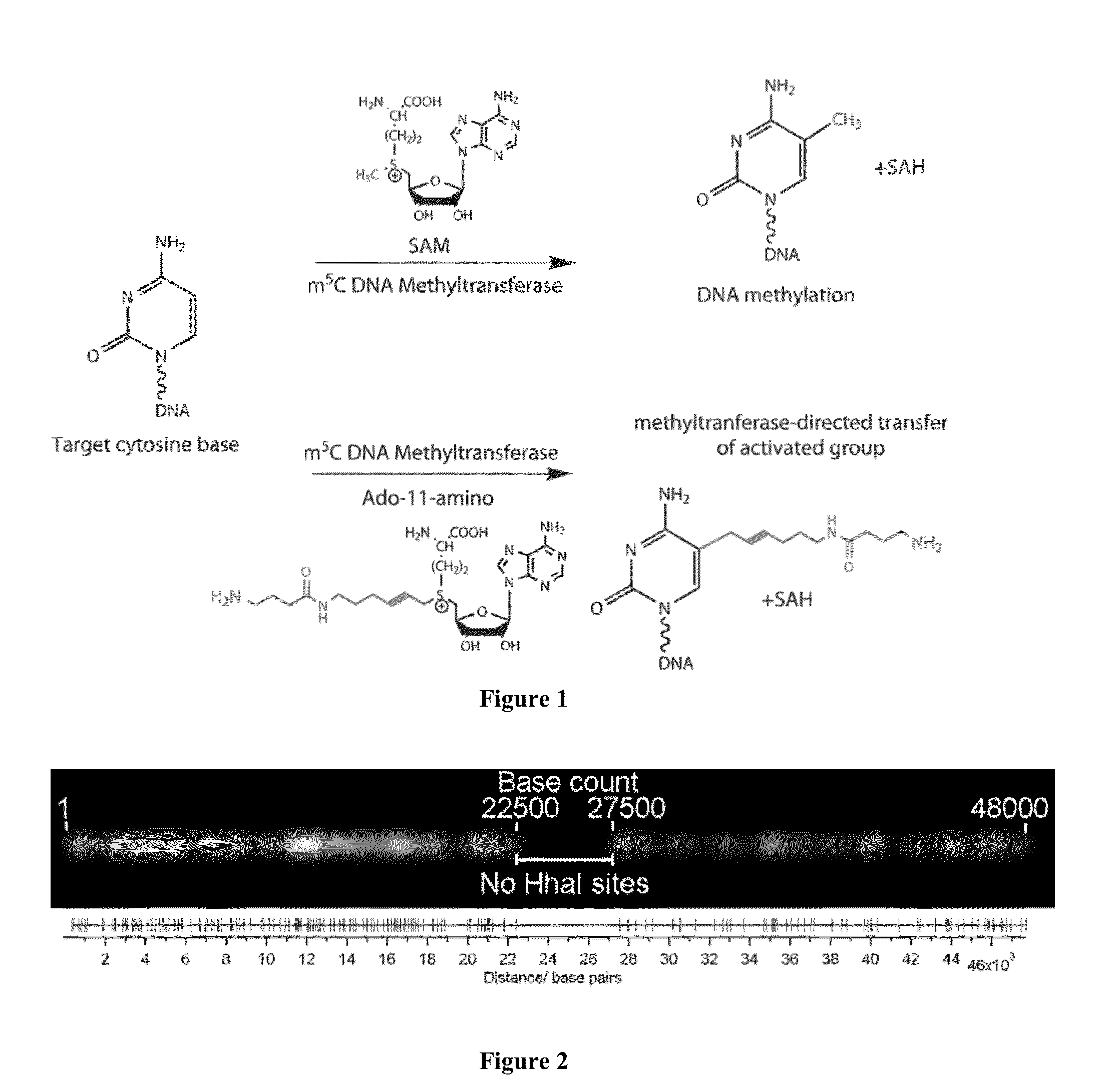
DNA Labeling Using Methyltransferase-Directed Transfer of Activated Groups (mTAG)
[0061]20 μg of X DNA (Fermentas) was incubated with M.HhaI (variant Q82A / Y254S / N304A) (equimolar amount to the target sites) and 20 M synthetic cofactor Ado-11-amino in 4001 of M.HhaI buffer (50 mM Tris.HCl pH 7.4, 15 mM NaCl, 0.01% 2-mercaptoethanol, 0.5 mM EDTA, 0.2 mg / ml BSA) for 30 min at 37° C. The completion of the modification reaction was verified by treating a 101 aliquot with R.Hin6I (Fermentas) and agarose gel electrophoresis. The modified DNA was then incubated with 187 g of Proteinase K (Fermentas) in the M.HhaI buffer supplemented with 0.025% SDS for 1 hour at 55° C. DNA was purified by passing through a 1.6 ml Sephacryl™ S-400 column in PBS buffer followed by isopropanol precipitation. Pellet was dissolved in 0.15 M NaHCO3 (pH 8.3) and incubated with a 75-fold molar access of ATTO-647N NHS ester (ATTO-TEC) for 6 h at room temperature. Fluorophore-labeled DNA was purified and redissolved i...
example 2
Coverslip Preparation
[0062]Coverslips were mounted in a Teflon rack and then washed by sonication in acetone, then 1M NaOH, followed by MilliQ-water (×2). Each sonication was carried out for 15 minutes. Polymethylmethacrylate (PMMA) (0.1% wt / vol) in chloroform was spin-coated (2000 rpm) onto the cleaned coverslips. The PMMA was subsequently annealed to the coverslips by baking at 120° C. for 1 h.
example 3
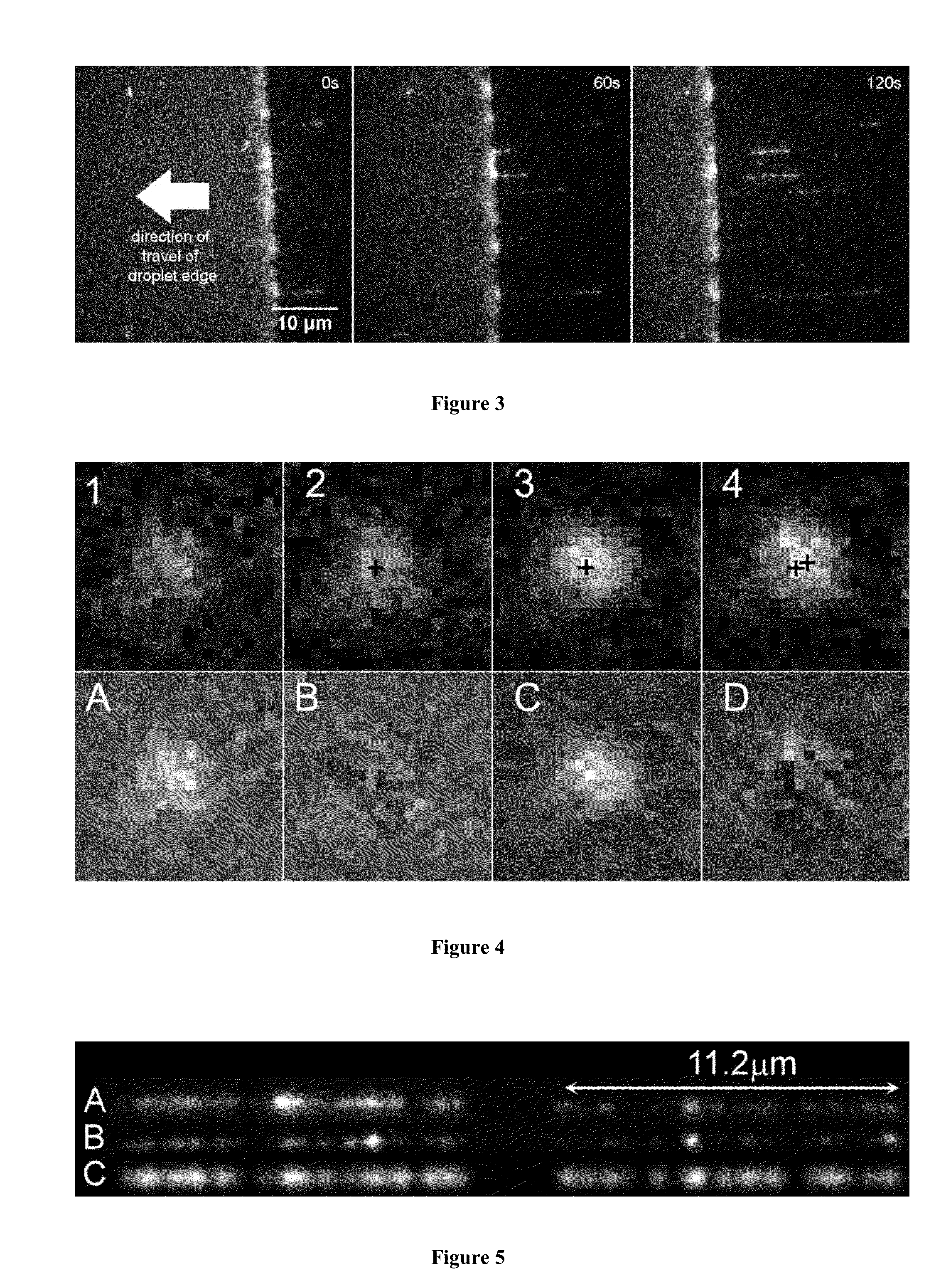
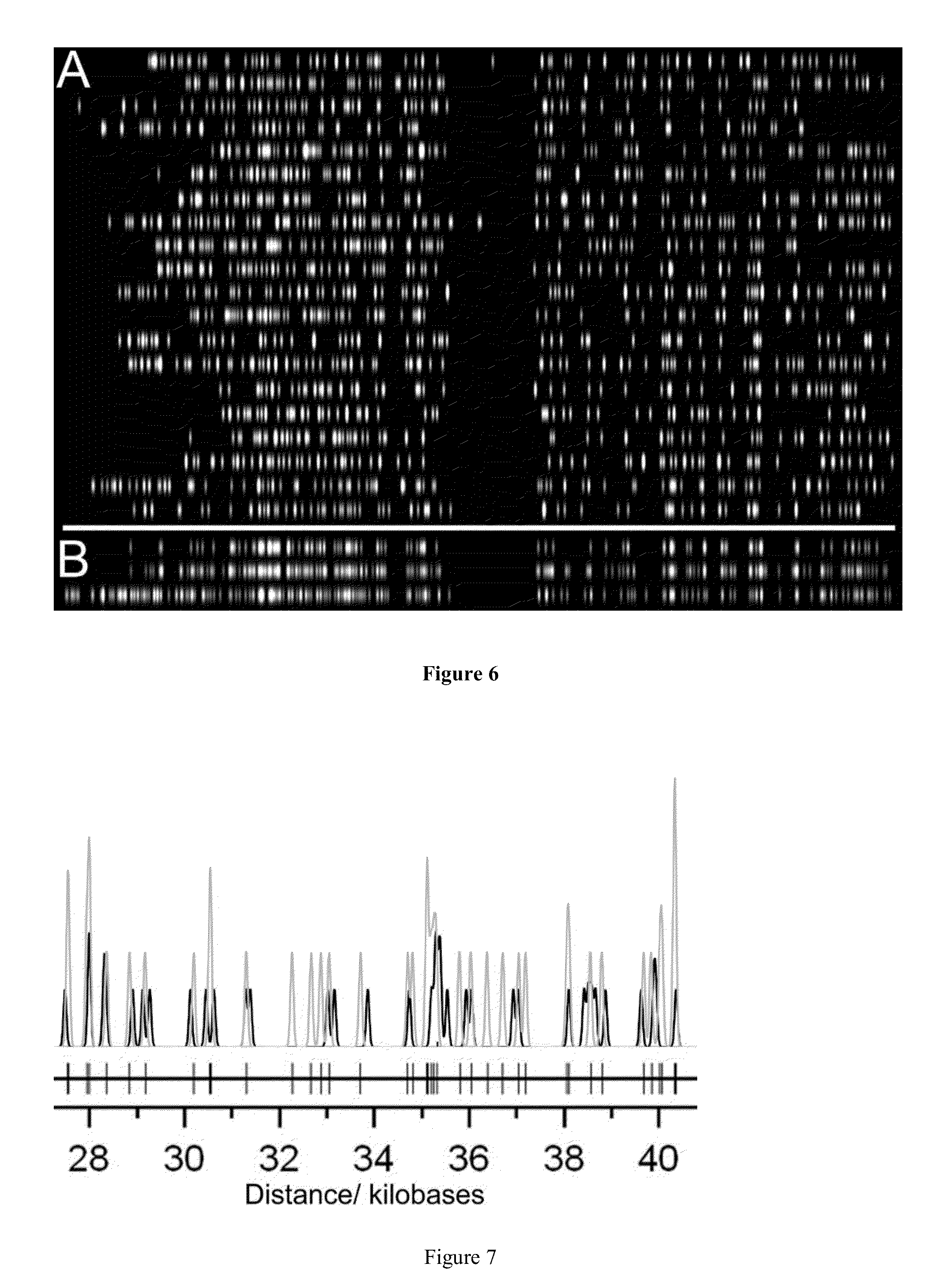
DNA Combing
[0063]Droplets of 1 uL volume, containing approximately 0.2 ug / ml of the labeled lambda DNA in 50 mM MES buffer at pH5.7 were deposited onto the PMMA-coated coverslips. The coverslips were placed on a heat block at 60° C. and droplets allowed to evaporate for 30 min.
PUM
| Property | Measurement | Unit |
|---|---|---|
| pH | aaaaa | aaaaa |
| pH | aaaaa | aaaaa |
| pH | aaaaa | aaaaa |
Abstract
Description
Claims
Application Information