Decellularized biomaterial from non-mammalian tissue

a biomaterial and non-mammalian technology, applied in the direction of drugs, peptide/protein ingredients, prosthesis, etc., can solve the problems of reducing the structural integrity of tissue, and reducing the antigenicity of biomaterials.

Active Publication Date: 2015-12-10
NEXTGEN BIOLOGICS INC
View PDF1 Cites 15 Cited by
  • Summary
  • Abstract
  • Description
  • Claims
  • Application Information

AI Technical Summary

Benefits of technology

[0020]Of particular importance for transplantation purposes, more generally, is the immunologically privileged state of the human neonate (e.g., fetal dermis) and the AM, a state mirrored by axolotl ECM, whereby immogenicity is rarely manifested. By virtue of the reduced immune response and the generally decreased inflammatory response as compared to adult humans, neonatal and axolotl skin healing alike are not characterized by accelerated tissue resorption, as is observed in adult human wound healing. Rather, the growth factor profile, enzymatic activity, structural composition and immunomodulating effect of urodele and neonate tissues alike favor an appropriately staged removal of structural scaffolding and tissue growth into the resulting negative void space, in addition to enhanced re-epithelialization, during wound healing. This results in an optimal wound healing environment and process. Also, the high concentration of antimicrobial peptides present in AM and urodele tissue further contributes to the favorable environment and enhanced re-epithelialization observed during wound healing.
[0022]In accordance with the invention, therefore, a biological scaffold biomaterial is provided that is the product of a process comprising (A) obtaining a tissue sample from a urodele, where the tissue comprises ECM, inclusive of the basement membrane, and (B) subjecting the tissue sample to a decellularization process that maintains the structural and functional integrity of the extracellular matrix, by virtue of retaining its fibrous and non-fibrous proteins, glycoaminoglycans (GAGs) and proteoglycans, while removing sufficient cellular components of the sample to reduce or eliminate antigenicity and immunogenicity for xenograft purposes. Also provided is methodology for using the urodele-derived biomaterial to enhance restoration of skin homeostasis, to reduce the severity, duration and associated damage caused by post-surgical inflammation, and to promote progression of natural healing and regeneration processes. In addition, biomaterial of the invention promotes the formation of remodeled tissue that is comparable in quality, function and compliance to undamaged human tissue.Decellularization
[0023]The biomaterial of the invention is produced by decellularizing a tissue sample obtained from a urodele. The primary constituent of the resulting urodele biomaterial is ECM, possibly with devitalized epithelial cells, which can retain moisture and otherwise protect the wound-healing environment.
[0027]According to one aspect of the invention, a biomaterial is provided. The material is produced by the process that includes (A) obtaining a tissue sample from a urodele, which tissue sample comprises extracellular matrix, and (B) decellularizing the sample to retain structural and functional integrity while removing sufficient cellular components of the sample to reduce or eliminate antigenicity of the biomaterial as a xenograft. In some embodiments, decellularizing comprises subjecting said tissue sample to an alkaline treatment. In embodiments, the process can further comprise subjecting said sample to sterilization. In embodiments, the process can further comprise devitalizing cells.
[0028]According to one aspect of the invention, a tissue graft is provided. The graft includes extracellular matrix components derived from a urodele. In embodiments, the extracellular matrix components are substantially free of components that induce an immune response when implanted as a xenograft. In embodiments, the extracellular matrix components are non-toxic.
[0038]According to one aspect of the invention, a method of preparing a biologic material is provided. The method involves (A) obtaining a tissue sample from a urodele, which tissue sample comprises extracellular matrix, and (B) decellularizing the sample to remove sufficient cellular components of the sample to reduce or eliminate antigenicity of the biomaterial as a xenograft. In embodiments, the method can further involve performing the decellularization in a manner to retain structural and functional integrity of the ECM sufficient to permit the ECM to be useful as a matrix upon and within which cells can grow. In any of the embodiments, the method can further involve homogenizing the ECM to form a particulate or powder. In some embodiments, the method can further involve reconstituting the powder as a gel. In any of the embodiments, the method can further involve sterilizing the ECM. In any of the embodiments, the method can further involve attaching the ECM to an agent xenogenic to a Urodele.

Problems solved by technology

In this regard, wound treatment and skin repair are areas of predominant focus, as the loss of skin integrity due to illness or injury can lead to chronic, life threatening complications.
The ability to restore biomechanical function of damaged tissue presents a true challenge.
The use of conventional tissue scaffold products is not without drawbacks, however.
Tissue harvesting from human donors can produce undesirable consequences such as donor site morbidity or infection associated with removal of skin for donation.
Disease transmission risk and intersample variation are additional drawbacks associated with biological scaffold products.
In addition, it may be difficult to obtain sufficient tissue components necessary to cover large areas of damaged tissue.
Furthermore, conventional biological and synthetic materials can be costly, not effective in many instances, and limited in availability.

Method used


Image

  • Decellularized biomaterial from non-mammalian tissue
  • Decellularized biomaterial from non-mammalian tissue
  • Decellularized biomaterial from non-mammalian tissue

Examples


example 1

Processing Axolotl Dermis

[0098]Axolotl dermis samples can be decellularized by preparing excised samples from healthy or healing axolotl dermal tissue and then subjecting the samples to hypo / hyperosmotic soaks for cell lysis, solvent dehydration, and oven drying. Specific processing of these grafts includes storage in 15-26% NaCl, multiple hypo / hyperosmotic soaks (utilizing NaCl solutions and water), and then solvent dehydration using ethanol, and then evaporation of the solvent either with air drying or oven drying at 37° C.

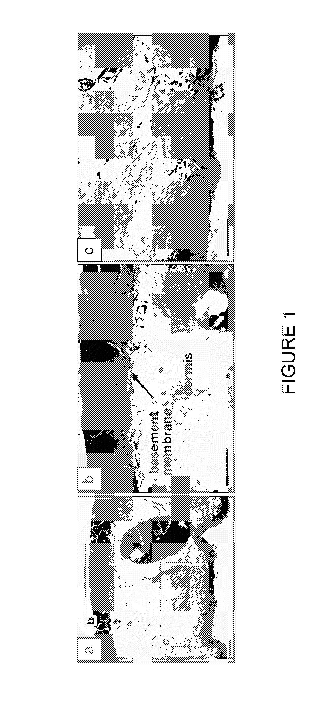
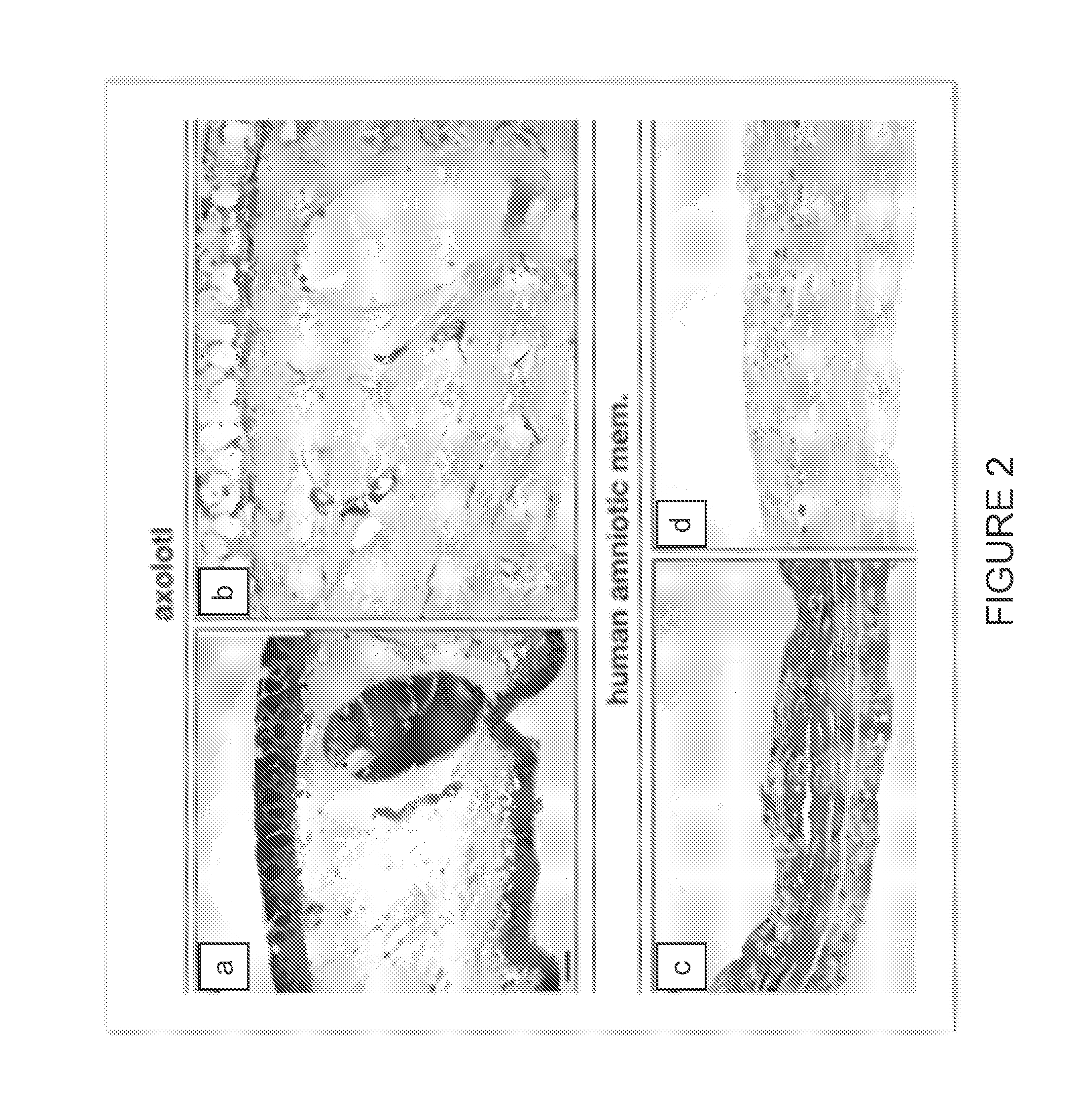
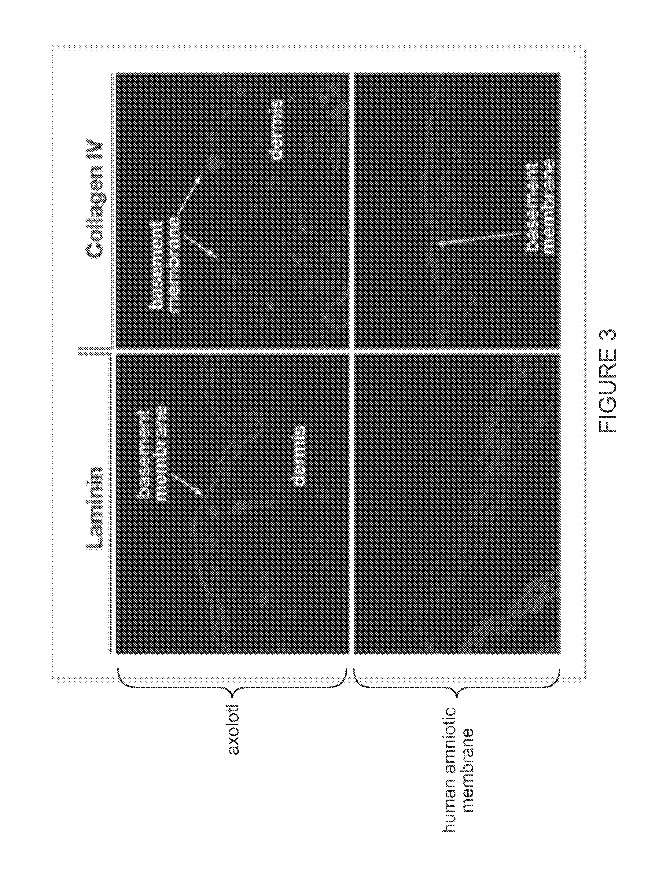
[0099]Histological examination of native axolotl dermal tissue was performed to identify the presence of the notable ECM elements, such as the basement membrane. See FIG. 1 and FIG. 4. Comparative histological and immunohistochemical analysis of native axolotl dermal tissue and human amniotic membrane was performed to compare the ECM structure and constituents, and to assess relative concentration and distribution of critical constituents. See FIG. 2, FIG. 3, an...

example 2

Splitting and Lamination of Acellular Dehydrated Axolotl Dermis

[0100]Decellularized dehydrated axolotl dermis can be split, via a mechanical splitter, to isolate heterogeneous matrix into homogenous sections. Isolated sections of desired thickness then can be rehydrated and lyophilized to obtain multilayered laminate structures of desired orientation with facial surface features. More specifically, dual-sided basement membrane structure, with interior open porous matrix obtained from the reticular dermis region of the dermal matrix, can be constructed to obtain desired facial surface properties. Alternatively, isolated native section can be used in native form for desired clinical outcome. For example, open porous homogenous matrix of the reticular dermis can be used to obtain augmentation of soft tissue structures.

[0101]A laminated custom construct with sulfated gags on both facial surface and collagen IV and laminin could be obtained for desirable dual-surface, anti-adhesion and a...

example 3

Preparation of Solubilized Acellular Dehydrated Axolotl Dermis, Pericardium, Fascia Lata, Periosteum, Peritoneum, or Dura Mater

[0102]Decellularized dehydrated axolotl native or isolated section of acellular urodele connectivue tissye matrix can be prepared by sectioning decellularized soft tissue structures into 1 cm2 sections and homogenizing the sections in a Warring blender (˜100 grams of tissue) in aqueous 1M glacier acetic acid for 30-60 seconds. Preparation of sponge can be obtained by the addition of varying volumes of water followed then neutralization and lypoholization of the slurry in a mold of desired geometric shape. The resultant porosity will correlate to the volume of water added to the matrix. Additionally, a selected range of bioactive extracts can be added to the slurry prior to neutralization, including particulated or small protein constituents extracted from digested human or urodele mineralized and nonmineralized connective tissues, such as demineralized bone ...


PUM

PropertyMeasurementUnit
dry weightaaaaaaaaaa
sizeaaaaaaaaaa
particle size distributionaaaaaaaaaa

Abstract

The growth factor profile, connective tissue matrix constituents, and immunoprivileged status of urodele extracellular matrix (ECM) and accompanying cutaneous tissue, plus the presence of antimicrobial peptides there, render urodele-derived tissue an ideal source for biological scaffolds for xenotransplantation. In particular, a biological scaffold biomaterial can be obtained by a process that entails (A) obtaining a tissue sample from a urodele, where the tissue comprises ECM, inclusive of the basement membrane, and (B) subjecting the tissue sample to a decellularization process that maintains the structural and functional integrity of the extracellular matrix, by virtue of retaining its fibrous and non-fibrous proteins, glycoaminoglycans (GAGs) and proteoglycans, while removing sufficient cellular components of the sample to reduce or eliminate antigenicity and immunogenicity for xenograft purposes. The resultant urodele-derived biomaterial can be used to enhance restoration of skin homeostasis, to reduce the severity, duration and associated damage caused by post-surgical inflammation, and to promote progression of natural healing and regeneration processes. In addition, the biomaterial promotes the formation of remodeled tissue that is comparable in quality, function, and compliance to undamaged human tissue.

Description

CROSS-REFERENCE TO RELATED APPLICATIONS[0001]This application claims the benefit under 35 USC §119(e) to U.S. provisional application 61 / 750,555, filed Jan. 9, 2013, the entire contents of which are incorporated herein by reference in their entirety.BACKGROUND OF THE INVENTION[0002]Tissue engineering efforts are ongoing to produce methods and materials for replacing biological functions, typically repairing or replacing whole tissues or portions thereof. In this regard, wound treatment and skin repair are areas of predominant focus, as the loss of skin integrity due to illness or injury can lead to chronic, life threatening complications.[0003]Wound healing involves complex interactions between cells, growth factors, and extracellular matrix (ECM) components to reconstitute tissue following injury. The wound healing process in adult mammalian tissue has been well characterized and can be broken down into three stages—inflammation, proliferation, and remodeling.[0004]Typically, in re...

Claims


Application Information

Patent Timeline
Patent Type & Authority Applications(United States)
IPC IPC(8): A61L27/36A61K9/70A61L27/58A61K35/12A61L27/54A61L27/38A61K38/17A61K35/65A61L27/60
CPCA61L27/3687A61L2300/252A61K9/70A61L27/58A61L27/60A61L27/54A61L27/3633A61L27/362A61L27/3691A61L27/38A61K38/1703A61K35/12A61L27/3625A61L27/3604A61L2430/40A61L2430/34A61L2300/64A61L2300/606A61L2300/604A61L2300/412A61K35/65A61L27/52C12N5/0625C12N2533/90C12N2533/92A61P17/02A61P41/00A61P43/00
Inventor EARLY, RYANNE
Owner NEXTGEN BIOLOGICS INC
Why Eureka
Try Eureka