Revision with a conventional hydraulic artificial urinary
sphincter poses greater risk for damaging small nerves and vessels than does the original procedure, tandem
cuff arrangements and combined placement with an erectile
prosthesis, for example, unlikely to prove satisfactory over a period of more than a few years.
By comparison, the hydraulic device appropriates much more space, is positioned in more intricate
anatomy, and is not amenable to redundancy.
While more recent artificial urinary sphincters are much improved in this regard, compressive degradation of the
urethra remains the single most problematic
sequela, often necessitating revision that requires remedial
surgery as well as the removal and replacement or resituation of the
cuff (see, for example, Rahman, N. U., Minor, T. X., Deng, D., and Lue, T. F. 2005.
The experience of Foley and subsequent developers of
artificial sphincters positioned along the bulbar
urethra made it clear that unless an
artificial sphincter, in this case urinary, could be positioned to encircle the native
sphincter with its epithelial lining adapted to withstand forcible closure, sustained compression would eventually, probably inevitably, result in urethral
atrophy and
erosion.
Synthetic or surgical, diligent maintenance notwithstanding, the treatment almost always gives rise to complications of
irritation if not infection that add annoyance to what has been an unwanted focus of attention in the first place, significantly degrading the
quality of life.
Surgical reconstruction is substantially limited to urinary and fecal waste diversion and unsuited to the creation of a permanent passageway from the
body surface into the medulla or medulla or
parenchyma of an internal organ such as the
spleen,
gall bladder,
kidney,
prostate,
uterus, and so on.
The surgical construction of a passageway as in a urostomy, which extends trauma and the risk of complications to uninvolved tissue, has been limited to relatively few organs and tissues.
The diversion of the internal thoracic (previously, the internal mammary), for example, can result in grave consequences (see, for example, Bintoudi, A., Malkotsi, T., Goutsaridou, F., Emmanoullidou, M., and Tsitouridis, I.
Broadly, the appropriation of any vessel of larger
caliber is likely to result in adverse consequences for its supply territory (see, for example, Zhu, Y. Y., Hayward, P. A., Hadinata, I. E., Matalanis, G., Buxton, B. F., Stewart, A. G., and Hare, D. L. 2013.
The central need for and the lack of safe and secure tissue perforating wound connectors is an obstruction to the implementation of such systems.
However, regardless of application thus, such means overcome the need to detain an otherwise
ambulatory patient in the clinic merely because a
catheter, infusion line, the tape securing it, or the solution used to promote antisepsis require frequent examination and changing or because more radical
surgery necessitates more time to heal.
The nondurability and in particular, the impermanence of a catheteric suprapubic cystostomy or a nephrostomy is due not only to
contamination or the accretion of crystalline matter but to
irritation and the
risk of infection at the body wall and at the renal and
urinary bladder or urocystic puncture wounds.
Here tissue irritation at the tissue interface is the initial factor limiting the duration of placement.
While avoiding the risk of rejection and the need for lifelong immunosuppressive medication,
surgery that harvests and reconfigures healthy
autologous tissue to support a different function, one for which the tissue is not adapted and ill suited, extends trauma and involves a loss of normal function at the donor site.
By contrast, urinary diversion using currently available synthetic, or alloplastic components, such as a catheter and hollow needle in a patient with a long term if not life long need for urinary diversion avoids significant trauma and the appropriation of
healthy tissue but does so at the expense of durability, complications that develop over time, necessitating periodic catheter replacement.
Due to the susceptibility to complications at the integumentary and internal structure entry wounds of conventional means such as
intravenous needles and
indwelling catheters for establishing connections inside the body, these are unsuited to use in a long term or life long automatic
ambulatory prosthetic disorder
response system.
Congenital deformities of the
gastrointestinal tract are often the result of
ischemia during development, and when the loss due to accidental trauma, mesentery will have been destroyed as well.
However, existing means for accomplishing urinary diversion which use
synthetic materials cannot be placed and then left unattended.
It has already been stated that the gut is segment by segment differentially adapted for the absorption of different nutrients and its lining unsuited for contact with much less the regular transport of
urine.
Thus, aside from the risk to both donor and recipient systems of much additional
dissection and prolonged
anesthesia to cover not one but two procedures, even if perfectly executed for catheters to be inserted through a
stoma, an ileal conduit or gut-derived neobladder will remain vulnerable to sequelae from frequent contact with
urine.
This will serve for urinary diversion and to pass through a calculus retrieval basket,
forceps, or scope, but does not allow positioning the distal tip of the catheter within the cortex or medulla.
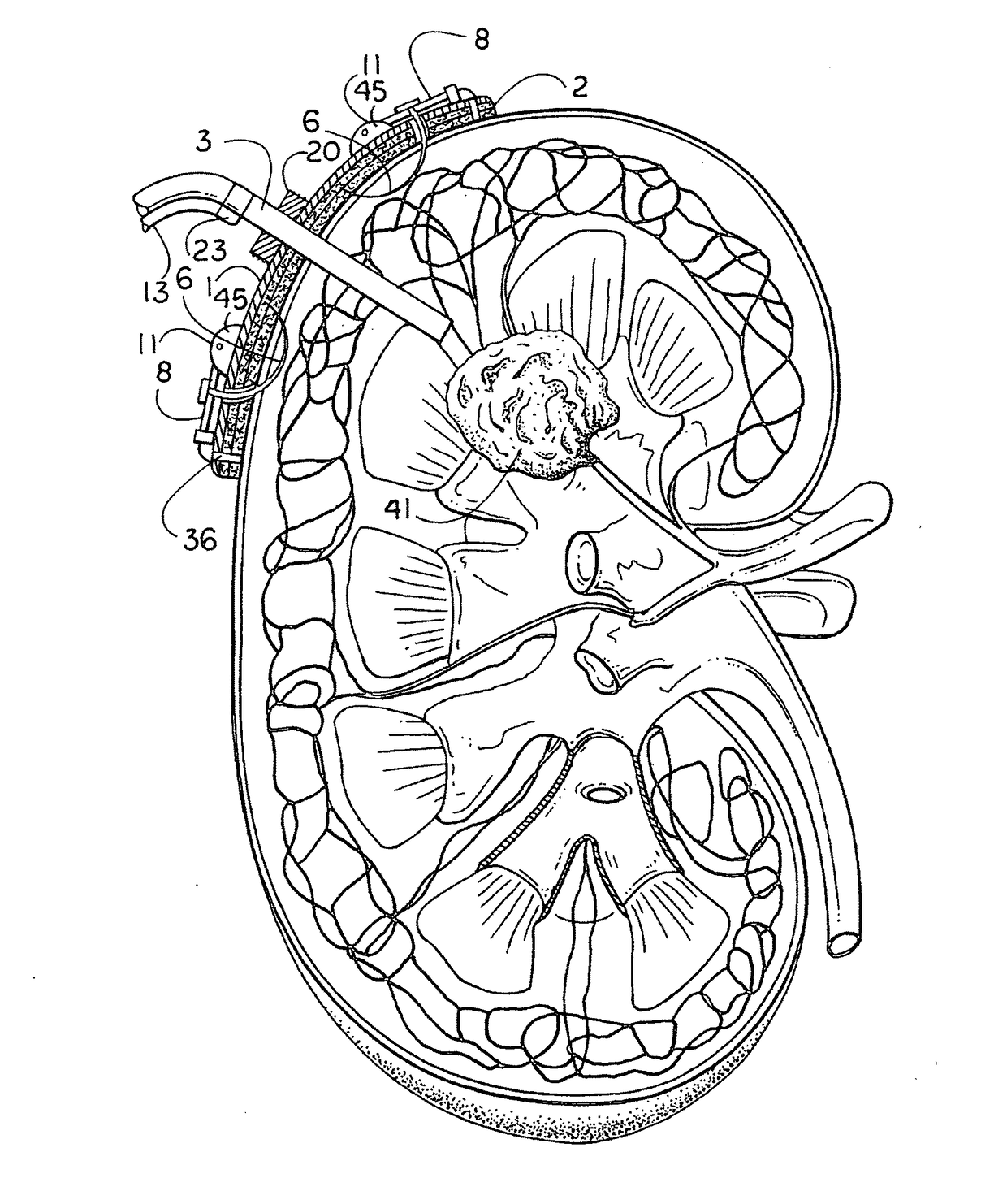
Conventional nephrostomy catheters can deliver drugs to the
pelvis or a calyx but are unintended for and incapable of the directly targeted delivery of drugs to a
lesion or region within the renal medulla and / or cortex.
None are connected at the
body surface or at the
point of entry into the
kidney as would allow the device to remain in place more than 4 weeks, and no surgical reconstruction can yield a permanent passage that would allow the functionality sought herein.
However, in a conventional
gastrostomy, the lack of a junction securely fastened to the tissue surrounding the small gastric entry wound risks distal retention-
balloon migration and gastric obstruction, which can lead to
vomiting, hematemesis, and aspiration pneumonia (see, for example, Than, M. M., Witherspoon, J., Tudor, G., and Saklani, A.
The reason is that the
balloon prevents the tube from pulling out through the gastric entry wound, but unattached and not firmly connected to the tissue surrounding the wound, cannot prevent distal migration or progressively more irritating movement between the sides of the catheter and the wound.
In some instances, adhesion will be jeopardized because of a potential for changes in
hardness of the subjacent tissue due to the
disease treated itself or an intercurrent degenerative process.
Otherwise, adhesives are broken down hydrolytically and enzymatically limiting the time these remain effective.


