Devices, treatments and methods to restore tissue elastic recoil

a tissue elastic and recoiling technology, applied in the field of tissues elastic recoiling, can solve the problems of difficulty in talking or basic breathing, reducing oxygen and carbon dioxide gas, and affecting lung function, so as to restore lung elastic recoil and restore lung tissue tension

Active Publication Date: 2019-10-10
FREE FLOW MEDICAL INC
View PDF10 Cites 4 Cited by
  • Summary
  • Abstract
  • Description
  • Claims
  • Application Information

AI Technical Summary

Benefits of technology

[0151]anchoring the pulmonary treatment device so as to assist in maintaining the torqued configuration.
[0670]In another aspect of the present invention, a lung airway straightening system is provided for enhancing breathing efficiency of a patient with an airway, the system comprising: an implantable device configured to impart tension on lung tissue, the implantable device including a proximal stabilizing end, and a distal tissue gathering end with a transition section connecting the two ends that includes at least one helical loop structure with a start and an end to the helical loop; furthermore, the device has a first delivery configuration and a second deployed configuration, the first configuration of the implantable device corresponds to a deliverable condition and a finite distance between the start and end of at least one of the helical loop structures, the second configuration is configured so the distance between the start and end of the same helical loop structures may be elastically strained longer to apply tension to lung tissue; wherein the implant comprises a spring element and wherein the implant is constrained to the delivery configuration during delivery and wherein the implant is configured to naturally recover from the constrained delivery configuration to the deployed configuration during deployment.

Problems solved by technology

Such symptoms may start as a minor impediment to daily life, but they often lead to difficulty in talking or basic breathing.
COPD reduces oxygen and carbon dioxide gas exchange which leads to circulatory problems, such as low oxygen levels in the blood, brain and heart muscles.
This negatively affects mental alertness and contributes to a very rapid heartbeat, due to increased strain on the heart.
However, about 24 million more people may have the disease and not know it.
Both smoke that the smoker inhales (through the filter) and the smoke from the burning end are toxic.
Nicotine also causes increased stickiness of blood platelets, which increases the risk of blood clotting.
Carbon monoxide combines irreversibly with hemoglobin so that oxygen cannot bind effectively.
This causes a strain on the heart muscle because it must pump more to provide the same amount of oxygen.
However, the inhalation of smoke ultimately destroys this elasticity and lung tissue itself.
The effect of tobacco smoke on lung elastin is extremely complicated, affecting many facets of connective tissue metabolism.
This has been shown by selective chemical oxidation to yield a relatively ineffective inhibitor that associates with elastase some 2000 times more slowly than the native protein.
This results in oxidant damage to lung cells and cellular components such as lipids, cofactors, and nucleic acids.
Endogenous antioxidant systems within the lung, such as ceruloplasmin, vitamin C, or methionine sulphoxide-peptide reductase, are adversely affected by cigarette smoke, lowering the lung's defense against oxidants.
The elastin maturation process is impaired by cigarette smoke.
Such damage affects the walls between the alveolar sacs.
This makes it more difficult for the capillaries to absorb enough oxygen and for the body to expel carbon dioxide, making it progressively harder to breathe.
In addition, the air sacs lose their elasticity making it harder to recoil and expel air.
The walls of the airways thicken and become swollen while making more mucus than normal which can clog the airways that lead to the air sacs.
Another common COPD symptom is air trapping which causes breathing disfunction as well as lobar and lung hyperinflation.
Both types of air trapping causes 1) lung gas congestion, preventing new oxygen from being inspired, 2) retainment of CO2 in the lung and blood stream (hypoxemia) and 3) crushing of better functioning lobes making them incapable of inspiration or expiration.
Ultimately, enzymes destroy and eliminate airways and alveoli tissue.
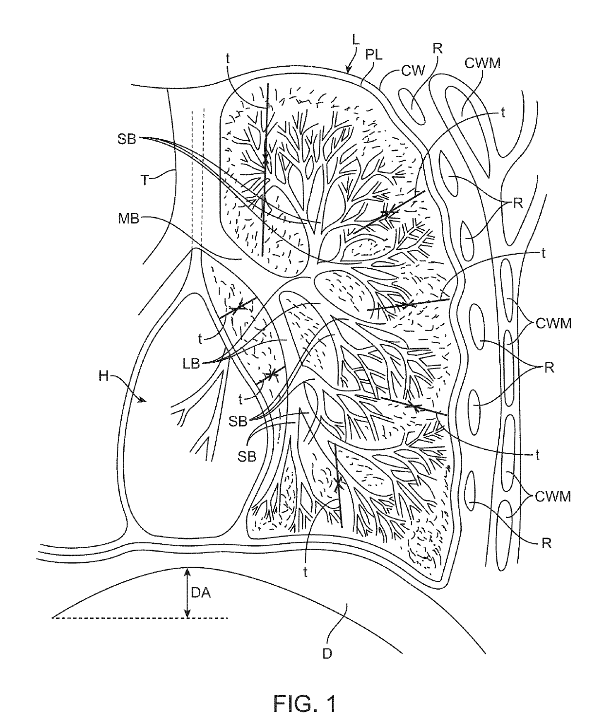
If they rupture, they allow air to escape into the pleural space between the lung and chest wall, which is normally holding the lungs expanded and pinned to the chest wall with vacuum, resulting in a spontaneous pneumothorax or collapse of the lung.
However, this surgery presents patients with high risk of surgery related morbidity and mortality.
Patients who already have distressed breathing due to the disease are further stressed with severe orthopedic trauma due to a sternotomy, which presents difficulty in reviving these patients from general anesthesia.
This reduces lung capacity that patients need to exchange gas.
LVRS is also not effective for homogenous disease, which is the type that most COPD patients suffer from.
Homogenous patients need therapy because they suffer from an insufficient lung capacity to exchange gas.
Removing more lung tissue only reduces their capacity.
Therefore, the surgery actually degrades these patient's ability to breathe.
Thus, the valves may actually degrade these patient's ability to breathe.
Another limitation with the valves is the fact that approximately 80% of patients present with additional flow paths that lead into the lobe in addition to the major airway tree that is typically shown in anatomy texts.
The valves are designed to block flow in airways but in the majority of patients, total blockage or perfect pneumatic isolation can never be achieved and the lobe never collapses.
Other times, the inflammation can cause long term morbidity and even mortality.
Other serious pulmonary side effects within 6 months after the procedure include repetitive COPD exacerbations, pneumonia, bronchitis, and hemoptysis.
However, such treatment again ultimately suffers from some of the same limitations as LVRS.
In particular, lung sealants destroy lung tissue and reduce lung capacity so they are not effective for homogenous disease, which is the type that most COPD patients suffer from.
Thus, these techniques actually degrade these patient's ability to breathe.
However, such bending and folding of the airways increases resistance to gas flow which blocks the airways from flowing efficiently to exchange gas.
Thus, there is limited inspiration and expiration in those regions which reduces the patient's capacity to breathe.
In addition, the coil design and dimensions provide a very small contact area which produces high pressure and compressive stress on the lung tissue.
This potentially allows for a kind of “cheese wire” cutting effect that limits the effective time that a treatment remains effective, even if initial results are positive.
However, due to the nature of the disease and the enzymatic destruction in COPD patients, substantial, thick walled airways are nearly absent beyond the 4th airway generation in patients with the requisite degree of disease that would require this type of intervention.
The typical disease related tissue destruction leaves only fragile segments of thin tissue in areas in contact with the coils and this can only accelerate the “cheese wire” effect which may reduce the potential for treatment success substantially.
Since the patient's entire cardiac pumping capacity is routed through the lungs and these vessels, the use of such coils on these airways would present the patient with extreme risk.
Devices such as the endobronchial coils and endobronchial valves that are mechanical structures suffer from fatigue related failure due to the high number of breathing cycles that these products endure and the nature of the flexure that lung airways present on these devices.
In many cases, device failure occurs where metallic or stiff biocompatible materials are placed in the lungs where coughing presents the devices with repeated high force flexure and airway collapse.
Another cause for device failure is tissue irritation and granular buildup of airway wall tissue and the formation of bacterial colonies that are commonly found on implanted polymers in the lung.
Most devices that have been previously proposed to treat COPD in the past have included one or more design flaws to cause granulation tissue formations or bacterial colonization's which are nearly impossible to remove or otherwise treat.
Compression of lung tissue can compress and block blood vessels leading to tissue necrosis and cell death, which in turn causes chronic air leaks and eventual lung collapse due to breaching of the vacuum seal between the lungs and chest wall.

Method used


Image

  • Devices, treatments and methods to restore tissue elastic recoil
  • Devices, treatments and methods to restore tissue elastic recoil
  • Devices, treatments and methods to restore tissue elastic recoil

Examples


Embodiment Construction

[0782]Specific embodiments of the disclosed device, delivery system, and method will now be described with reference to the drawings. Nothing in this detailed description is intended to imply that any particular component, feature, or step is essential to the invention.

Anatomical Changes in COPD

[0783]FIG. 1 illustrates a healthy lung L of a patient. As shown, the lung L includes a tracheobronchial tree which is the anatomical and functional segment of the respiratory system that conducts air from the larger upper airways to the lung parenchyma. It is comprised of the trachea T and various intrapulmonary airways, including the bronchi, bronchioles and terminal bronchioles. The trachea and bronchi have cartilaginous walls which makes them thick, fibrous and this allows them to maintain patency during breathing. Bronchi undergo multiple divisions and eventually give rise to the terminal bronchioles, which by definition, lack cartilage. The most distal respiratory bronchioles and alveol...


PUM


Abstract

Pulmonary treatment devices, systems and methods of use are provided which take into account the vast tissue damage of advanced COPD sufferers and provide treatments designed specifically to treat the particularly compromised lung tissues that are present in these patients. These treatments reduce trapped air volume, tension lung tissue and enhance lung elastic recoil. A variety of embodiments are provided, including pulmonary treatment devices that move portions of lung tissue around a rotational axis into a torqued configuration, anchoring such tissue in place for improved breathing mechanics.

Description

CROSS-REFERENCE TO RELATED APPLICATIONS[0001]This application is a continuation of PCT Application No. PCT / US18 / 67160, filed Dec. 21, 2018, (Attorney Docket No. 52086-703601), which claims priority to Provisional No. 62 / 609,761, filed Dec. 22, 2017, (Attorney Docket No. 52086-703.101); Provisional No. 62 / 651,573, filed Apr. 2, 2018, (Attorney Docket No. 52086-703.102); Provisional No. 62 / 714,411, filed Aug. 3, 2018, (Attorney Docket No. 52086-703.103); Provisional No. 62 / 720,004, filed Aug. 20, 2018, (Attorney Docket No. 52086-703.104); and 62 / 749,005, filed Oct. 22, 2018, (Attorney Docket No. 52086-703.105), the entire contents of which are incorporated herein by reference.BACKGROUND OF THE INVENTION[0002]Chronic obstructive pulmonary disease (COPD) is a common progressive, debilitating lung disease that is often fatal. COPD patients are diagnosed with either emphysema, chronic bronchitis or more commonly, a combination of both. The symptoms of COPD include a persistent cough, part...

Claims


Application Information

Patent Timeline
Patent Type & Authority Applications(United States)
IPC IPC(8): A61B17/12A61B17/00
CPCA61B17/12136A61B2017/00557A61B17/12104A61B2017/00539A61B2017/00867A61B2017/00809A61B17/0057A61B2017/00615A61B1/00085A61B1/01A61B1/267A61F2/848A61F2/88A61F2/95A61M16/0406A61B1/00148A61M16/00A61F2/04A61F2/02A61B17/12036A61B17/1214A61B2017/1205
Inventor MATHIS, MARK L.LAU, MICHAEL W.MITZ, KEVINBRAXTAN, RYAN
Owner FREE FLOW MEDICAL INC
Why Eureka
Try Eureka